J Yeungnam Med Sci.
2023 Oct;40(4):321-327. 10.12701/jyms.2023.00976.
State-of-the-art update for diagnosing diabetic foot osteomyelitis: a narrative review
- Affiliations
-
- 1Department of Orthopaedic Surgery, Yeungnam University Hospital, Daegu, Korea
- 2Department of Orthopaedic Surgery, Yeungnam University College of Medicine, Daegu, Korea
- KMID: 2547354
- DOI: http://doi.org/10.12701/jyms.2023.00976
Abstract
- Recently, the International Working Group on the Diabetic Foot and the Infectious Diseases Society of America divided diabetic foot disease into diabetic foot infection (DFI) and diabetic foot osteomyelitis (DFO). DFI is usually diagnosed clinically, while numerous methods exist to diagnose DFO. In this narrative review, the authors aim to summarize the updated data on the diagnosis of DFO. An extensive literature search using “diabetic foot [MeSH]” and “osteomyelitis [MeSH]” or “diagnosis” was performed using PubMed and Google Scholar in July 2023. The possibility of DFO is based on inflammatory clinical signs, including the probe-to-bone (PTB) test. Elevated inflammatory biochemical markers, especially erythrocyte sedimentation rate, are beneficial. Distinguishing abnormal findings of plain radiographs is also a first-line approach. Moreover, sophisticated modalities, including magnetic resonance imaging and nuclear medicine imaging, are helpful if doubt remains after a first-line diagnosis. Transcutaneous bone biopsy, which does not pass through the wound, is necessary to avoid contaminating the sample. This review focuses on the current diagnostic techniques for DFOs with an emphasis on the updates. To obtain the correct therapeutic results, selecting a proper option is necessary. Based on these numerous diagnosis modalities and indications, the proper choice of diagnostic tool can have favorable treatment outcomes.
Keyword
Figure
-
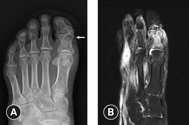
Fig. 1. Diabetic foot osteomyelitis at left first proximal phalanx. (A) Plain X-ray shows an erosive, destructive lesion on the proximal phalanx of the first toe (arrow). (B) A lesion on the plain X-ray does not appear serious, but a high signal intensity is marked on the T2 fat suppressed magnetic resonance imaging.

Fig. 2. Osteomyelitis at the right second metatarsal bone. Positron emission tomography/computed tomography image illustrates diffusely increased fluorodeoxyglucose accumulation along the bone.
Cited by 1 articles
-
Unveiling the challenges of diabetic foot infections: diagnosis, pathogenesis, treatment, and rehabilitation
Chul Hyun Park
J Yeungnam Med Sci. 2023;40(4):319-320. doi: 10.12701/jyms.2023.01011.
Reference
-
References
1. Carracher AM, Marathe PH, Close KL. International diabetes federation 2017. J Diabetes. 2018; 10:353–6.
Article2. Woo I, Park J, Seok H, Kim TG, Moon JS, Chung SM, et al. The fate of antibiotic impregnated cement space in treatment for forefoot osteomyelitis. J Clin Med. 2022; 11:1976.
Article3. Pop-Busui R, Boulton AJ, Feldman EL, Bril V, Freeman R, Malik RA, et al. Diabetic neuropathy: a position statement by the American Diabetes Association. Diabetes Care. 2017; 40:136–54.
Article4. Lavery LA, Peters EJ, Armstrong DG, Wendel CS, Murdoch DP, Lipsky BA. Risk factors for developing osteomyelitis in patients with diabetic foot wounds. Diabetes Res Clin Pract. 2009; 83:347–52.
Article5. Sohrabi K, Belczyk R. Surgical treatment of diabetic foot and ankle osteomyelitis. Clin Podiatr Med Surg. 2022; 39:307–19.
Article6. Nteleki B, Njokweni M. Want to avoid DFUs? A multidisciplinary team approach works best. J Wound Care. 2015; 24(5 Suppl 2):8–14.
Article7. Berendt AR, Peters EJ, Bakker K, Embil JM, Eneroth M, Hinchliffe RJ, et al. Diabetic foot osteomyelitis: a progress report on diagnosis and a systematic review of treatment. Diabetes Metab Res Rev. 2008; 24(Suppl 1):S145–61.
Article8. Lipsky BA, Aragón-Sánchez J, Diggle M, Embil J, Kono S, Lavery L, et al. IWGDF guidance on the diagnosis and management of foot infections in persons with diabetes. Diabetes Metab Res Rev. 2016; 32(Suppl 1):45–74.
Article9. Lipsky BA, Berendt AR, Deery HG, Embil JM, Joseph WS, Karchmer AW, et al. Diagnosis and treatment of diabetic foot infections. Plast Reconstr Surg. 2006; 117(7 Suppl):212S–238S.
Article10. Lipsky BA, Senneville É, Abbas ZG, Aragón-Sánchez J, Diggle M, Embil JM, et al. Guidelines on the diagnosis and treatment of foot infection in persons with diabetes (IWGDF 2019 update). Diabetes Metab Res Rev. 2020; 36(Suppl 1):e3280.
Article11. Schaper NC, van Netten JJ, Apelqvist J, Bus SA, Fitridge R, Game F, et al. Practical guidelines on the prevention and management of diabetes-related foot disease (IWGDF 2023 update). Diabetes Metab Res Rev. 2023 May 27 [Epub]. https://doi.org/10.1002/dmrr.3657.
Article12. Boulton AJ, Armstrong DG, Hardman MJ, Malone M, Embil JM, Attinger CE, et al. Diagnosis and management of diabetic foot infections. Arlington (VA): American Diabetes Association;2020.13. Aragón-Sánchez J, Lipsky BA. Modern management of diabetic foot osteomyelitis: the when, how and why of conservative approaches. Expert Rev Anti Infect Ther. 2018; 16:35–50.
Article14. Giurato L, Meloni M, Izzo V, Uccioli L. Osteomyelitis in diabetic foot: a comprehensive overview. World J Diabetes. 2017; 8:135–42.
Article15. Senneville EM, Lipsky BA, van Asten SA, Peters EJ. Diagnosing diabetic foot osteomyelitis. Diabetes Metab Res Rev. 2020; 36(Suppl 1):e3250.
Article16. Edelson GW, Armstrong DG, Lavery LA, Caicco G. The acutely infected diabetic foot is not adequately evaluated in an inpatient setting. Arch Intern Med. 1996; 156:2373–8.
Article17. Grayson ML, Gibbons GW, Balogh K, Levin E, Karchmer AW. Probing to bone in infected pedal ulcers. A clinical sign of underlying osteomyelitis in diabetic patients. JAMA. 1995; 273:721–3.
Article18. Markanday A. Diagnosing diabetic foot osteomyelitis: narrative review and a suggested 2-step score-based diagnostic pathway for clinicians. Open Forum Infect Dis. 2014; 1:ofu060.
Article19. Lavery LA, Armstrong DG, Peters EJ, Lipsky BA. Probe-to-bone test for diagnosing diabetic foot osteomyelitis: reliable or relic? Diabetes Care. 2007; 30:270–4.20. Lam K, van Asten SA, Nguyen T, La Fontaine J, Lavery LA. Diagnostic accuracy of probe to bone to detect osteomyelitis in the diabetic foot: a systematic review. Clin Infect Dis. 2016; 63:944–8.
Article21. Michail M, Jude E, Liaskos C, Karamagiolis S, Makrilakis K, Dimitroulis D, et al. The performance of serum inflammatory markers for the diagnosis and follow-up of patients with osteomyelitis. Int J Low Extrem Wounds. 2013; 12:94–9.
Article22. Butalia S, Palda VA, Sargeant RJ, Detsky AS, Mourad O. Does this patient with diabetes have osteomyelitis of the lower extremity? JAMA. 2008; 299:806–13.
Article23. Vangaveti VN, Heyes O, Jhamb S, Haleagrahara N, Malabu UH. Usefulness of procalcitonin in diagnosing diabetic foot osteomyelitis: a pilot study. Wounds. 2021; 33:192–6.
Article24. Caruso P, Maiorino MI, Scappaticcio L, Porcellini C, Matrone R, Cirillo P, et al. Biochemical predictors of diabetic foot osteomyelitis: a potential diagnostic role for parathormone. Diabetes Metab Res Rev. 2023; 39:e3590.
Article25. Dinh T, Snyder G, Veves A. Current techniques to detect foot infection in the diabetic patient. Int J Low Extrem Wounds. 2010; 9:24–30.26. Alazraki N, Dalinka MK, Berquist TH, Daffner RJ, De Smet AA, el-Khoury GY, et al. Imaging diagnosis of osteomyelitis in patients with diabetes mellitus. American College of Radiology. ACR Appropriateness Criteria. Radiology. 2000; 215(Suppl):303–10.27. Tan PL, Teh J. MRI of the diabetic foot: differentiation of infection from neuropathic change. Br J Radiol. 2007; 80:939–48.
Article28. Aragón-Sánchez J, Lipsky BA, Lázaro-Martínez JL. Diagnosing diabetic foot osteomyelitis: is the combination of probe-to-bone test and plain radiography sufficient for high-risk inpatients? Diabet Med. 2011; 28:191–4.
Article29. Donovan A, Schweitzer ME. Use of MR imaging in diagnosing diabetes-related pedal osteomyelitis. Radiographics. 2010; 30:723–36.
Article30. Lauri C, Leone A, Cavallini M, Signore A, Giurato L, Uccioli L. Diabetic foot infections: the diagnostic challenges. J Clin Med. 2020; 9:1779.
Article31. Wukich DK, Schaper NC, Gooday C, Bal A, Bem R, Chhabra A, et al. Guidelines on the diagnosis and treatment of active Charcot neuro-osteoarthropathy in persons with diabetes mellitus (IWGDF 2023). Diabetes Metab Res Rev. 2023 May 23 [Epub]. https://doi.org/10.1002/dmrr.3646.
Article32. La Fontaine J, Bhavan K, Jupiter D, Lavery LA, Chhabra A. Magnetic resonance imaging of diabetic foot osteomyelitis: imaging accuracy in biopsy-proven disease. J Foot Ankle Surg. 2021; 60:17–20.
Article33. Ertugrul BM, Lipsky BA, Savk O. Osteomyelitis or Charcot neuro-osteoarthropathy?: differentiating these disorders in diabetic patients with a foot problem. Diabet Foot Ankle. 2013; 4:21855.
Article34. Lázaro-Martínez JL, Tardáguila-García A, García-Klepzig JL. Diagnostic and therapeutic update on diabetic foot osteomyelitis. Endocrinol Diabetes Nutr. 2017; 64:100–8.
Article35. Daneshvar K, Anwander H. Diagnostic imaging of diabetic foot disorders. Foot Ankle Clin. 2022; 27:513–27.
Article36. Lipsky BA, Berendt AR, Cornia PB, Pile JC, Peters EJ, Armstrong DG, et al. 2012 Infectious Diseases Society of America clinical practice guideline for the diagnosis and treatment of diabetic foot infections. Clin Infect Dis. 2012; 54:e132–73.37. Sella EJ, Grosser DM. Imaging modalities of the diabetic foot. Clin Podiatr Med Surg. 2003; 20:729–40.
Article38. Nawaz A, Torigian DA, Siegelman ES, Basu S, Chryssikos T, Alavi A. Diagnostic performance of FDG-PET, MRI, and plain film radiography (PFR) for the diagnosis of osteomyelitis in the diabetic foot. Mol Imaging Biol. 2010; 12:335–42.
Article39. Maurer AH, Millmond SH, Knight LC, Mesgarzadeh M, Siegel JA, Shuman CR, et al. Infection in diabetic osteoarthropathy: use of indium-labeled leukocytes for diagnosis. Radiology. 1986; 161:221–5.
Article40. Keenan AM, Tindel NL, Alavi A. Diagnosis of pedal osteomyelitis in diabetic patients using current scintigraphic techniques. Arch Intern Med. 1989; 149:2262–6.
Article41. Newman LG, Waller J, Palestro CJ, Hermann G, Klein MJ, Schwartz M, et al. Leukocyte scanning with 111In is superior to magnetic resonance imaging in diagnosis of clinically unsuspected osteomyelitis in diabetic foot ulcers. Diabetes Care. 1992; 15:1527–30.
Article42. Unal SN, Birinci H, Baktiroğlu S, Cantez S. Comparison of Tc-99m methylene diphosphonate, Tc-99m human immune globulin, and Tc-99m-labeled white blood cell scintigraphy in the diabetic foot. Clin Nucl Med. 2001; 26:1016–21.
Article43. Lauri C, Glaudemans AW, Campagna G, Keidar Z, Muchnik Kurash M, Georga S, et al. Comparison of white blood cell scintigraphy, FDG PET/CT and MRI in suspected diabetic foot infection: results of a large retrospective multicenter study. J Clin Med. 2020; 9:1645.
Article44. Termaat MF, Raijmakers PG, Scholten HJ, Bakker FC, Patka P, Haarman HJ. The accuracy of diagnostic imaging for the assessment of chronic osteomyelitis: a systematic review and meta-analysis. J Bone Joint Surg Am. 2005; 87:2464–71.
Article45. Treglia G, Sadeghi R, Annunziata S, Caldarella C, Bertagna F, Giovanella L. Diagnostic performance of fluorine-18-fluorodeoxyglucose positron emission tomography in the postchemotherapy management of patients with seminoma: systematic review and meta-analysis. Biomed Res Int. 2014; 2014:852681.
Article46. Senneville E, Gaworowska D, Topolinski H, Devemy F, Nguyen S, Singer B, et al. Outcome of patients with diabetes with negative percutaneous bone biopsy performed for suspicion of osteomyelitis of the foot. Diabet Med. 2012; 29:56–61.47. Cecilia-Matilla A, Lázaro-Martínez JL, Aragón-Sánchez J, García-Morales E, García-Álvarez Y, Beneit-Montesinos JV. Histopathologic characteristics of bone infection complicating foot ulcers in diabetic patients. J Am Podiatr Med Assoc. 2013; 103:24–31.
Article48. Elamurugan TP, Jagdish S, Kate V, Chandra Parija S. Role of bone biopsy specimen culture in the management of diabetic foot osteomyelitis. Int J Surg. 2011; 9:214–6.
Article49. Couturier A, Chabaud A, Desbiez F, Descamps S, Petrosyan E, Letertre-Gilbert P, et al. Comparison of microbiological results obtained from per-wound bone biopsies versus transcutaneous bone biopsies in diabetic foot osteomyelitis: a prospective cohort study. Eur J Clin Microbiol Infect Dis. 2019; 38:1287–91.
Article50. Tardáguila-García A, Sanz-Corbalán I, García-Morales E, García-Álvarez Y, Molines-Barroso RJ, Lázaro-Martínez JL. Diagnostic accuracy of bone culture versus biopsy in diabetic foot osteomyelitis. Adv Skin Wound Care. 2021; 34:204–8.
Article
- Full Text Links
-
- Actions
-
Cited
- CITED
-
- Close
- Share
-
- Similar articles
-
- Clinical Analysis of the Conservative Treatment for Diabetic Foot Osteomyelitis
- Management of diabetic foot ulcers: a narrative review
- Diagnosis and Management of Diabetic Foot
- A Case of Vancomycin-induced Pancytopenia in the Diabetic Patient with Infected Foot Ulcer
- Management and rehabilitation of moderate-to-severe diabetic foot infection: a narrative review
