Unilateral Oculomotor Nerve Palsy as a Rare Manifestation of Isolated Pre-Communicating Segment of Posterior Cerebral Artery Thrombosis
- Affiliations
-
- 1Section of Endovascular Neurosurgery, Department of Neurosurgery, Ghaem Hospital, Mashhad University of Medical Sciences, Mashhad, Iran
- 2Department of Neurosurgery, Mazandaran University of Medical Sciences, Sari, Iran
- 3Department of Interventional Neuroradiology, Rothschild Foundation Hospital, Paris, France
- KMID: 2547282
- DOI: http://doi.org/10.5469/neuroint.2023.00283
Abstract
- Ipsilateral mydriasis (IM) is usually not acute. However, the acute occurrence of unilateral dilated pupil may result in acute ischemic stroke. Herein, we present 3 patients with IM, lateral eye deviation, ptosis, and contralateral hemiparesis due to isolated occlusion of the pre-communicating segment of the posterior cerebral artery with preservation of the posterior communicating artery, which was successfully treated by emergent mechanical thrombectomy. In a 3-month follow-up, all patients were independent without any neurological deficits.
Keyword
Figure
-
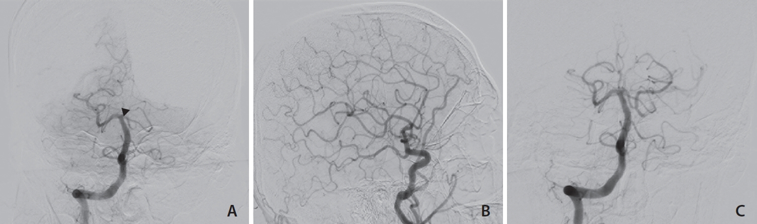
Fig. 1. Patient 1. (A) Cerebral angiogram shows left isolated pre-communicating (P1) occlusion (black arrowhead). (B) Ipsilateral internal carotid artery injection shows post-communicating posterior cerebral artery (PCA) filling by the left posterior communicating artery. (C) Post-thrombectomy angiogram shows patent P1 and PCA.
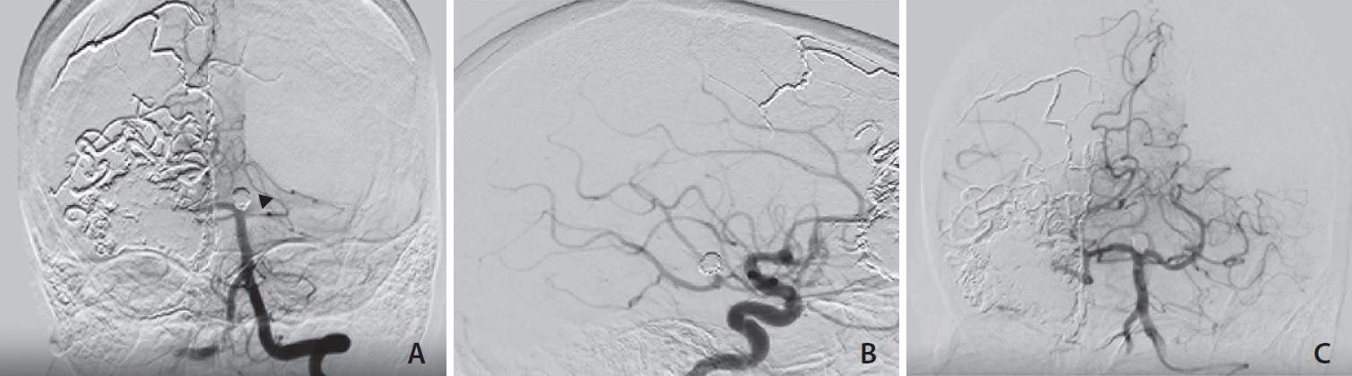
Fig. 2. Patient 2. (A) Post-operative angiography reveals isolated left pre-communicating (P1) occlusion in vertebral injection (black arrowhead). (B) Ipsilateral internal carotid artery injection shows post-communicating posterior cerebral artery (PCA) filling by left posterior communicating artery. (C) Post-thrombectomy angiogram shows patent P1 and PCA in vertebral injection.

Fig. 3. Patient 3. (A) Initial vertebral injection angiogram shows right pre-communicating segment occlusion (black arrowhead). (B) Right carotidal injection shows the right posterior communicating (PCOM) artery. (C) Right vertebral injection also demonstrates patent basilar artery and PCOM artery.
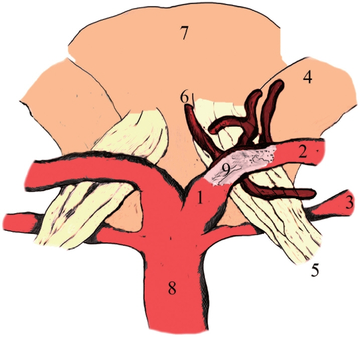
Fig. 4. The illustration shows the pre-communicating (P1) segment of posterior cerebral artery branches (thalamoperforating arteries) supplying the oculomotor nerve. The occlusion of the P1 could affect the arterial supply of the oculomotor nerve in acute ischemic stroke. 1, P1 segment; 2, P2 anterior segment; 3, superior cerebellar artery; 4, cerebellar peduncle; 5, oculomotor nerve; 6, thalamoperforating arteries; 7, mesencephalon; 8, basilar artery; 9, a clot in the P1 segment.
Reference
-
1. Lv X, Jiang C, Li Y, Yang X, Wu Z. Isolated oculomotor nerve palsy in interventional neuroradiology. Eur J Radiol. 2010; 74:441–444.
Article2. Clusmann H, Schaller C, Schramm J. Fixed and dilated pupils after trauma, stroke, and previous intracranial surgery: management and outcome. J Neurol Neurosurg Psychiatry. 2001; 71:175–181.
Article3. Hinduja A, Samant R, Feng D, Hannawi Y. Herniation despite decompressive hemicraniectomy in large hemispherical ischemic strokes. J Stroke Cerebrovasc Dis. 2018; 27:418–424.
Article4. Santos T, Morais H, Oliveira G, Barros P. Isolated oculomotor nerve palsy: a rare manifestation of internal carotid artery dissection. BMJ Case Rep. 2014; 2014:bcr2014205413.
Article5. Voss YL, Weber R, Nordmeyer H, Chapot R. Acute- onset oculomotor paresis attributed to isolated P1 occlusion successfully treated by mechanical thrombectomy. Stroke Vasc Interv Neurol. 2023; 3:e000682.
Article6. Maus V, Rogozinski S, Borggrefe J, Barnikol UB, Saklak M, Mpotsaris A. Clinical presentation of posterior cerebral artery occlusions - clinical rationale for a more aggressive therapeutic strategy? eNeurologicalSci. 2021; 25:100368.
Article7. Altenbernd J, Forsting M, Weber W, Fischer S. Thrombectomy of symptomatic isolated occlusions of posterior cerebral arteries in segment P1 and P2 in acute stroke treatment. Acta Radiol. 2022; 63:802–809.
Article8. Kaya AH, Dagcinar A, Ulu MO, Topal A, Bayri Y, Ulus A, et al. The perforating branches of the P1 segment of the posterior cerebral artery. J Clin Neurosci. 2010; 17:80–84.
Article9. Hendrix P, Griessenauer CJ, Foreman P, Shoja MM, Tubbs RS. Blood supply of the cranial nerves. In: Tubbs RS, Rizk E, Shoja MM, Loukas M, Barbaro N, Spinner RJ. Nerves and nerve injuries. Vol. 1, History, embryology, anatomy, imaging, and diagnostics. Academic Press, 2015;427-438.10. Krisht A, Barnett DW, Barrow DL, Bonner G. The blood supply of the intracavernous cranial nerves: an anatomic study. Neurosurgery. 1994; 34:275–279; discussion 279.
- Full Text Links
-
- Actions
-
Cited
- CITED
-
- Close
- Share
-
- Similar articles
-
- Pituitary Apoplexy Presenting as Isolated Oculomotor Nerve Palsy
- Oculomotor Nerve Palsy in Internal Carotid-Posterior Communicating Artery Aneurysm
- Improvement of Unilateral Oculomotor Nerve Palsy after Clipping of Internal Carotid-posterior Communicating Artery Aneurysm
- Oculomotor Nerve Palsy Associated with Rupture of Middle Cerebral Artery Aneurysm
- Ruptured Anterior Communicating Artery Aneurysm Causing Bilateral Oculomotor Nerve Palsy: A Case Report
