J Korean Med Sci.
2023 Apr;38(16):e159. 10.3346/jkms.2023.38.e159.
Associations of Particulate Matter Exposures With Brain Gray Matter Thickness and White Matter Hyperintensities: Effect Modification by Low-Grade Chronic Inflammation
- Affiliations
-
- 1Department of Preventive Medicine, Yonsei University College of Medicine, Seoul, Korea
- 2Institute for Environmental Research, Yonsei University College of Medicine, Seoul, Korea
- 3Institute of Human Complexity and Systems Science, Yonsei University, Incheon, Korea
- 4Department of Neurology, Gachon University Gil Medical Center, Incheon, Korea
- 5Department of Radiology, Severance Hospital, Yonsei University College of Medicine, Seoul, Korea
- 6Department of Occupational and Environmental Medicine, Wonju Severance Christian Hospital, Wonju College of Medicine, Yonsei University, Wonju, Korea
- 7Department of Cancer Control and Population Health, Graduate School of Cancer Science and Policy, National Cancer Center, Goyang, Korea
- KMID: 2541755
- DOI: http://doi.org/10.3346/jkms.2023.38.e159
Abstract
- Background
Numerous studies have shown the effect of particulate matter exposure on brain imaging markers. However, little evidence exists about whether the effect differs by the level of low-grade chronic systemic inflammation. We investigated whether the level of c-reactive protein (CRP, a marker of systemic inflammation) modifies the associations of particulate matter exposures with brain cortical gray matter thickness and white matter hyperintensities (WMH).
Methods
We conducted a cross-sectional study of baseline data from a prospective cohort study including adults with no dementia or stroke. Long-term concentrations of particulate matter ≤ 10 µm in diameter (PM10) and ≤ 2.5 µm (PM2.5) at each participant’s home address were estimated. Global cortical thickness (n = 874) and WMH volumes (n = 397) were estimated from brain magnetic resonance images. We built linear and logistic regression models for cortical thickness and WMH volumes (higher versus lower than median), respectively. Significance of difference in the association between the CRP group (higher versus lower than median) was expressed as P for interaction.
Results
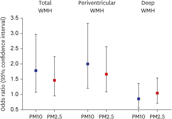
Particulate matter exposures were significantly associated with a reduced global cortical thickness only in the higher CRP group among men (P for interaction = 0.015 for PM10 and 0.006 for PM2.5). A 10 μg/m3 increase in PM10 was associated with the higher volumes of total WMH (odds ratio, 1.78; 95% confidence interval, 1.07–2.97) and periventricular WMH (2.00; 1.20–3.33). A 1 μg/m3 increase in PM2.5 was associated with the higher volume of periventricular WMH (odds ratio, 1.66; 95% confidence interval, 1.08–2.56). These associations did not significantly differ by the level of high sensitivity CRP.
Conclusion
Particulate matter exposures were associated with a reduced global cortical thickness in men with a high level of chronic inflammation. Men with a high level of chronic inflammation may be susceptible to cortical atrophy attributable to particulate matter exposures.
Keyword
Figure
Reference
-
1. Clifford A, Lang L, Chen R, Anstey KJ, Seaton A. Exposure to air pollution and cognitive functioning across the life course--a systematic literature review. Environ Res. 2016; 147:383–398. PMID: 26945620.2. Peters R, Ee N, Peters J, Booth A, Mudway I, Anstey KJ. Air pollution and dementia: a systematic review. J Alzheimers Dis. 2019; 70(S1):S145–S163. PMID: 30775976.3. Wilker EH, Preis SR, Beiser AS, Wolf PA, Au R, Kloog I, et al. Long-term exposure to fine particulate matter, residential proximity to major roads and measures of brain structure. Stroke. 2015; 46(5):1161–1166. PMID: 25908455.4. Wilker EH, Martinez-Ramirez S, Kloog I, Schwartz J, Mostofsky E, Koutrakis P, et al. Fine particulate matter, residential proximity to major roads, and markers of small vessel disease in a memory study population. J Alzheimers Dis. 2016; 53(4):1315–1323. PMID: 27372639.5. Casanova R, Wang X, Reyes J, Akita Y, Serre ML, Vizuete W, et al. A voxel-based morphometry study reveals local brain structural alterations associated with ambient fine particles in older women. Front Hum Neurosci. 2016; 10:495. PMID: 27790103.6. Chen JC, Wang X, Wellenius GA, Serre ML, Driscoll I, Casanova R, et al. Ambient air pollution and neurotoxicity on brain structure: Evidence from women’s health initiative memory study. Ann Neurol. 2015; 78(3):466–476. PMID: 26075655.7. Power MC, Lamichhane AP, Liao D, Xu X, Jack CR, Gottesman RF, et al. The association of long-term exposure to particulate matter air pollution with brain MRI findings: the ARIC study. Environ Health Perspect. 2018; 126(2):027009. PMID: 29467108.8. Cho J, Noh Y, Kim SY, Sohn J, Noh J, Kim W, et al. Long-term ambient air pollution exposures and brain imaging markers in Korean adults: the Environmental Pollution-Induced Neurological EFfects (EPINEF) study. Environ Health Perspect. 2020; 128(11):117006. PMID: 33215932.9. Crous-Bou M, Gascon M, Gispert JD, Cirach M, Sánchez-Benavides G, Falcon C, et al. ALFA Study. Impact of urban environmental exposures on cognitive performance and brain structure of healthy individuals at risk for Alzheimer’s dementia. Environ Int. 2020; 138:105546. PMID: 32151419.10. Block ML, Calderón-Garcidueñas L. Air pollution: mechanisms of neuroinflammation and CNS disease. Trends Neurosci. 2009; 32(9):506–516. PMID: 19716187.11. Chen C, Xun P, Kaufman JD, Hayden KM, Espeland MA, Whitsel EA, et al. Erythrocyte omega-3 index, ambient fine particle exposure, and brain aging. Neurology. 2020; 95(8):e995–1007. PMID: 32669395.12. Gabay C, Kushner I. Acute-phase proteins and other systemic responses to inflammation. N Engl J Med. 1999; 340(6):448–454. PMID: 9971870.13. Du Clos TW. Function of C-reactive protein. Ann Med. 2000; 32(4):274–278. PMID: 10852144.14. Lee MS, Eum KD, Fang SC, Rodrigues EG, Modest GA, Christiani DC. Oxidative stress and systemic inflammation as modifiers of cardiac autonomic responses to particulate air pollution. Int J Cardiol. 2014; 176(1):166–170. PMID: 25074558.15. Huang W, Zhu T, Pan X, Hu M, Lu SE, Lin Y, et al. Air pollution and autonomic and vascular dysfunction in patients with cardiovascular disease: interactions of systemic inflammation, overweight, and gender. Am J Epidemiol. 2012; 176(2):117–126. PMID: 22763390.16. Brook RD, Rajagopalan S, Pope CA 3rd, Brook JR, Bhatnagar A, Diez-Roux AV, et al. Particulate matter air pollution and cardiovascular disease: An update to the scientific statement from the American Heart Association. Circulation. 2010; 121(21):2331–2378. PMID: 20458016.17. Jang H, Kim W, Cho J, Sohn J, Noh J, Seo G, et al. Cohort profile: the Environmental-Pollution-Induced Neurological EFfects (EPINEF) study: a multicenter cohort study of Korean adults. Epidemiol Health. 2021; 43:e2021067. PMID: 34607405.18. Kim SY, Song I. National-scale exposure prediction for long-term concentrations of particulate matter and nitrogen dioxide in South Korea. Environ Pollut. 2017; 226:21–29. PMID: 28399503.19. Jeon S, Yoon U, Park JS, Seo SW, Kim JH, Kim ST, et al. Fully automated pipeline for quantification and localization of white matter hyperintensity in brain magnetic resonance image. Int J Imaging Syst Technol. 2011; 21(2):193–200.20. Noh Y, Seo SW, Jeon S, Lee JM, Kim JH, Kim GH, et al. White matter hyperintensities are associated with amyloid burden in APOE4 non-carriers. J Alzheimers Dis. 2014; 40(4):877–886. PMID: 24577457.21. Choi JW, Lee KO, Jang YJ, Kim HK, Seo T, Roh YJ, et al. High mean platelet volume is associated with cerebral white matter hyperintensities in non-stroke individuals. Yonsei Med J. 2023; 64(1):35–41. PMID: 36579377.22. Zheng F, Xie W. High-sensitivity C-reactive protein and cognitive decline: the English longitudinal study of ageing. Psychol Med. 2018; 48(8):1381–1389. PMID: 29108529.23. Ferrucci L, Fabbri E. Inflammageing: chronic inflammation in ageing, cardiovascular disease, and frailty. Nat Rev Cardiol. 2018; 15(9):505–522. PMID: 30065258.24. Chung HK, Kim JH, Choi A, Ahn CW, Kim YS, Nam JS. Antioxidant-rich dietary intervention improves cardiometabolic profiles and arterial stiffness in elderly Koreans with metabolic syndrome. Yonsei Med J. 2022; 63(1):26–33. PMID: 34913281.25. Altman DG, Bland JM. Interaction revisited: the difference between two estimates. BMJ. 2003; 326(7382):219. PMID: 12543843.26. Ritchie SJ, Cox SR, Shen X, Lombardo MV, Reus LM, Alloza C, et al. Sex differences in the adult human brain: evidence from 5216 UK biobank participants. Cereb Cortex. 2018; 28(8):2959–2975. PMID: 29771288.27. Saito I, Maruyama K, Eguchi E. C-reactive protein and cardiovascular disease in East asians: a systematic review. Clin Med Insights Cardiol. 2015; 8(Suppl 3):35–42. PMID: 25698882.28. Giordano G, Tait L, Furlong CE, Cole TB, Kavanagh TJ, Costa LG. Gender differences in brain susceptibility to oxidative stress are mediated by levels of paraoxonase-2 expression. Free Radic Biol Med. 2013; 58:98–108. PMID: 23376469.29. Costa LG, Cole TB, Coburn J, Chang YC, Dao K, Roque P. Neurotoxicants are in the air: convergence of human, animal, and in vitro studies on the effects of air pollution on the brain. BioMed Res Int. 2014; 2014:736385. PMID: 24524086.30. Varatharaj A, Galea I. The blood-brain barrier in systemic inflammation. Brain Behav Immun. 2017; 60:1–12. PMID: 26995317.31. Elwood E, Lim Z, Naveed H, Galea I. The effect of systemic inflammation on human brain barrier function. Brain Behav Immun. 2017; 62:35–40. PMID: 27810376.32. Shou Y, Huang Y, Zhu X, Liu C, Hu Y, Wang H. A review of the possible associations between ambient PM2.5 exposures and the development of Alzheimer’s disease. Ecotoxicol Environ Saf. 2019; 174:344–352. PMID: 30849654.33. Sweeney MD, Sagare AP, Zlokovic BV. Blood-brain barrier breakdown in Alzheimer disease and other neurodegenerative disorders. Nat Rev Neurol. 2018; 14(3):133–150. PMID: 29377008.34. Furlong MA, Alexander GE, Klimentidis YC, Raichlen DA. Association of air pollution and physical activity with brain volumes. Neurology. 2021; 98(4):e416–e426. PMID: 34880089.35. Blair GW, Thrippleton MJ, Shi Y, Hamilton I, Stringer M, Chappell F, et al. Intracranial hemodynamic relationships in patients with cerebral small vessel disease. Neurology. 2020; 94(21):e2258–e2269. PMID: 32366534.36. ten Dam VH, van den Heuvel DM, de Craen AJ, Bollen EL, Murray HM, Westendorp RG, et al. Decline in total cerebral blood flow is linked with increase in periventricular but not deep white matter hyperintensities. Radiology. 2007; 243(1):198–203. PMID: 17329688.37. DeCarli C, Fletcher E, Ramey V, Harvey D, Jagust WJ. Anatomical mapping of white matter hyperintensities (WMH): exploring the relationships between periventricular WMH, deep WMH, and total WMH burden. Stroke. 2005; 36(1):50–55. PMID: 15576652.38. Griffanti L, Jenkinson M, Suri S, Zsoldos E, Mahmood A, Filippini N, et al. Classification and characterization of periventricular and deep white matter hyperintensities on MRI: a study in older adults. Neuroimage. 2018; 170:174–181. PMID: 28315460.39. Low A, Mak E, Rowe JB, Markus HS, O’Brien JT. Inflammation and cerebral small vessel disease: a systematic review. Ageing Res Rev. 2019; 53:100916. PMID: 31181331.40. Prins ND, van Dijk EJ, den Heijer T, Vermeer SE, Jolles J, Koudstaal PJ, et al. Cerebral small-vessel disease and decline in information processing speed, executive function and memory. Brain. 2005; 128(Pt 9):2034–2041. PMID: 15947059.41. van den Heuvel DM, ten Dam VH, de Craen AJ, Admiraal-Behloul F, Olofsen H, Bollen EL, et al. Increase in periventricular white matter hyperintensities parallels decline in mental processing speed in a non-demented elderly population. J Neurol Neurosurg Psychiatry. 2006; 77(2):149–153. PMID: 16421114.42. Debette S, Bombois S, Bruandet A, Delbeuck X, Lepoittevin S, Delmaire C, et al. Subcortical hyperintensities are associated with cognitive decline in patients with mild cognitive impairment. Stroke. 2007; 38(11):2924–2930. PMID: 17885256.43. Liu CC, Liu CC, Kanekiyo T, Xu H, Bu G. Apolipoprotein E and Alzheimer disease: risk, mechanisms and therapy. Nat Rev Neurol. 2013; 9(2):106–118. PMID: 23296339.44. Cacciaglia R, Molinuevo JL, Falcón C, Sánchez-Benavides G, Gramunt N, Brugulat-Serrat A, et al. APOE-ε4 risk variant for Alzheimer’s disease modifies the association between cognitive performance and cerebral morphology in healthy middle-aged individuals. Neuroimage Clin. 2019; 23:101818. PMID: 30991302.
- Full Text Links
- Actions
-
Cited
- CITED
-
- Close
- Share
- Similar articles
-
- Association of Particulate Matter With ENT Diseases
- Fine Particulate Matter and Urology: Emphasis on the Lower Urinary Tract
- Relative Signal Intensity Changes of Frontal and Occipital White Matters on T2 Weighted Axial MR Image: Correlation with Age
- Association of Low Blood Pressure with White Matter Hyperintensities in Elderly Individuals with Controlled Hypertension
- Perinatal Hypoxic-lschemic Brain Injury: MR Findings


