J Korean Neurosurg Soc.
2023 Mar;66(2):113-120. 10.3340/jkns.2022.0130.
Artificial Intelligence for Neurosurgery : Current State and Future Directions
- Affiliations
-
- 1Department of Neurosurgery, Ajou University College of Medicine, Suwon, Korea
- 2Department of Neurosurgery, Yonsei University College of Medicine, Seoul, Korea
- 3Department of Neurosurgery, Spine and Spinal Cord Institute, Severance Hospital, Yonsei University College of Medicine, Seoul, Korea
- KMID: 2539872
- DOI: http://doi.org/10.3340/jkns.2022.0130
Abstract
- Artificial intelligence (AI) is a field of computer science that equips machines with human-like intelligence and enables them to learn, reason, and solve problems when presented with data in various formats. Neurosurgery is often at the forefront of innovative and disruptive technologies, which have similarly altered the course of acute and chronic diseases. In diagnostic imaging, such as X-rays, computed tomography, and magnetic resonance imaging, AI is used to analyze images. The use of robots in the field of neurosurgery is also increasing. In neurointensive care units, AI is used to analyze data and provide care to critically ill patients. Moreover, AI can be used to predict a patient’s prognosis. Several AI applications have already been introduced in the field of neurosurgery, and many more are expected in the near future. Ultimately, it is our responsibility to keep pace with this evolution to provide meaningful outcomes and personalize each patient’s care. Rather than blindly relying on AI in the future, neurosurgeons should gain a thorough understanding of it and use it to enhance their patient care.
Keyword
Figure
-
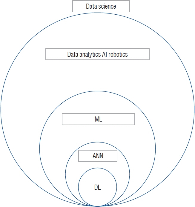
Fig. 1. To define the relationship between the words in data science. AI : artificial intelligence, ML : machine learning, ANN : artificial neural network, DL : deep learning.
Cited by 1 articles
-
Artificial Intelligence-Enhanced Neurocritical Care for Traumatic Brain Injury : Past, Present and Future
Kyung Ah Kim, Hakseung Kim, Eun Jin Ha, Byung C. Yoon, Dong-Joo Kim
J Korean Neurosurg Soc. 2024;67(5):493-509. doi: 10.3340/jkns.2023.0195.
Reference
-
References
1. Aerts HJ, Velazquez ER, Leijenaar RT, Parmar C, Grossmann P, Carvalho S, et al. Decoding tumour phenotype by noninvasive imaging using a quantitative radiomics approach. Nat Commun. 5:4006. 2014.2. Bakkar N, Kovalik T, Lorenzini I, Spangler S, Lacoste A, Sponaugle K, et al. Artificial intelligence in neurodegenerative disease research: use of IBM Watson to identify additional RNA-binding proteins altered in amyotrophic lateral sclerosis. Acta Neuropathol. 135:227–247. 2018.3. Ball T, González-Martínez J, Zemmar A, Sweid A, Chandra S, VanSickle D, et al. Robotic applications in cranial neurosurgery: current and future. Oper Neurosurg (Hagerstown). 21:371–379. 2021.4. Bickle J. Precis of philosophy and neuroscience: a ruthlessly reductive account. Phenom Cogn Sci. 4:231–238. 2005.5. Brain Trauma Foundation; American Association of Neurological Surgeons; Congress of Neurological Surgeons; Joint Section on Neurotrauma and Critical Care; AANS/CNS, Bratton SL, et al. Guidelines for the management of severe traumatic brain injury. II. Hyperosmolar therapy. J Neurotrauma. 24(Suppl 1):S14–S20. 2007.6. Buchlak QD, Esmaili N, Leveque JC, Bennett C, Farrokhi F, Piccardi M. Machine learning applications to neuroimaging for glioma detection and classification: an artificial intelligence augmented systematic review. J Clin Neurosci. 89:177–198. 2021.7. Burström G, Buerger C, Hoppenbrouwers J, Nachabe R, Lorenz C, Babic D, et al. Machine learning for automated 3-dimensional segmentation of the spine and suggested placement of pedicle screws based on intraoperative cone-beam computer tomography. J Neurosurg Spine. 31:147–154. 2019.8. Chumnanvej S, Pillai BM, Chalongwongse S, Suthakorn J. Endonasal endoscopic transsphenoidal approach robot prototype: a cadaveric trial. Asian J Surg. 44:345–351. 2021.9. Chwialkowski MP, Shile PE, Pfeifer D, Parkey RW, Peshock RM. Automated localization and identification of lower spinal anatomy in magnetic resonance images. Comput Biomed Res. 24:99–117. 1991.10. Claassen J, Doyle K, Matory A, Couch C, Burger KM, Velazquez A, et al. Detection of brain activation in unresponsive patients with acute brain injury. N Engl J Med. 380:2497–2505. 2019.11. Goyal A, Ngufor C, Kerezoudis P, McCutcheon B, Storlie C, Bydon M. Can machine learning algorithms accurately predict discharge to nonhome facility and early unplanned readmissions following spinal fusion? Analysis of a national surgical registry. J Neurosurg Spine. 31:568–578. 2019.12. Graziano Michael SA. Consciousness and the social brain. New York: Oxford University Press;2013.13. Haladjian HH, Montemayor C. Artificial consciousness and the consciousness-attention dissociation. Conscious Cogn. 45:210–225. 2016.14. Hale AT, Stonko DP, Wang L, Strother MK, Chambless LB. Machine learning analyses can differentiate meningioma grade by features on magnetic resonance imaging. Neurosurg Focus. 45:E4. 2018.15. Hernandes Rocha TA, Elahi C, Cristina da Silva N, Sakita FM, Fuller A, Mmbaga BT, et al. A traumatic brain injury prognostic model to support in-hospital triage in a low-income country: a machine learningbased approach. J Neurosurg. 132:1961–1969. 2019.16. Hinton GE, Osindero S, Teh YW. A fast learning algorithm for deep belief nets. Neural Comput. 18:1527–1554. 2006.17. Hopkins BS, Yamaguchi JT, Garcia R, Kesavabhotla K, Weiss H, Hsu WK, et al. Using machine learning to predict 30-day readmissions after posterior lumbar fusion: an NSQIP study involving 23,264 patients. J Neurosurg Spine. 32:399–406. 2020.18. Huang KT, Silva MA, See AP, Wu KC, Gallerani T, Zaidi HA, et al. A computer vision approach to identifying the manufacturer and model of anterior cervical spinal hardware. J Neurosurg Spine. 31:844–850. 2019.19. Kalagara S, Eltorai AEM, Durand WM, DePasse JM, Daniels AH. Machine learning modeling for predicting hospital readmission following lumbar laminectomy. J Neurosurg Spine. 30:344–352. 2018.20. Korez R, Putzier M, Vrtovec T. A deep learning tool for fully automated measurements of sagittal spinopelvic balance from X-ray images: performance evaluation. Eur Spine J. 29:2295–2305. 2020.21. Lee MH, Kim J, Kim ST, Shin HM, You HJ, Choi JW, et al. Prediction of IDH1 mutation status in glioblastoma using machine learning technique based on quantitative radiomic data. World Neurosurg. 125:e688–e696. 2019.22. Marcus HJ, Hughes-Hallett A, Kwasnicki RM, Darzi A, Yang GZ, Nandi D. Technological innovation in neurosurgery: a quantitative study. J Neurosurg. 123:174–181. 2015.23. McDonald RJ, Schwartz KM, Eckel LJ, Diehn FE, Hunt CH, Bartholmai BJ, et al. The effects of changes in utilization and technological advancements of cross-sectional imaging on radiologist workload. Acad Radiol. 22:1191–1198. 2015.24. Minsky M, Papert S. Perceptrons: an introduction to computational geometry. Cambridge: MIT Press;1969.25. Mohamed AR, Sainath TN, Dahl G, Ramabhadran B, Hinton GE, Picheny MA. Deep Belief Networks using discriminative features for phone recognition. In : 2011 IEEE International Conference on Acoustics, Speech and Signal Processing (ICASSP); 2012 Nov 1; Prague Computer Science. IEEE Signal Processing Magazine. 5060–5063. 2011.26. Muhlestein WE, Akagi DS, Davies JM, Chambless LB. Predicting inpatient length of stay after brain tumor surgery: developing machine learning ensembles to improve predictive performance. Neurosurgery. 85:384–393. 2019.27. Paliwal N, Jaiswal P, Tutino VM, Shallwani H, Davies JM, Siddiqui AH, et al. Outcome prediction of intracranial aneurysm treatment by flow diverters using machine learning. Neurosurg Focus. 45:E7. 2018.28. Philipp LR, Matias CM, Thalheimer S, Mehta SH, Sharan A, Wu C. Robot-assisted stereotaxy reduces target error: a meta-analysis and metaregression of 6056 trajectories. Neurosurgery. 88:222–233. 2021.29. Raj R, Luostarinen T, Pursiainen E, Posti JP, Takala RSK, Bendel S, et al. Machine learning-based dynamic mortality prediction after traumatic brain injury. Sci Rep. 9:17672. 2019.30. Ratner M. FDA backs clinician-free AI imaging diagnostic tools. Nat Biotechnol. 36:673–674. 2018.31. Rosenblatt F. The perceptron: a probabilistic model for information storage and organization in the brain. Psychol Rev. 65:386–408. 1958.32. Scherer M, Cordes J, Younsi A, Sahin YA, Götz M, Möhlenbruch M, et al. Development and validation of an automatic segmentation algorithm for quantification of intracerebral hemorrhage. Stroke. 47:2776–2782. 2016.33. Searle J. Chinese room argument. Scholarpedia. 4:3100. 2009.34. Senders JT, Staples P, Mehrtash A, Cote DJ, Taphoorn MJB, Reardon DA, et al. An online calculator for the prediction of survival in glioblastoma patients using classical statistics and machine learning. Neurosurgery. 86:E184–E192. 2020.35. Siccoli A, de Wispelaere MP, Schröder ML, Staartjes VE. Machine learning-based preoperative predictive analytics for lumbar spinal stenosis. Neurosurg Focus. 46:E5. 2019.36. Staartjes VE, Serra C, Muscas G, Maldaner N, Akeret K, van Niftrik CHB, et al. Utility of deep neural networks in predicting gross-total resection after transsphenoidal surgery for pituitary adenoma: a pilot study. Neurosurg Focus. 45:E12. 2018.37. Staartjes VE, Zattra CM, Akeret K, Maldaner N, Muscas G, Bas van Niftrik CH, et al. Neural network-based identification of patients at high risk for intraoperative cerebrospinal fluid leaks in endoscopic pituitary surgery. J Neurosurg. 133:329–335. 2020.38. Starr PA, Martin AJ, Ostrem JL, Talke P, Levesque N, Larson PS. Subthalamic nucleus deep brain stimulator placement using high-field interventional magnetic resonance imaging and a skull-mounted aiming device: technique and application accuracy. J Neurosurg. 112:479–490. 2010.39. Tanioka S, Ishida F, Nakano F, Kawakita F, Kanamaru H, Nakatsuka Y, et al. Machine learning analysis of matricellular proteins and clinical variables for early prediction of delayed cerebral ischemia after aneurysmal subarachnoid hemorrhage. Mol Neurobiol. 56:7128–7135. 2019.40. Tunthanathip T, Sae-Heng S, Oearsakul T, Sakarunchai I, Kaewborisutsakul A, Taweesomboonyat C. Machine learning applications for the prediction of surgical site infection in neurological operations. Neurosurg Focus. 47:E7. 2019.41. Turing AM. Computing machinery and intelligence. Mind. 59:433–460. 1950.42. Urbizu A, Martin BA, Moncho D, Rovira A, Poca MA, Sahuquillo J, et al. Machine learning applied to neuroimaging for diagnosis of adult classic Chiari malformation: role of the basion as a key morphometric indicator. J Neurosurg. 129:779–791. 2018.43. Voter AF, Meram E, Garrett JW, Yu JJ. Diagnostic accuracy and failure mode analysis of a deep learning algorithm for the detection of intracranial hemorrhage. J Am Coll Radiol. 18:1143–1152. 2021.44. Wagner K, Vaz-Guimaraes F, Camstra K, Lam S. Endoscope-assisted hemispherotomy: translation of technique from cadaveric anatomical feasibility study to clinical implementation. J Neurosurg Pediatr. 23:178–186. 2018.45. Wang L, Alexander CA. Big data in medical applications and health care. Current Research in Medicine. 6:1–8. 2015.46. Welter ML, Schüpbach M, Czernecki V, Karachi C, Fernandez-Vidal S, Golmard JL, et al. Optimal target localization for subthalamic stimulation in patients with Parkinson disease. Neurology. 82:1352–1361. 2014.47. Zlojutro A, Rey D, Gardner L. A decision-support framework to optimize border control for global outbreak mitigation. Sci Rep. 9:2216. 2019.
- Full Text Links
-
- Actions
-
Cited
- CITED
-
- Close
- Share
-
- Similar articles
-
- Application of Artificial Intelligence in Thoracic Radiology: A Narrative Review
- Role of artificial intelligence in diagnosing Barrett’s esophagus-related neoplasia
- Artificial Intelligence Applications in Diabetic Retinopathy: What We Have Now and What to Expect in the Future
- Automated Bone Age Assessment Using Artificial Intelligence: The Future of Bone Age Assessment
- Artificial Intelligence in Pathology
