Endocrinol Metab.
2024 Jun;39(3):416-424. 10.3803/EnM.2023.1913.
Artificial Intelligence Applications in Diabetic Retinopathy: What We Have Now and What to Expect in the Future
- Affiliations
-
- 1Department of Ophthalmology, Kangbuk Samsung Hospital, Sungkyunkwan University School of Medicine, Seoul, Korea
- 2Biomedical Institute for Convergence (BICS), Sungkyunkwan University, Suwon, Korea
- KMID: 2556633
- DOI: http://doi.org/10.3803/EnM.2023.1913
Abstract
- Diabetic retinopathy (DR) is a major complication of diabetes mellitus and is a leading cause of vision loss globally. A prompt and accurate diagnosis is crucial for ensuring favorable visual outcomes, highlighting the need for increased access to medical care. The recent remarkable advancements in artificial intelligence (AI) have raised high expectations for its role in disease diagnosis and prognosis prediction across various medical fields. In addition to achieving high precision comparable to that of ophthalmologists, AI-based diagnosis of DR has the potential to improve medical accessibility, especially through telemedicine. In this review paper, we aim to examine the current role of AI in the diagnosis of DR and explore future directions.
Keyword
Figure
-
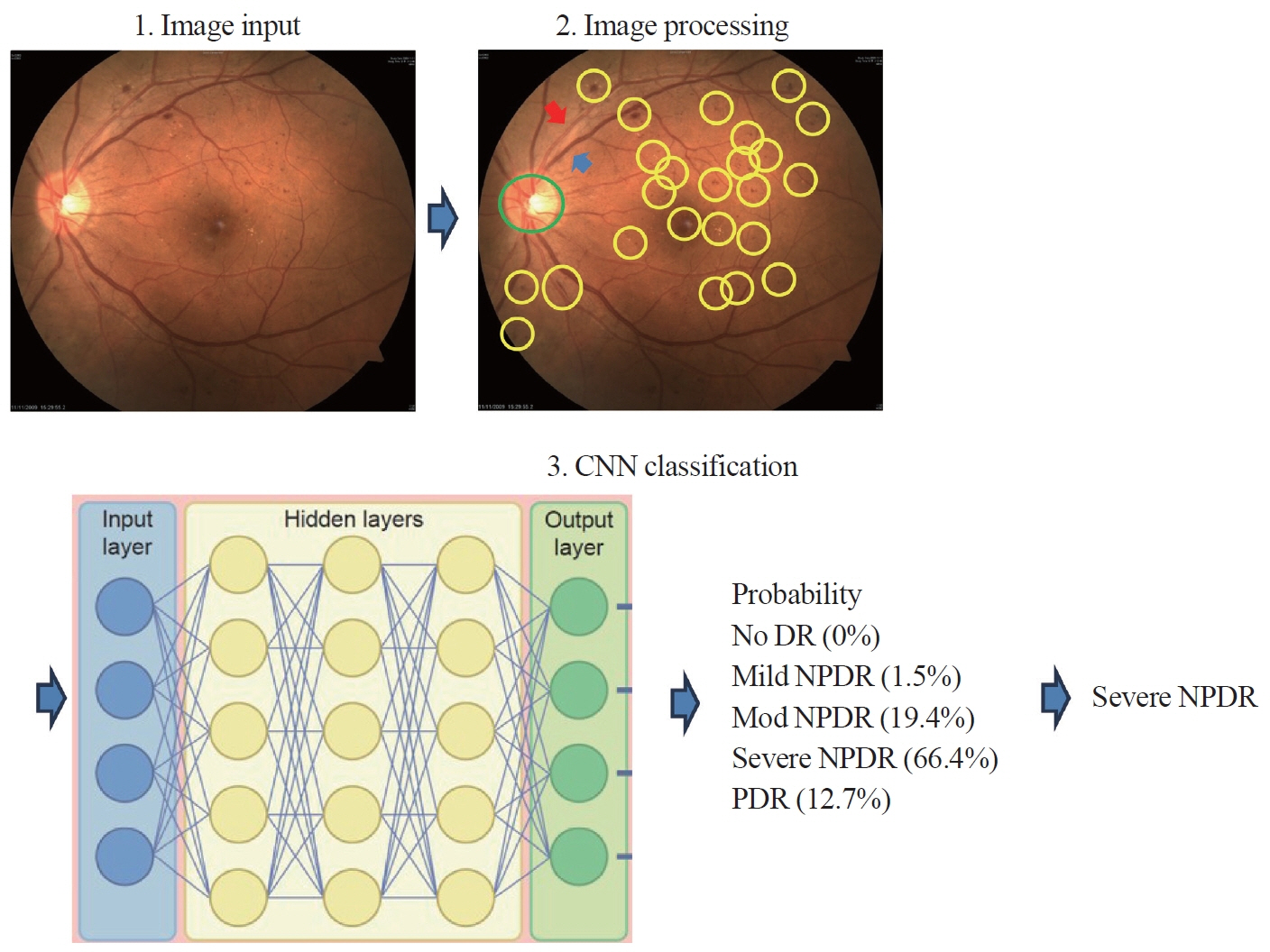
Fig. 1. A diagram illustrating the classification of diabetic retinopathy using Convolutional Neural Network (CNN). DR, diabetic retinopathy; NPDR, nonproliferative diabetic retinopathy; PDR, proliferative diabetic retinopathy.
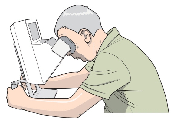
Fig. 2. Home optical coherence tomography.
Reference
-
1. GBD 2021 Diabetes Collaborators. Global, regional, and national burden of diabetes from 1990 to 2021, with projections of prevalence to 2050: a systematic analysis for the Global Burden of Disease Study 2021. Lancet. 2023; 402:203–34.2. Kim KS, Choi CH, Lee DY, Kim EJ. Epidemiological study on diabetes mellitus among rural Korean. J Korean Diabetes. 1972; 1:17–24.3. Jung CH, Son JW, Kang S, Kim WJ, Kim HS, Kim HS, et al. Diabetes fact sheets in Korea, 2020: an appraisal of current status. Diabetes Metab J. 2021; 45:1–10.
Article4. Bourne RR, Jonas JB, Bron AM, Cicinelli MV, Das A, Flaxman SR, et al. Prevalence and causes of vision loss in highincome countries and in Eastern and Central Europe in 2015: magnitude, temporal trends and projections. Br J Ophthalmol. 2018; 102:575–85.
Article5. Song SJ, Han K, Choi KS, Ko SH, Rhee EJ, Park CY, et al. Trends in diabetic retinopathy and related medical practices among type 2 diabetes patients: results from the National Insurance Service Survey 2006-2013. J Diabetes Investig. 2018; 9:173–8.
Article6. Jiang F, Jiang Y, Zhi H, Dong Y, Li H, Ma S, et al. Artificial intelligence in healthcare: past, present and future. Stroke Vasc Neurol. 2017; 2:230–43.
Article7. Khan A, Sohail A, Zahoora U, Qureshi AS. A survey of the recent architectures of deep convolutional neural networks. Artif Intell Rev. 2020; 53:5455–516.
Article8. LeCun Y, Bengio Y, Hinton G. Deep learning. Nature. 2015; 521:436–44.
Article9. Early Treatment Diabetic Retinopathy Study Research Group. Grading diabetic retinopathy from stereoscopic color fundus photographs: an extension of the modified Airlie House classification. ETDRS report number 10. Ophthalmology. 1991; 98(5 Suppl):786–806.10. Xu K, Feng D, Mi H. Deep convolutional neural networkbased early automated detection of diabetic retinopathy using fundus image. Molecules. 2017; 22:2054.
Article11. Esfahani MT, Ghaderi M, Kafiyeh R. Classification of diabetic and normal fundus images using new deep learning method. Leonardo Electron J Pract Technol. 2018; 17:233–48.12. Quellec G, Charriere K, Boudi Y, Cochener B, Lamard M. Deep image mining for diabetic retinopathy screening. Med Image Anal. 2017; 39:178–93.
Article13. Jiang H, Yang K, Gao M, Zhang D, Ma H, Qian W. An interpretable ensemble deep learning model for diabetic retinopathy disease classification. Annu Int Conf IEEE Eng Med Biol Soc. 2019; 2019:2045–8.
Article14. Abramoff MD, Lou Y, Erginay A, Clarida W, Amelon R, Folk JC, et al. Improved automated detection of diabetic retinopathy on a publicly available dataset through integration of deep learning. Invest Ophthalmol Vis Sci. 2016; 57:5200–6.
Article15. Shamsan A, Senan EM, Ahmad Shatnawi HS. Predicting of diabetic retinopathy development stages of fundus images using deep learning based on combined features. PLoS One. 2023; 18:e0289555.
Article16. Johnson MW. Etiology and treatment of macular edema. Am J Ophthalmol. 2009; 147:11–21.
Article17. Klein R, Klein BE, Moss SE, Davis MD, DeMets DL. The Wisconsin epidemiologic study of diabetic retinopathy. IV. Diabetic macular edema. Ophthalmology. 1984; 91:1464–74.
Article18. Ixcamey M, Palma C. Diabetic macular edema. Dis Mon. 2021; 67:101138.
Article19. Early Treatment Diabetic Retinopathy Study Research Group. Treatment techniques and clinical guidelines for photocoagulation of diabetic macular edema: Early Treatment Diabetic Retinopathy Study Report Number 2. Ophthalmology. 1987; 94:761–74.20. Sadda SR, Tan O, Walsh AC, Schuman JS, Varma R, Huang D. Automated detection of clinically significant macular edema by grid scanning optical coherence tomography. Ophthalmology. 2006; 113:1187.
Article21. Nayak J, Bhat PS, Acharya UR. Automatic identification of diabetic maculopathy stages using fundus images. J Med Eng Technol. 2009; 33:119–29.
Article22. Shahriari MH, Sabbaghi H, Asadi F, Hosseini A, Khorrami Z. Artificial intelligence in screening, diagnosis, and classification of diabetic macular edema: a systematic review. Surv Ophthalmol. 2023; 68:42–53.
Article23. Wang Z, Li Z, Li K, Mu S, Zhou X, Di Y. Performance of artificial intelligence in diabetic retinopathy screening: a systematic review and meta-analysis of prospective studies. Front Endocrinol (Lausanne). 2023; 14:1197783.
Article24. Dai L, Sheng B, Chen T, Wu Q, Liu R, Cai C, et al. A deep learning system for predicting time to progression of diabetic retinopathy. Nat Med. 2024; 30:584–94.
Article25. Prahs P, Radeck V, Mayer C, Cvetkov Y, Cvetkova N, Helbig H, et al. OCT-based deep learning algorithm for the evaluation of treatment indication with anti-vascular endothelial growth factor medications. Graefes Arch Clin Exp Ophthalmol. 2018; 256:91–8.
Article26. Ye X, Gao K, He S, Zhong X, Shen Y, Wang Y, et al. Artificial intelligence-based quantification of central macular fluid volume and VA prediction for diabetic macular edema using OCT images. Ophthalmol Ther. 2023; 12:2441–52.
Article27. Abramoff MD, Lavin PT, Birch M, Shah N, Folk JC. Pivotal trial of an autonomous AI-based diagnostic system for detection of diabetic retinopathy in primary care offices. NPJ Digit Med. 2018; 1:39.28. Ipp E, Liljenquist D, Bode B, Shah VN, Silverstein S, Regillo CD, et al. Pivotal evaluation of an artificial intelligence system for autonomous detection of referrable and visionthreatening diabetic retinopathy. JAMA Netw Open. 2021; 4:e2134254.
Article29. Kauppi T, Kamarainen JK, Lensu L, Kalesnykiene V, Sorri I, Uusitalo H, et al. Constructing benchmark databases and protocols for medical image analysis: diabetic retinopathy. Comput Math Methods Med. 2013; 2013:368514.
Article30. Andersen JK, Grauslund J, Savarimuthu TR. Comparing objective functions for segmentation and detection of microaneurysms in retinal images. Proc Mach Learn Res. 2020; 121:19–32.31. Ibrahim H, Liu X, Rivera SC, Moher D, Chan AW, Sydes MR, et al. Reporting guidelines for clinical trials of artificial intelligence interventions: the SPIRIT-AI and CONSORTAI guidelines. Trials. 2021; 22:11.
Article32. Yang Q, Liu Y, Chen T, Tong Y. Federated machine learning: concept and applications. ACM Trans Intell Syst Technol. 2019; 10:1–19.33. Kairouz P, McMahan HB, Avent B, Bellet A, Bennis M, Bhagoji AN, et al. Advances and open problems in federated learning. Found Trends Mach Learn. 2021; 14:1–210.
Article34. Abramoff MD, Tobey D, Char DS. Lessons learned about autonomous AI: finding a safe, efficacious, and ethical path through the development process. Am J Ophthalmol. 2020; 214:134–42.
Article35. Xie Y, Nguyen QD, Hamzah H, Lim G, Bellemo V, Gunasekeran DV, et al. Artificial intelligence for teleophthalmologybased diabetic retinopathy screening in a national programme: an economic analysis modelling study. Lancet Digit Health. 2020; 2:e240. –9.
Article36. Fuller SD, Hu J, Liu JC, Gibson E, Gregory M, Kuo J, et al. Five-year cost-effectiveness modeling of primary care-based, nonmydriatic automated retinal image analysis screening among low-income patients with diabetes. J Diabetes Sci Technol. 2022; 16:415–27.
Article37. Bogunovic H, Waldstein SM, Schlegl T, Langs G, Sadeghipour A, Liu X, et al. Prediction of anti-VEGF treatment requirements in neovascular AMD using a machine learning approach. Invest Ophthalmol Vis Sci. 2017; 58:3240–8.
Article38. Lee J, Lee J, Cho S, Song J, Lee M, Kim SH, et al. Development of decision support software for deep learning-based automated retinal disease screening using relatively limited fundus photograph data. Electronics. 2021; 10:163.
Article39. Cheung CY, Xu D, Cheng CY, Sabanayagam C, Tham YC, Yu M, et al. A deep-learning system for the assessment of cardiovascular disease risk via the measurement of retinal-vessel calibre. Nat Biomed Eng. 2021; 5:498–508.
Article40. Cheung CY, Tay WT, Ikram MK, Ong YT, De Silva DA, Chow KY, et al. Retinal microvascular changes and risk of stroke: the Singapore Malay Eye Study. Stroke. 2013; 44:2402–8.41. Poplin R, Varadarajan AV, Blumer K, Liu Y, McConnell MV, Corrado GS, et al. Prediction of cardiovascular risk factors from retinal fundus photographs via deep learning. Nat Biomed Eng. 2018; 2:158–64.
Article42. Zhang K, Liu X, Xu J, Yuan J, Cai W, Chen T, et al. Deeplearning models for the detection and incidence prediction of chronic kidney disease and type 2 diabetes from retinal fundus images. Nat Biomed Eng. 2021; 5:533–45.
Article43. Yip W, Ong PG, Teo BW, Cheung CY, Tai ES, Cheng CY, et al. Retinal vascular imaging markers and incident chronic kidney disease: a prospective cohort study. Sci Rep. 2017; 7:9374.
Article44. Simo R, Ciudin A, Simo-Servat O, Hernandez C. Cognitive impairment and dementia: a new emerging complication of type 2 diabetes: the diabetologist’s perspective. Acta Diabetol. 2017; 54:417–24.45. Cheung CY, Ikram MK, Chen C, Wong TY. Imaging retina to study dementia and stroke. Prog Retin Eye Res. 2017; 57:89–107.
Article46. Ahn S, Shin J, Song SJ, Yoon WT, Sagong M, Jeong A, et al. Neurologic dysfunction assessment in Parkinson disease based on fundus photographs using deep learning. JAMA Ophthalmol. 2023; 141:234–40.
Article47. Quazi S. Artificial intelligence and machine learning in precision and genomic medicine. Med Oncol. 2022; 39:120.
Article48. Alowais SA, Alghamdi SS, Alsuhebany N, Alqahtani T, Alshaya AI, Almohareb SN, et al. Revolutionizing healthcare: the role of artificial intelligence in clinical practice. BMC Med Educ. 2023; 23:689.
Article49. Huang C, Clayton EA, Matyunina LV, McDonald LD, Benigno BB, Vannberg F, et al. Machine learning predicts individual cancer patient responses to therapeutic drugs with high accuracy. Sci Rep. 2018; 8:16444.
Article50. Sheu YH, Magdamo C, Miller M, Das S, Blacker D, Smoller JW. AI-assisted prediction of differential response to antidepressant classes using electronic health records. NPJ Digit Med. 2023; 6:73.
Article51. Faes L, Islam M, Bachmann LM, Lienhard KR, Schmid MK, Sim DA. False alarms and the positive predictive value of smartphone-based hyperacuity home monitoring for the progression of macular disease: a prospective cohort study. Eye (Lond). 2021; 35:3035–40.
Article52. Kaiser PK, Wang YZ, He YG, Weisberger A, Wolf S, Smith CH. Feasibility of a novel remote daily monitoring system for age-related macular degeneration using mobile handheld devices: results of a pilot study. Retina. 2013; 33:1863–70.53. Chew EY, Clemons TE, Harrington M, Bressler SB, Elman MJ, Kim JE, et al. Effectiveness of different monitoring modalities in the detection of neovascular age-related macular degeneration: the Home study, report number 3. Retina. 2016; 36:1542–7.54. AREDS2-HOME Study Research Group; Chew EY, Clemons TE, Bressler SB, Elman MJ, Danis RP, et al. Randomized trial of a home monitoring system for early detection of choroidal neovascularization home monitoring of the eye (HOME) study. Ophthalmology. 2014; 121:535–44.55. Holekamp NM. Moving from clinic to home: what the future holds for ophthalmic telemedicine. Am J Ophthalmol. 2018; 187:xxviii–xxxv.
Article56. Blinder KJ, Calhoun C, Maguire MG, Glassman AR, Mein CE, Baskin DE, et al. Home OCT imaging for newly diagnosed neovascular age-related macular degeneration: a feasibility study. Ophthalmol Retina. 2024; 8:376–87.57. Bhaskaranand M, Cuadros J, Ramachandra C, Bhat S, Nittala MG, Sadda SR, et al. EyeArt+EyePACS: automated retinal image analysis for diabetic retinopathy screening in a telemedicine system. In: Proceedings of the Ophthalmic Medical Image Analysis International Workshop; 2015 Oct 9; Munich, Germany. Iowa City: University of Iowa; 2015. p. 105-12. https://pubs.lib.uiowa.edu/omia/article/27670/galley/135961/view.58. Betzler BK, Chen H, Cheng CY, Lee CS, Ning G, Song SJ, et al. Large language models and their impact in ophthalmology. Lancet Digit Health. 2023; 5:e917. –24.
Article59. Lee P, Bubeck S, Petro J. Benefits, limits, and risks of GPT4 as an AI chatbot for medicine. N Engl J Med. 2023; 388:1233–9.
Article60. Gopalakrishnan N, Joshi A, Chhablani J, Yadav NK, Reddy NG, Rani PK, et al. Recommendations for initial diabetic retinopathy screening of diabetic patients using large language model-based artificial intelligence in real-life case scenarios. Int J Retina Vitreous. 2024; 10:11.
Article
- Full Text Links
-
- Actions
-
Cited
- CITED
-
- Close
- Share
-
- Similar articles
-
- The Clinical Applications of Multifocal Electroretinogram in Diabetic Retinopathy
- The principles of artificial intelligence and its applications in dentistry
- The Value of Non-Clinical Applications of Artificial Intelligence in Radiology Should Be Noted
- Artificial Intelligence in Pathology
- The Role of medical doctor in the era of artificial intelligence
