Ann Surg Treat Res.
2023 Mar;104(3):164-169. 10.4174/astr.2023.104.3.164.
Endovenous radiofrequency ablation using a new bipolar electrode in a canine model: a new endovenous radiofrequency electrode
- Affiliations
-
- 1Department of Radiology, Konkuk University School of Medicine, Seoul, Korea
- 2Department of Radiology, Seoul Sun Vascular Pain Center, Seoul, Korea
- 3Department of Radiology, Cheil Orthopedic Hospital, Seoul, Korea
- 4Department of Thoracic and Cardiovascular Surgery, Konkuk University School of Medicine, Seoul, Korea
- KMID: 2539695
- DOI: http://doi.org/10.4174/astr.2023.104.3.164
Abstract
- Purpose
This study aimed to determine the effectiveness and safety of a newly developed endovenous radiofrequency (RF) catheter compared with that of the existing RF catheter in a canine model.
Methods
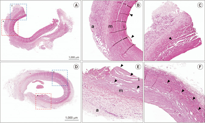
Seven dogs underwent ablation using 1 control catheter (ClosureFAST, CF; Covidien) and 1 experimental catheter (VENISTAR, VS; STARmed Co., Ltd.) in the femoral and cephalic veins. The ablated vein was evaluated macroscopically (2,3,5-triphenyltetrazolium chloride staining, TTC), microscopically (hematoxylin and eosin staining), and ultrasonographically. Vessel injury score was used to evaluate the ablating effect objectively. Veins from 1 dog were evaluated on the day of ablation, while in the remaining 6 dogs, the ablated veins were evaluated 2 weeks later.
Results
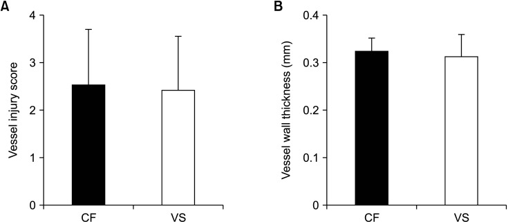
A total of 23 veins (CF, 11 veins; VS, 12 veins) were ablated in 7 dogs. Non–TTC-stained vein wall areas were identified in all ablated veins. No significant difference was observed in the mean vessel injury score (2.54 ± 1.16 vs. 2.42 ± 1.13, P = 0.656) and the mean vessel wall thickness (0.32 ± 0.03 mm vs. 0.31 ± 0.05 mm, P = 0.212) between CF and VS. There was no blood flow in all veins ablated with VS, whereas there was remaining blood flow in 1 vein ablated with CF. Perivenous complication was not observed.
Conclusion
Endovenous RF ablation using a newly developed VS RF catheter seems to provide comparable occlusion rate and degree of vein wall injury without perivenous adverse events compared to the most commonly used RF catheter (CF).
Figure
Reference
-
1. García-Madrid C, Pastor Manrique JO, Gómez-Blasco F, Sala Planell E. Update on endovenous radio-frequency closure ablation of varicose veins. Ann Vasc Surg. 2012; 26:281–291. PMID: 22197525.2. Goodyear SJ, Nyamekye IK. Radiofrequency ablation of varicose veins: best practice techniques and evidence. Phlebology. 2015; 30(2 Suppl):9–17. PMID: 26556697.3. Sullivan TM, Ainsworth SD, Langan EM, Taylor S, Snyder B, Cull D, et al. Effect of endovascular stent strut geometry on vascular injury, myointimal hyperplasia, and restenosis. J Vasc Surg. 2002; 36:143–149. PMID: 12096272.4. Baek JH, Lee JH, Valcavi R, Pacella CM, Rhim H, Na DG. Thermal ablation for benign thyroid nodules: radiofrequency and laser. Korean J Radiol. 2011; 12:525–540. PMID: 21927553.5. Goldberg SN. Radiofrequency tumor ablation: principles and techniques. Eur J Ultrasound. 2001; 13:129–147. PMID: 11369525.6. Goldberg SN, Gazelle GS, Solbiati L, Rittman WJ, Mueller PR. Radiofrequency tissue ablation: increased lesion diameter with a perfusion electrode. Acad Radiol. 1996; 3:636–644. PMID: 8796727.7. Scheffer HJ, Vogel JA, van den Bos W, Neal RE 2nd, van Lienden KP, Besselink MG, et al. The influence of a metal stent on the distribution of thermal energy during irreversible electroporation. PLoS One. 2016; 11:e0148457. PMID: 26844550.8. Jin J, Teng G, Feng Y, Wu Y, Jin Q, Wang Y, et al. Magnetic resonance imaging of acute reperfused myocardial infarction: intraindividual comparison of ECIII-60 and Gd-DTPA in a swine model. Cardiovasc Intervent Radiol. 2007; 30:248–256. PMID: 17216375.9. Ikponmwosa A, Abbott C, Graham A, Homer-Vanniasinkam S, Gough MJ. The impact of different concentrations of sodium tetradecyl sulphate and initial balloon denudation on endothelial cell loss and tunica media injury in a model of foam sclerotherapy. Eur J Vasc Endovasc Surg. 2010; 39:366–371. PMID: 20080424.10. Zikorus AW, Mirizzi MS. Evaluation of setpoint temperature and pullback speed on vein adventitial temperature during endovenous radiofrequency energy delivery in an in-vitro model. Vasc Endovascular Surg. 2004; 38:167–174. PMID: 15064848.
- Full Text Links
- Actions
-
Cited
- CITED
-
- Close
- Share
- Similar articles
-
- Endovenous Radiofrequency Ablation using Stent-type Electrode for Varicose Veins: an Experimental Study in Goats
- Second-generation treatment of varicose veins: endovenous thermal ablation by laser or radiofrequency ablation
- Result of Cox Maze Procedure with Bipolar Radiofrequency Electrode and Cryoablator for Persistent Atrial Fibrillation: Compared with Cut-sew Technique
- Radiofrequency Thermal Ablation of Hepatocellular Carcinomas
- Post-Endovenous Ablation Retained Guidewire Causing Severe Headache in Patient with Varicose Veins


