Perspectives on single-nucleus RNA sequencing in different cell types and tissues
- Affiliations
-
- 1Department of Microbiology, College of Medicine, The Catholic University of Korea, Seoul, Korea
- 2Department of Biomedicine and Health Sciences, Graduate School, The Catholic University of Korea, Seoul, Korea
- 3Department of Medical Life Sciences, College of Medicine, The Catholic University of Korea, Seoul, Korea
- 4Precision Medicine Research Center, College of Medicine, The Catholic University of Korea, Seoul, Korea
- KMID: 2539036
- DOI: http://doi.org/10.4132/jptm.2022.12.19
Abstract
- Single-cell RNA sequencing has become a powerful and essential tool for delineating cellular diversity in normal tissues and alterations in disease states. For certain cell types and conditions, there are difficulties in isolating intact cells for transcriptome profiling due to their fragility, large size, tight interconnections, and other factors. Single-nucleus RNA sequencing (snRNA-seq) is an alternative or complementary approach for cells that are difficult to isolate. In this review, we will provide an overview of the experimental and analysis steps of snRNA-seq to understand the methods and characteristics of general and tissue-specific snRNA-seq data. Knowing the advantages and limitations of snRNA-seq will increase its use and improve the biological interpretation of the data generated using this technique.
Keyword
Figure
-

Fig. 1 Summary of the single-nucleus RNA sequencing (snRNA-seq) experimental process. (A) Representative cell types and tissues fit for snRNA-seq–based transcriptome profiling. (B) Experimental workflow to isolate intact nuclei for snRNA-seq. Frozen tissue is dissected, chemically and mechanically lysed, and then filtered to obtain a single-nucleus suspension. Sucrose gradient centrifugation or flow cytometry analysis is used for nuclei enrichment (Optional). After reverse transcription and amplification, a cDNA library is constructed for sequencing. (C) Representative image of extracted nuclei stained with Trypan blue. High-quality (blue arrowhead) and poor-quality (red arrowhead) nuclei are marked. Scale bar = 20 μm. FACS, fluorescence-activated cell sorting; FSC, forward scatter; SSC, side scatter.
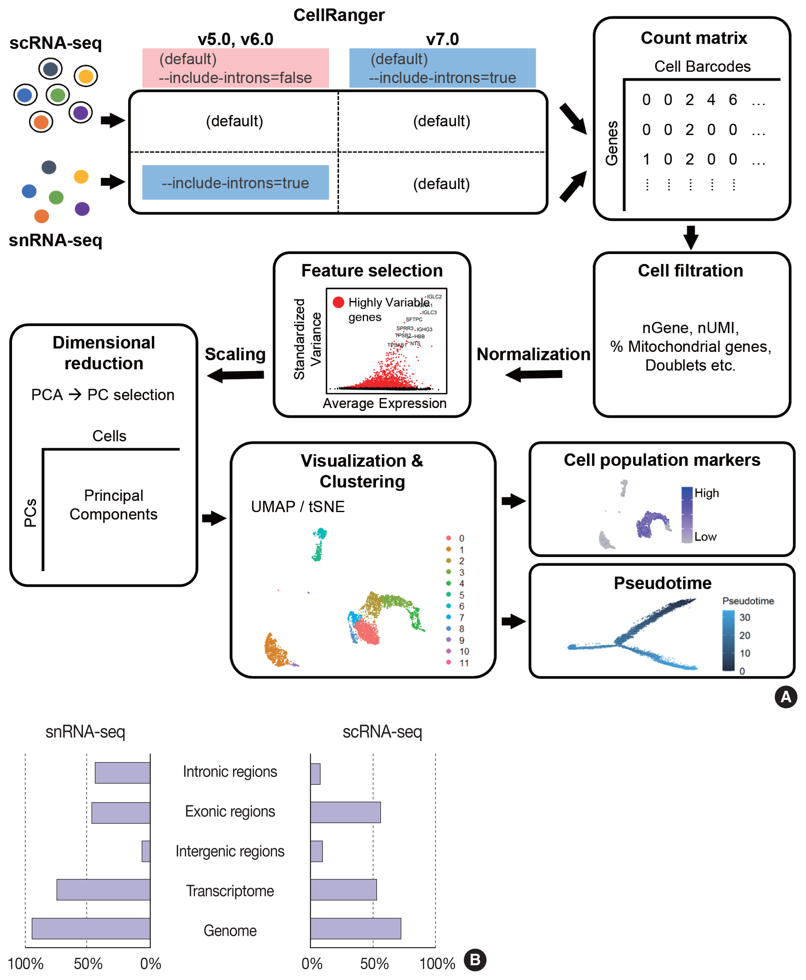
Fig. 2 Summary of single-nucleus RNA sequencing (snRNA-seq) and single-cell RNA sequencing (scRNA-seq) analyses. (A) Schematic workflow of snRNA-seq and scRNA-seq analysis processes. (B) Distribution of confidently mapped snRNA-seq and scRNA-seq reads. Transcriptome, the fraction of reads mapped to the exons of an annotated transcript. Genome, fraction of reads mapped to exonic and non-exonic loci. PC, principal cells; PCA, principal component analysis; UMAP, uniform manifold approximation and projection.

Fig. 3 Comparison of cell types detected by single-nucleus RNA sequencing (snRNA-seq) and single-cell RNA sequencing (scRNA-seq). (A) Uniform manifold approximation and projection (UMAP) plots of snRNA-seq and scRNA-seq data for the human kidney. A bar plot representing the percentages of annotated nuclei and cell identities. AMB, ambiguous; CD, collecting duct; DT, distal tubule; IC, intercalated cells; LH, loop of Henle; LOH (AL), loop of Henle, ascending limb; LOH (DL), loop of Henle, distal limb; NK, natural killer; PC, principal cells; PT, proximal tubule. (B) UMAP plots of snRNA-seq and scRNA-seq data for lung tumors from a lung cancer patient.
Reference
-
References
1. Kolodziejczyk AA, Kim JK, Svensson V, Marioni JC, Teichmann SA. The technology and biology of single-cell RNA sequencing. Mol Cell. 2015; 58:610–20.
Article2. Zeisel A, Munoz-Manchado AB, Codeluppi S, et al. Brain structure. Cell types in the mouse cortex and hippocampus revealed by single-cell RNA-seq. Science. 2015; 347:1138–42.
Article3. Habib N, Avraham-Davidi I, Basu A, et al. Massively parallel single-nucleus RNA-seq with DroNc-seq. Nat Methods. 2017; 14:955–8.
Article4. Lake BB, Ai R, Kaeser GE, et al. Neuronal subtypes and diversity revealed by single-nucleus RNA sequencing of the human brain. Science. 2016; 352:1586–90.
Article5. Andrews TS, Atif J, Liu JC, et al. Single-cell, single-nucleus, and spatial RNA sequencing of the human liver identifies cholangiocyte and mesenchymal heterogeneity. Hepatol Commun. 2022; 6:821–40.
Article6. Petrany MJ, Swoboda CO, Sun C, et al. Single-nucleus RNA-seq identifies transcriptional heterogeneity in multinucleated skeletal myofibers. Nat Commun. 2020; 11:6374.7. Sun W, Dong H, Balaz M, et al. snRNA-seq reveals a subpopulation of adipocytes that regulates thermogenesis. Nature. 2020; 587:98–102.
Article8. Tucker NR, Chaffin M, Fleming SJ, et al. Transcriptional and cellular diversity of the human heart. Circulation. 2020; 142:466–82.
Article9. Wu H, Kirita Y, Donnelly EL, Humphreys BD. Advantages of single-nucleus over single-cell RNA sequencing of adult kidney: rare cell types and novel cell states revealed in fibrosis. J Am Soc Nephrol. 2019; 30:23–32.
Article10. Denisenko E, Guo BB, Jones M, et al. Systematic assessment of tissue dissociation and storage biases in single-cell and single-nucleus RNA-seq workflows. Genome Biol. 2020; 21:130.
Article11. Yamawaki TM, Lu DR, Ellwanger DC, et al. Systematic comparison of high-throughput single-cell RNA-seq methods for immune cell profiling. BMC Genomics. 2021; 22:66.
Article12. Slyper M, Porter CB, Ashenberg O, et al. A single-cell and single-nucleus RNA-Seq toolbox for fresh and frozen human tumors. Nat Med. 2020; 26:792–802.
Article13. Bakken TE, Hodge RD, Miller JA, et al. Single-nucleus and single-cell transcriptomes compared in matched cortical cell types. PLoS One. 2018; 13:e0209648.
Article14. Eraslan G, Drokhlyansky E, Anand S, et al. Single-nucleus cross-tissue molecular reference maps toward understanding disease gene function. Science. 2022; 376:eabl4290.
Article15. Zhou Y, Song WM, Andhey PS, et al. Human and mouse single-nucleus transcriptomics reveal TREM2-dependent and TREM2-independent cellular responses in Alzheimer’s disease. Nat Med. 2020; 26:131–42.
Article16. La Manno G, Soldatov R, Zeisel A, et al. RNA velocity of single cells. Nature. 2018; 560:494–8.
Article17. Hay SB, Ferchen K, Chetal K, Grimes HL, Salomonis N. The Human Cell Atlas bone marrow single-cell interactive web portal. Exp Hematol. 2018; 68:51–61.
Article18. Ullrich S, Guigo R. Dynamic changes in intron retention are tightly associated with regulation of splicing factors and proliferative activity during B-cell development. Nucleic Acids Res. 2020; 48:1327–40.
Article19. Wong JJ, Ritchie W, Ebner OA, et al. Orchestrated intron retention regulates normal granulocyte differentiation. Cell. 2013; 154:583–95.
Article20. Xie X, Shi Q, Wu P, et al. Single-cell transcriptome profiling reveals neutrophil heterogeneity in homeostasis and infection. Nat Immunol. 2020; 21:1119–33.
Article21. Barthelson RA, Lambert GM, Vanier C, Lynch RM, Galbraith DW. Comparison of the contributions of the nuclear and cytoplasmic compartments to global gene expression in human cells. BMC Genomics. 2007; 8:340.
Article22. Grindberg RV, Yee-Greenbaum JL, McConnell MJ, et al. RNA-sequencing from single nuclei. Proc Natl Acad Sci U S A. 2013; 110:19802–7.
Article23. Lake BB, Codeluppi S, Yung YC, et al. A comparative strategy for single-nucleus and single-cell transcriptomes confirms accuracy in predicted cell-type expression from nuclear RNA. Sci Rep. 2017; 7:6031.
Article24. Gupta A, Shamsi F, Altemose N, et al. Characterization of transcript enrichment and detection bias in single-nucleus RNA-seq for mapping of distinct human adipocyte lineages. Genome Res. 2022; 32:242–57.
Article25. Krishnaswami SR, Grindberg RV, Novotny M, et al. Using single nuclei for RNA-seq to capture the transcriptome of postmortem neurons. Nat Protoc. 2016; 11:499–524.
Article26. Lovatt D, Ruble BK, Lee J, et al. Transcriptome in vivo analysis (TIVA) of spatially defined single cells in live tissue. Nat Methods. 2014; 11:190–6.
Article27. Citri A, Pang ZP, Sudhof TC, Wernig M, Malenka RC. Comprehensive qPCR profiling of gene expression in single neuronal cells. Nat Protoc. 2011; 7:118–27.
Article28. Qiu S, Luo S, Evgrafov O, et al. Single-neuron RNA-Seq: technical feasibility and reproducibility. Front Genet. 2012; 3:124.
Article29. Lovatt D, Bell T, Eberwine J. Single-neuron isolation for RNA analysis using pipette capture and laser capture microdissection. Cold Spring Harb Protoc. 2015; 2015:pdb prot072439.
Article30. Ngai J. BRAIN 2.0: Transforming neuroscience. Cell. 2022; 185:4–8.
Article31. Ecker JR, Geschwind DH, Kriegstein AR, et al. The BRAIN Initiative Cell Census Consortium: lessons learned toward generating a comprehensive brain cell atlas. Neuron. 2017; 96:542–57.
Article32. Thrupp N, Sala Frigerio C, Wolfs L, et al. Single-nucleus RNA-seq is not suitable for detection of microglial activation genes in humans. Cell Rep. 2020; 32:108189.
Article33. Bertram JF, Douglas-Denton RN, Diouf B, Hughson MD, Hoy WE. Human nephron number: implications for health and disease. Pediatr Nephrol. 2011; 26:1529–33.
Article34. Park J, Shrestha R, Qiu C, et al. Single-cell transcriptomics of the mouse kidney reveals potential cellular targets of kidney disease. Science. 2018; 360:758–63.
Article35. Hansen J, Sealfon R, Menon R, et al. A reference tissue atlas for the human kidney. Sci Adv. 2022; 8:eabn4965.
Article36. Wu H, Malone AF, Donnelly EL, et al. Single-cell transcriptomics of a human kidney allograft biopsy specimen defines a diverse inflammatory response. J Am Soc Nephrol. 2018; 29:2069–80.
Article37. O’Sullivan ED, Mylonas KJ, Hughes J, Ferenbach DA. Complementary roles for single-nucleus and single-cell RNA sequencing in kidney disease research. J Am Soc Nephrol. 2019; 30:712–3.
Article38. Wen F, Tang X, Xu L, Qu H. Comparison of single-nucleus and single-cell transcriptomes in hepatocellular carcinoma tissue. Mol Med Rep. 2022; 26:339.
Article39. Hwang WL, Jagadeesh KA, Guo JA, et al. Single-nucleus and spatial transcriptome profiling of pancreatic cancer identifies multicellular dynamics associated with neoadjuvant treatment. Nat Genet. 2022; 54:1178–91.
Article
- Full Text Links
-
- Actions
-
Cited
- CITED
-
- Close
- Share
-
- Similar articles
-
- Single-cell and spatial sequencing application in pathology
- Applications of Single-Cell Omics Technologies for Induced Pluripotent Stem Cell-Based Cardiovascular Research
- A semi-automatic cell type annotation method for single-cell RNA sequencing dataset
- Strategy of Patient-Specific Therapeutics in Cardiovascular Disease Through Single-Cell RNA Sequencing
- Functional annotation of lung cancer‒associated genetic variants by cell type‒specific epigenome and long-range chromatin interactome
