Evaluation of the cell viability and antimicrobial effects of orthodontic bands coated with silver or zinc oxide nanoparticles: An in vitro study
- Affiliations
-
- 1Department of Orthodontics, School of Dentistry, Tehran University of Medical Sciences, Tehran, Iran
- 2Dental Research Center, Dentistry Research Institute, Tehran University of Medical Sciences, Tehran, Iran
- 3School of Chemistry, College of Science, University of Tehran, Tehran, Iran
- 4Department of Dental Biomaterials, School of Dentistry, Tehran University of Medical Sciences, Tehran, Iran
- KMID: 2538710
- DOI: http://doi.org/10.4041/kjod22.091
Abstract
Objective
We aimed to evaluate the cell viability and antimicrobial effects of orthodontic bands coated with silver or zinc oxide nanoparticles (nanoAg and nano-ZnO, respectively).
Methods
In this experimental study, 30 orthodontic bands were divided into three groups (n = 10 each): control (uncoated band), Ag (silver-coated band), and ZnO (zinc oxide-coated band). The electrostatic spray-assisted vapor deposition method was used to coat orthodontic bands with nano-Ag or nano-ZnO. The biofilm inhibition test was used to assess the antimicrobial effectiveness of nano-Ag and nano-ZnO against Streptococcus mutans, Lactobacillus acidophilus, and Candida albicans. Biocompatibility tests were conducted using the 3-(4, 5-dimethylthiazol-2-yl)-2, 5-diphenyltetrazolium bromide assay. The groups were compared using oneway analysis of variance with a post-hoc test.
Results
The Ag group showed a significantly higher reduction in the number of L. acidophilus, C. albicans, and S.mutans colonies than the ZnO group (p = 0.015, 0.003, and 0.005, respectively). Compared with the control group, the Ag group showed a 2-log 10 reduction in all the microorganisms' replication ability, but only S. mutants showed a 2-log10 reduction in replication ability in the ZnO group. The lowest mean cell viability was observed in the Ag group, but the difference between the groups was insignificant (p > 0.05).
Conclusions
Coating orthodontic bands with nanoZnO or nano-Ag induced antimicrobial effects against oral pathogens. Among the nanoparticles, nano-Ag showed the best antimicrobial activity and nanoZnO showed the highest biocompatibility.
Figure
-
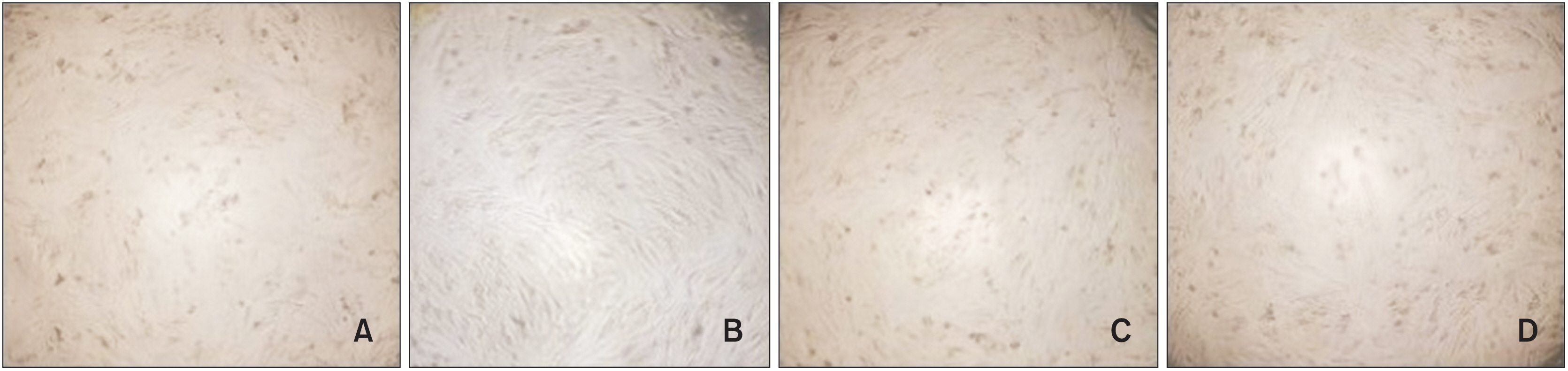
Figure 1 Cells after the addition of eluents but before 3-(4, 5-dimethylthiazol-2-yl)-2, 5-diphenyltetrazolium bromide assay. A, Uncontacted cells with the band. B, Contacted cells with the nano-Ag–coated band. C, Contacted cells with the nano-ZnO–coated band. D, Contacted cells with the uncoated band.

Figure 2 Characterization of synthesized Ag nanoparticles. A, Transmission electron microscopy image of Ag nanoparticles in optimal conditions (scale bar: 20 nm). B, Field emission scanning electron microscopy image of Ag nanoparticles in optimal conditions (scale bar: 200 nm). C, X-ray diffraction graph of Ag nanoparticles.

Figure 3 Characterization of synthesized ZnO nanoparticles. A, Transmission electron microscopy image of ZnO nanoparticles in optimal conditions (scale bar: 20 nm). B, Field emission scanning electron microscopy image of ZnO nanoparticles in optimal conditions (scale bar: 200 nm). C, X-ray diffraction graph of ZnO nanoparticles.

Figure 4 Band surface coated with ZnO nanoparticles. A, Field emission scanning electron microscopy image. B, Energy-dispersive X-ray spectroscopy analysis. L, length. L1, L2 and L3 were randomly selected.
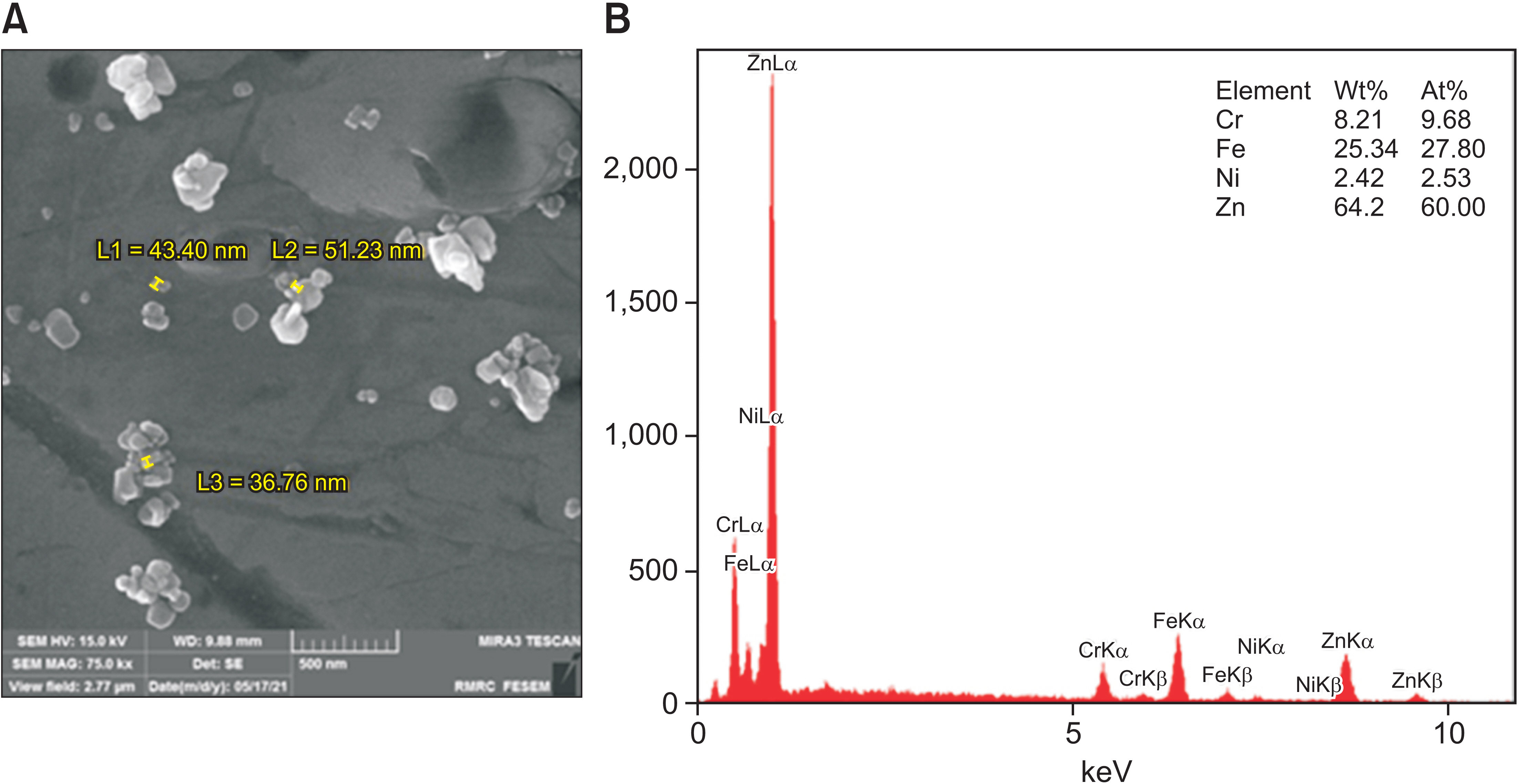
Figure 5 Band surface coated with Ag nanoparticles. A, Field emission scanning electron microscopy image. B, Energy-dispersive X-ray spectroscopy analysis. L, length. L1, L2 and L3 were randomly selected.
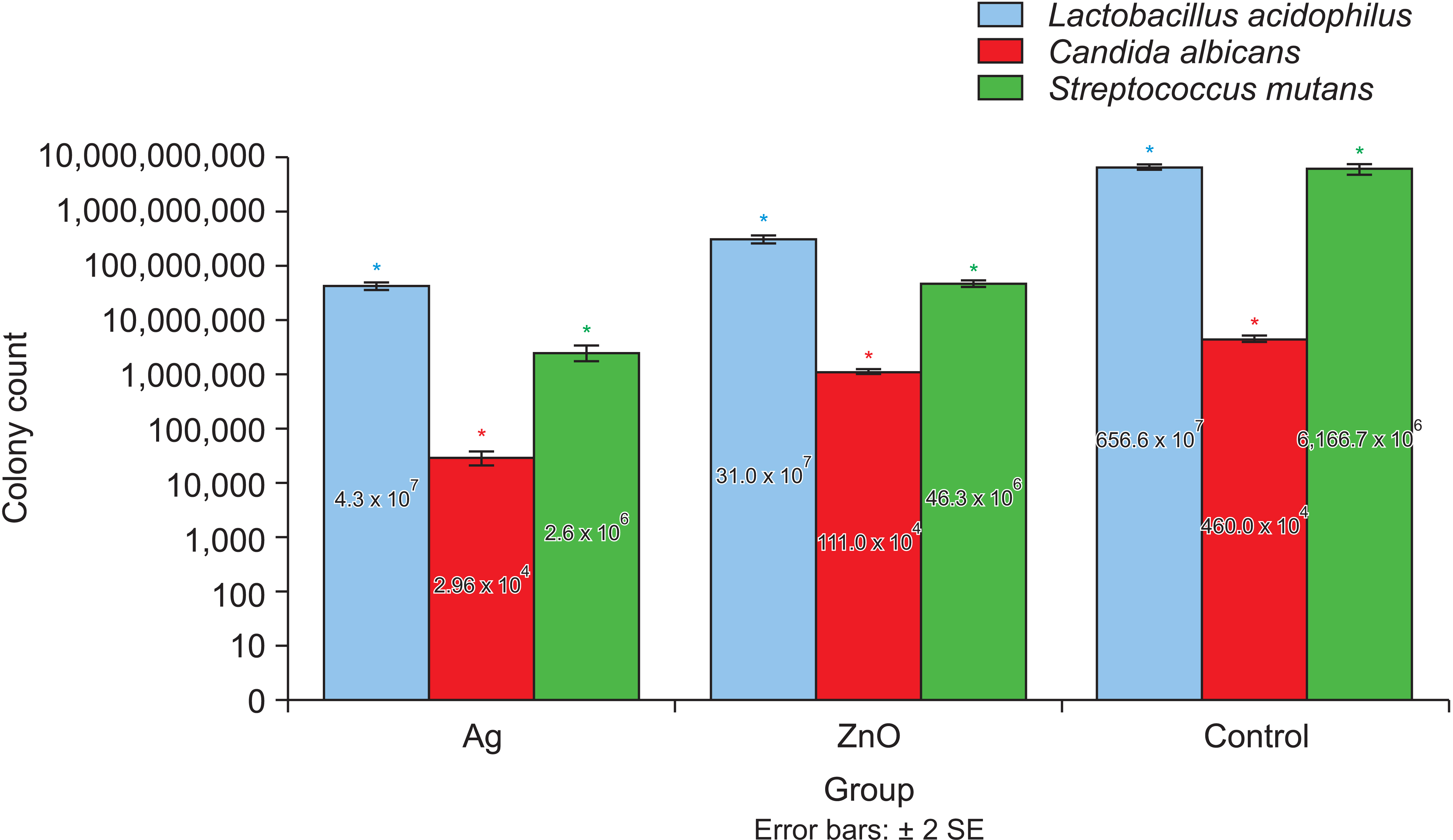
Figure 6 Colony count of the microorganisms for the three groups (colony forming units/mL). SE, standard error. *Significantly different with p < 0.05.
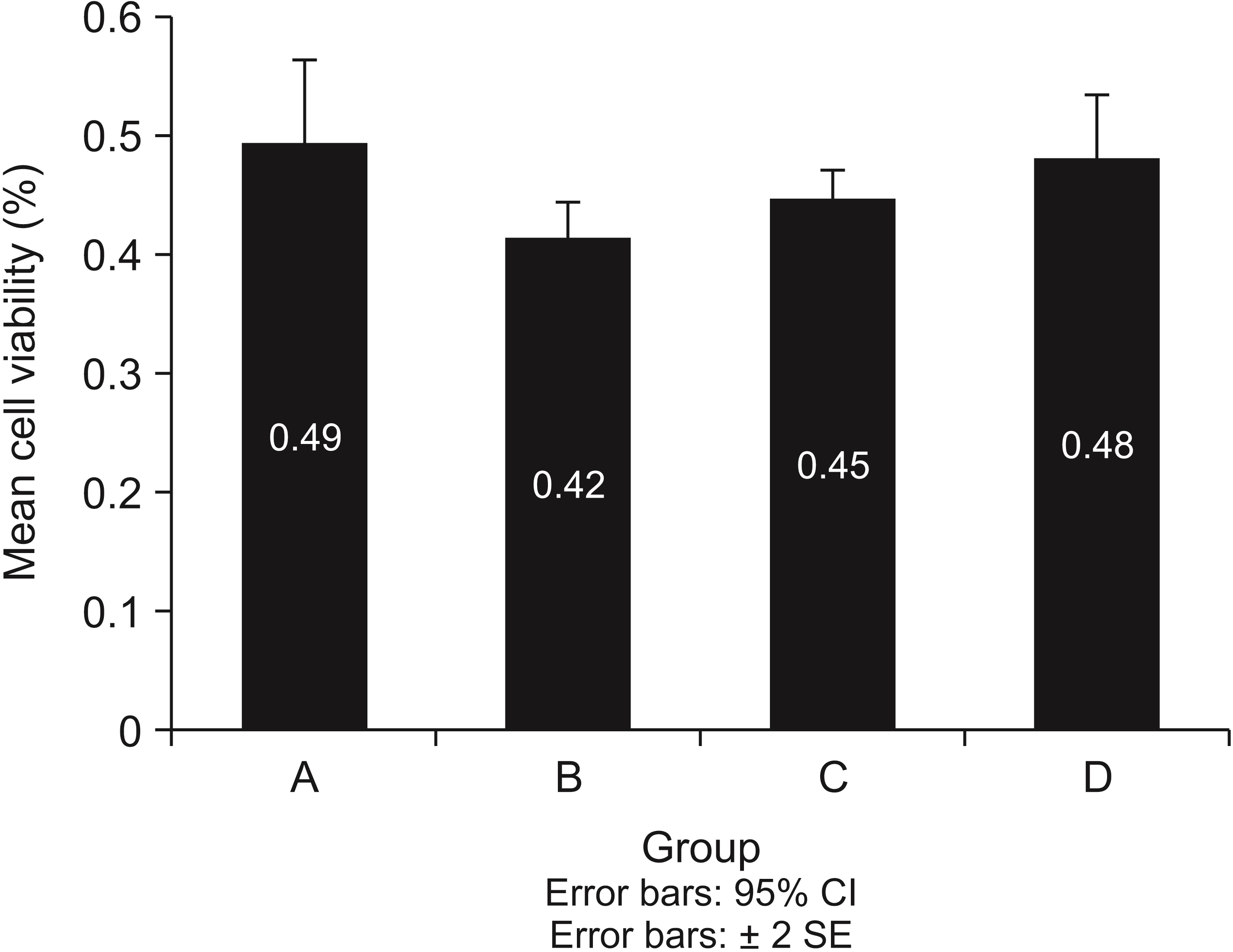
Figure 7 Mean cell viability in each group. A, Uncontacted cells with the band. B, Contacted cells with the nano-Ag–coated band. C, Contacted cells with the nano-ZnO–coated band. D, Contacted cells with the uncoated band. CI, confidence interval; SE, standard error.
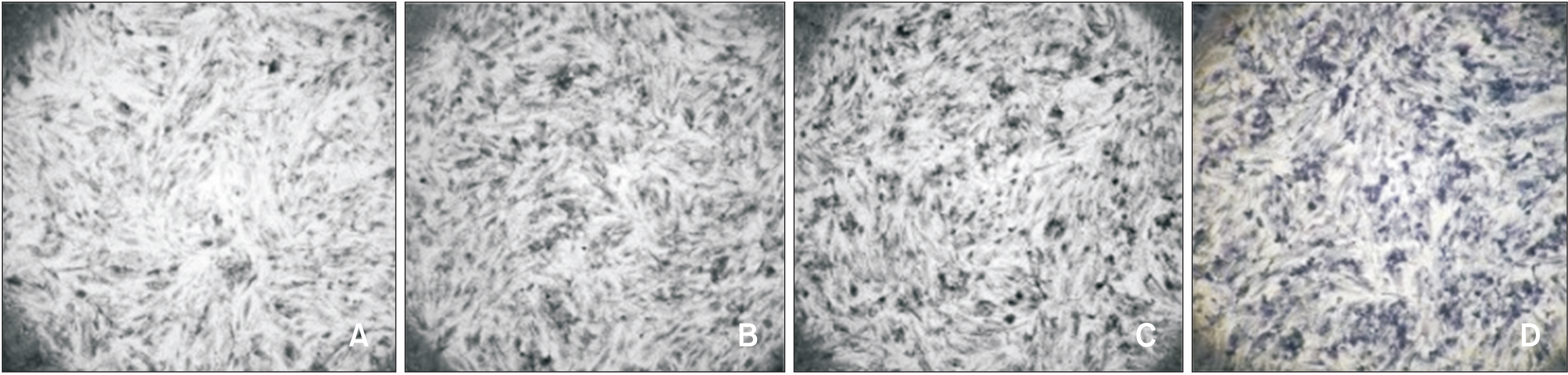
Figure 8 Monolayer culture of the 10459 human gingival fibroblast (HGF) cell line was used for indirect contact assay. A, The control sample consists of a confluent layer of fibroblast cells. Most of the cells are spindle-shaped, which is considered normal. B, The band coated with Ag nanoparticles showed no change in cell morphology following contact with the 10459 HGF confluent layer. C, The 10459 HGF cells in contact with the band coated with ZnO nanoparticles also showed normal morphology. D, The 10459 HGF cells in contact with the uncoated band are spindle-shaped. A, Uncontacted cells with the band; B, Contacted cells with the nano-Ag–coated band; C, Contacted cells with nano-ZnO–coated band; and D, Contacted cells with the uncoated band.
Reference
-
1. Antonio-Zancajo L, Montero J, Albaladejo A, Oteo-Calatayud MD, Alvarado-Lorenzo A. 2020; Pain and oral-health-related quality of life in orthodontic patients during initial therapy with conventional, low-friction, and lingual brackets and aligners (Invisalign): a prospective clinical study. J Clin Med. 9:2088. DOI: 10.3390/jcm9072088. PMID: 32635196. PMCID: PMC7408790. PMID: ae922d85ec61459999b0394a5d12238d.
Article2. Erbe C, Hornikel S, Schmidtmann I, Wehrbein H. 2011; Quantity and distribution of plaque in orthodontic patients treated with molar bands. J Orofac Orthop. 72:13–20. DOI: 10.1007/s00056-010-0001-4. PMID: 21484542.
Article3. Anhoury P, Nathanson D, Hughes CV, Socransky S, Feres M, Chou LL. 2002; Microbial profile on metallic and ceramic bracket materials. Angle Orthod. 72:338–43. DOI: 10.1043/0003-3219(2002)072<0338:MPOMAC>2.0.CO;2. PMID: 12169034.4. Manuelli M, Marcolina M, Nardi N, Bertossi D, De Santis D, Ricciardi G, et al. 2019; Oral mucosal complications in orthodontic treatment. Minerva Stomatol. 68:84–8. DOI: 10.23736/S0026-4970.18.04127-4. PMID: 30854838.
Article5. Bishara SE, Ostby AW. 2008; White spot lesions: formation, prevention, and treatment. Semin Orthod. 14:174–82. DOI: 10.1053/j.sodo.2008.03.002.
Article6. Walsh LJ, Healey DL. 2019; Prevention and caries risk management in teenage and orthodontic patients. Aust Dent J. 64 Suppl 1:S37–45. DOI: 10.1111/adj.12671. PMID: 31144319.
Article7. Lacerda Rangel Esper MÂ, Junqueira JC, Uchoa AF, Bresciani E, Navarro RS, Nara de Souza Rastelli A, et al. 2019; Photodynamic inactivation of planktonic cultures and Streptococcus mutans biofilms for prevention of white spot lesions during orthodontic treatment: an in vitro investigation. Am J Orthod Dentofacial Orthop. 155:243–53. Erratum in: Am J Orthod Dentofacial Orthop 2019;155:458. DOI: 10.1016/j.ajodo.2018.03.027. PMID: 30712696.8. Beyth N, Houri-Haddad Y, Baraness-Hadar L, Yudovin-Farber I, Domb AJ, Weiss EI. 2008; Surface antimicrobial activity and biocompatibility of incorporated polyethylenimine nanoparticles. Biomaterials. 29:4157–63. DOI: 10.1016/j.biomaterials.2008.07.003. PMID: 18678404.
Article9. Lim BS, Lee SJ, Lee JW, Ahn SJ. 2008; Quantitative analysis of adhesion of cariogenic streptococci to orthodontic raw materials. Am J Orthod Dentofacial Orthop. 133:882–8. DOI: 10.1016/j.ajodo.2006.07.027. PMID: 18538253.
Article10. Shah AG, Shetty PC, Ramachandra CS, Bhat NS, Laxmikanth SM. 2011; In vitro assessment of photocatalytic titanium oxide surface modified stainless steel orthodontic brackets for antiadherent and antibacterial properties against Lactobacillus acidophilus. Angle Orthod. 81:1028–35. DOI: 10.2319/021111-101.1. PMID: 22007663. PMCID: PMC8903869.11. De Stefani A, Bruno G, Preo G, Gracco A. 2020; Application of nanotechnology in orthodontic materials: a state-of-the-art review. Dent J (Basel). 8:126. DOI: 10.3390/dj8040126. PMID: 33182424. PMCID: PMC7712537. PMID: edbfccbb4482466e9c6ea6eba21d3ea4.
Article12. Doudi M, Naghsh N, Heiedarpour A. 2011; The effect of silver nanoparticles on gram-negative bacilli resistant to extended-spectrum β-lactamase enzymes. Med Lab J. 5:44–51.13. Lloyd JR. 2003; Microbial reduction of metals and radionuclides. FEMS Microbiol Rev. 27:411–25. DOI: 10.1016/S0168-6445(03)00044-5. PMID: 12829277.
Article14. Bürgers R, Eidt A, Frankenberger R, Rosentritt M, Schweikl H, Handel G, et al. 2009; The anti-adherence activity and bactericidal effect of microparticulate silver additives in composite resin materials. Arch Oral Biol. 54:595–601. DOI: 10.1016/j.archoralbio.2009.03.004. PMID: 19375069.
Article15. Spacciapoli P, Buxton D, Rothstein D, Friden P. 2001; Antimicrobial activity of silver nitrate against periodontal pathogens. J Periodontal Res. 36:108–13. DOI: 10.1034/j.1600-0765.2001.360207.x. PMID: 11327077.
Article16. Ohira T, Yamamoto O, Iida Y, Nakagawa ZE. 2008; Antibacterial activity of ZnO powder with crystallographic orientation. J Mater Sci Mater Med. 19:1407–12. DOI: 10.1007/s10856-007-3246-8. PMID: 17914627.
Article17. Afonso Camargo SE, Mohiuddeen AS, Fares C, Partain JL, Carey PH 4th, Ren F, et al. 2020; Anti-bacterial properties and biocompatibility of novel SiC coating for dental ceramic. J Funct Biomater. 11:33. DOI: 10.3390/jfb11020033. PMID: 32443691. PMCID: PMC7353563. PMID: 114fa56d7f834778a4ccad9da19491fe.
Article18. Miles AA, Misra SS, Irwin JO. 1938; The estimation of the bactericidal power of the blood. J Hyg (Lond). 38:732–49. DOI: 10.1017/S002217240001158X. PMID: 20475467. PMCID: PMC2199673.
Article19. International Organization for Standardization (ISO). 2009. ISO 10993-5:2009. Biological evaluation of medical devices- part 5: tests for in vitro cytotoxicity. ISO Copyright Office;Geneva:20. Tavassoli-Hojjati S, Haghgoo R, Mehran M, Niktash A. 2012; Evaluation of the effect of fluoride gel and varnish on the demineralization resistance of enamel: an in vitro. J Iran Dent Assoc. 24:28–34.21. Farhadian N, Usefi Mashoof R, Khanizadeh S, Ghaderi E, Farhadian M, Miresmaeili A. 2016; Streptococcus mutans counts in patients wearing removable retainers with silver nanoparticles vs those wearing conventional retainers: a randomized clinical trial. Am J Orthod Dentofacial Orthop. 149:155–60. Erratum in: Am J Orthod Dentofacial Orthop 2017;151:11. DOI: 10.1016/j.ajodo.2015.07.031. PMID: 26827971.22. Arash V, Keikhaee F, Rabiee SM, Rajabnia R, Khafri S, Tavanafar S. 2016; Evaluation of antibacterial effects of silver-coated stainless steel orthodontic brackets. J Dent (Tehran). 13:49–54. PMID: 27536328. PMCID: PMC4983565. PMID: 93db76e397d94eb4b16f3f304c3d33e2.23. Ghorbanzadeh R, Pourakbari B, Bahador A. 2015; Effects of baseplates of orthodontic appliances with in situ generated silver nanoparticles on cariogenic bacteria: a randomized, double-blind cross-over clinical trial. J Contemp Dent Pract. 16:291–8. DOI: 10.5005/jp-journals-10024-1678. PMID: 26067732.
Article24. Poosti M, Ramazanzadeh B, Zebarjad M, Javadzadeh P, Naderinasab M, Shakeri MT. 2013; Shear bond strength and antibacterial effects of orthodontic composite containing TiO2 nanoparticles. Eur J Orthod. 35:676–9. DOI: 10.1093/ejo/cjs073. PMID: 23264617.
Article25. Jedrychowski JR, Caputo AA, Kerper S. 1983; Antibacterial and mechanical properties of restorative materials combined with chlorhexidines. J Oral Rehabil. 10:373–81. DOI: 10.1111/j.1365-2842.1983.tb00133.x. PMID: 6355413.
Article26. Bulut H, Türkün M, Türkün LS, Işiksal E. 2007; Evaluation of the shear bond strength of 3 curing bracket bonding systems combined with an antibacterial adhesive. Am J Orthod Dentofacial Orthop. 132:77–83. DOI: 10.1016/j.ajodo.2005.06.040. PMID: 17628254.
Article27. Kachoei M, Divband B, Rahbar M, Esmaeilzadeh M, Ghanizadeh M, Alam M. 2021; A novel developed bioactive composite resin containing silver/zinc oxide (Ag/ZnO) nanoparticles as an antimicrobial material against Streptococcus mutans, Lactobacillus, and Candida albicans. Evid Based Complement Alternat Med. 2021:4743411. DOI: 10.1155/2021/4743411. PMID: 34697547. PMCID: PMC8541865.28. Garmasheva I, Kovalenko N, Voychuk S, Ostapchuk A, Livins'ka O, Oleschenko L. 2016; Lactobacillus species mediated synthesis of silver nanoparticles and their antibacterial activity against opportunistic pathogens in vitro. Bioimpacts. 6:219–23. DOI: 10.15171/bi.2016.29. PMID: 28265538. PMCID: PMC5326670.
Article29. Sharma VK, Yngard RA, Lin Y. 2009; Silver nanoparticles: green synthesis and their antimicrobial activities. Adv Colloid Interface Sci. 145:83–96. DOI: 10.1016/j.cis.2008.09.002. PMID: 18945421.
Article30. Chen X, Schluesener HJ. 2008; Nanosilver: a nanoproduct in medical application. Toxicol Lett. 176:1–12. DOI: 10.1016/j.toxlet.2007.10.004. PMID: 18022772.
Article31. Hernández-Sierra JF, Ruiz F, Pena DC, Martínez-Gutiérrez F, Martínez AE, Guillén Ade J, et al. 2008; The antimicrobial sensitivity of Streptococcus mutans to nanoparticles of silver, zinc oxide, and gold. Nanomedicine. 4:237–40. DOI: 10.1016/j.nano.2008.04.005. PMID: 18565800.
Article32. Cieplik F, Aparicio C, Kreth J, Schmalz G. 2022; Development of standard protocols for biofilm-biomaterial interface testing. JADA Found Sci. 1:100008. DOI: 10.1016/j.jfscie.2022.100008.
Article33. Kasraei S, Sami L, Hendi S, Alikhani MY, Rezaei-Soufi L, Khamverdi Z. 2014; Antibacterial properties of composite resins incorporating silver and zinc oxide nanoparticles on Streptococcus mutans and Lactobacillus. Restor Dent Endod. 39:109–14. DOI: 10.5395/rde.2014.39.2.109. PMID: 24790923. PMCID: PMC3978100.
Article34. Hailan SY, Al-Khatieeb MM. 2019; Antimicrobial efficacy of silver, zinc oxide, and titanium dioxide nanoparticles incorporated in orthodontic bonding agent. J Baghdad Coll Dent. 31:10–6. DOI: 10.26477/jbcd.v31i3.2693. PMID: 2adb939a2c1747d28c8cc28b100e6b21.
Article35. Ahrari F, Eslami N, Rajabi O, Ghazvini K, Barati S. 2015; The antimicrobial sensitivity of Streptococcus mutans and Streptococcus sangius to colloidal solutions of different nanoparticles applied as mouthwashes. Dent Res J (Isfahan). 12:44–9. DOI: 10.4103/1735-3327.150330. PMID: 25709674. PMCID: PMC4336971.
Article36. Prabha RD, Kandasamy R, Sivaraman US, Nandkumar MA, Nair PD. 2016; Antibacterial nanosilver coated orthodontic bands with potential implications in dentistry. Indian J Med Res. 144:580–6. DOI: 10.4103/0971-5916.200895. PMID: 28256467. PMCID: PMC5345305.37. Halimi SU, Bakar NFA, Ismail SN, Hashib SA. 2014; Electrospray deposition of titanium dioxide (TiO2) nanoparticles. AIP Conf Proc. 1586:57. DOI: 10.1063/1.4866730.
Article38. Bondarenko O, Juganson K, Ivask A, Kasemets K, Mortimer M, Kahru A. 2013; Toxicity of Ag, CuO and ZnO nanoparticles to selected environmentally relevant test organisms and mammalian cells in vitro: a critical review. Arch Toxicol. 87:1181–200. DOI: 10.1007/s00204-013-1079-4. PMID: 23728526. PMCID: PMC3677982.
Article39. Kim S, Choi JE, Choi J, Chung KH, Park K, Yi J, et al. 2009; Oxidative stress-dependent toxicity of silver nanoparticles in human hepatoma cells. Toxicol In Vitro. 23:1076–84. DOI: 10.1016/j.tiv.2009.06.001. PMID: 19508889.
Article40. Lesniak A, Salvati A, Santos-Martinez MJ, Radomski MW, Dawson KA, Åberg C. 2013; Nanoparticle adhesion to the cell membrane and its effect on nanoparticle uptake efficiency. J Am Chem Soc. 135:1438–44. DOI: 10.1021/ja309812z. PMID: 23301582.
Article41. Shahi S, Özcan M, Maleki Dizaj S, Sharifi S, Al-Haj Husain N, Eftekhari A, et al. 2019; A review on potential toxicity of dental material and screening their biocompatibility. Toxicol Mech Methods. 29:368–77. DOI: 10.1080/15376516.2019.1566424. PMID: 30642212.
Article
- Full Text Links
-
- Actions
-
Cited
- CITED
-
- Close
- Share
-
- Similar articles
-
- Skin corrosion and irritation test of sunscreen nanoparticles using reconstructed 3D human skin model
- Antibacterial properties of composite resins incorporating silver and zinc oxide nanoparticles on Streptococcus mutans and Lactobacillus
- Silver Nanoparticles as a Smart Antimicrobial Agent
- In vitro antimicrobial effect of the tissue conditioner containing silver nanoparticles
- An experimental study on the cytotoxicity of various orthodontic bands
