Digital technique in diagnosis and restoration of maxillary anterior implant: a case report
- Affiliations
-
- 1Department of Prosthodontics, School of Dentistry, Chonnam National University, Gwangju, Republic of Korea
- KMID: 2538523
- DOI: http://doi.org/10.14368/jdras.2022.38.4.249
Abstract
- The implant prosthesis of anterior maxilla requires careful consideration in planning. In order to satisfy both esthetic and functional needs of a patient, fusion of intra-oral scan in Cone-beam computed tomography (CBCT) and facial scan can be considered. Bony structures and soft tissues captured in CBCT and occlusal surfaces of intra oral scan were incorporated into personal characteristics from facial scan. The patient had insufficient buccal bone on maxillary anterior area. The maxillary implants could not be placed on the most ideal position. However, the “top down” approach completed by computer-generated arranging of teeth in implant planning and surgery with surgical guide resulted in esthetically and functionally satisfying result regardless of the limitation. Careful diagnosis with digital technique and the usage of surgical guide resulted in successful surgery and esthetic restoration. The temporary fixed prostheses were designed, restored and evaluated. The patient was not satisfied with the first design of temporary prosthesis, which showed uneven space distribution between teeth due to the position of maxillary implant. The design was modified by changing proximal emergence contours and line angle to alter the perceived since of incisors. The patient was satisfied with the new design of provisional restoration. A digital occlusion analyzer (Arcus Digma II, KaVo, Leutkirch, Germany) was used to measure inherent condylar guidance and anterior guidance of a patient to provide a definitive prosthesis.
Keyword
Figure
-
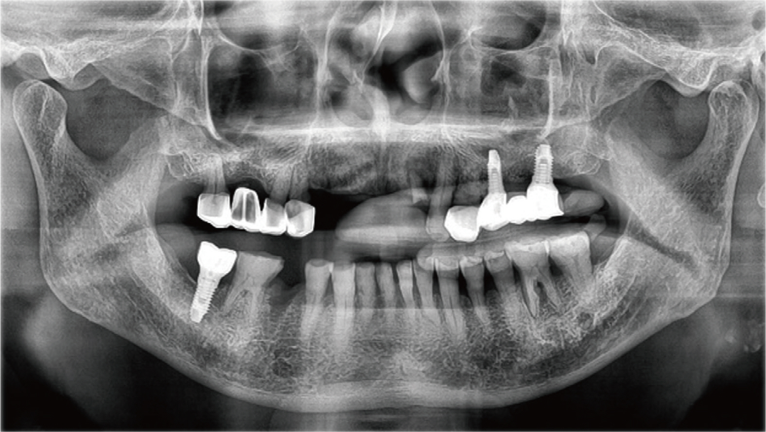
Fig. 1 Initial panoramic radiograph.
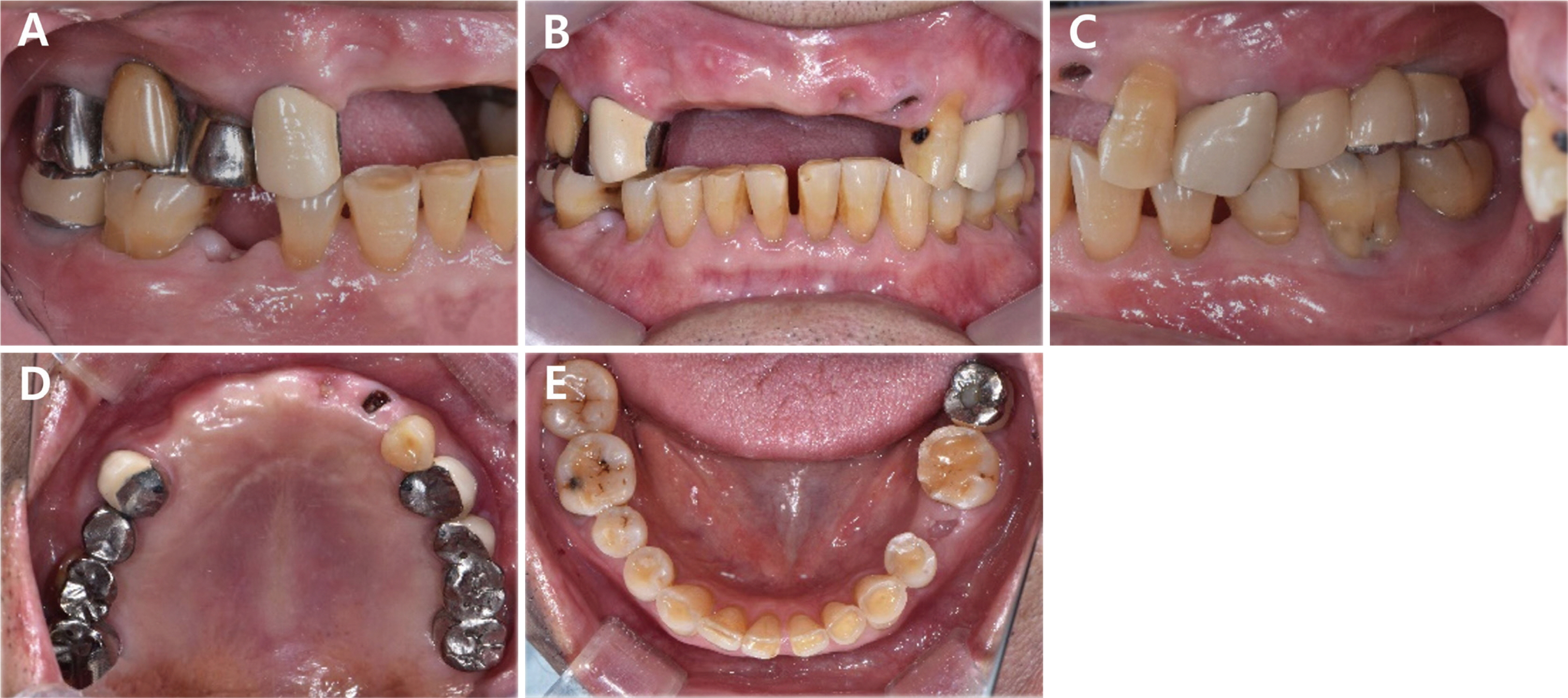
Fig. 2 Initial intraoral photographs. (A) Right lateral view, (B) Frontal view, (C) Left lateral view, (D) Maxillary occlusal view, (E) Mandibular occlusal view.

Fig. 3 Digital facebow transfer. (A) Digital facebow: right view, (B) Digital facebow: front view, (C) Digital facebow: left view.
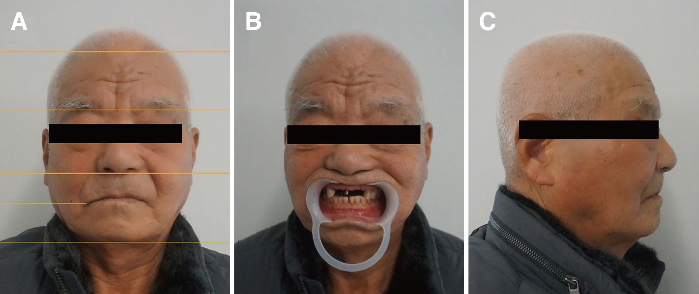
Fig. 4 Extra-oral photographs for esthetic analysis. (A) Front view, showing hairline, glabella, subnasale, lip line, and menton, (B) Front view with lip retractor, (C) Lateral view.

Fig. 5 Design of Surgical Guide with facial scan, CBCT (Dicom file), and intra oral scan. (A) Digital wax up on CAD software and virtual implant placement, (B) Front view: Superimposition of wax up, intra oral scan, CBCT (dicom file), and facial scan, (C) Digital wax up of maxillary anterior area and placement of implant fixture, (D) Lateral view: superimposition of surgical guide, intra oral scan, CBCT (dicom file), a facial scan.
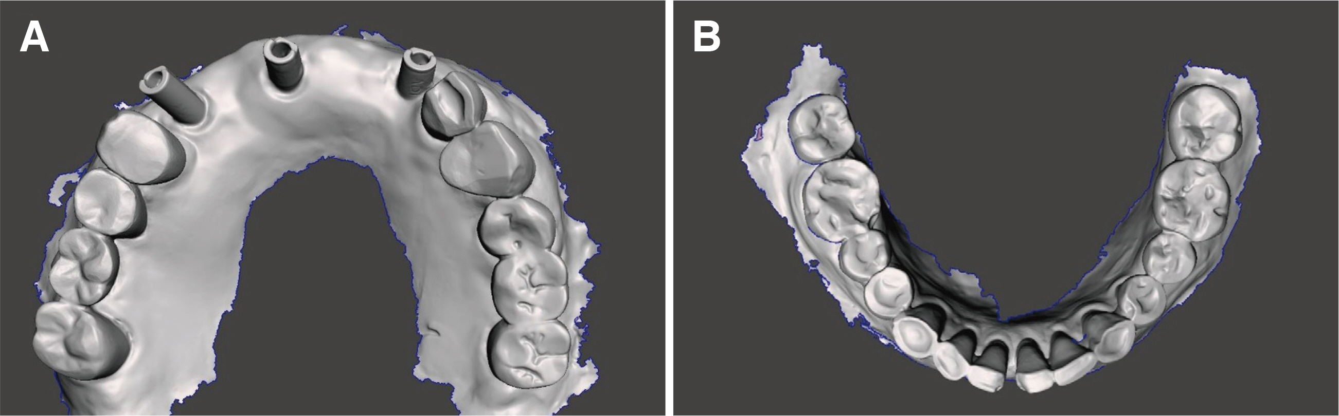
Fig. 6 Impression taking with intraoral scan. (A) Digital impression taking with scanbody, (B) Intra oral scan of mandible.

Fig. 7 Provisional restoration. (A) CAD design of provisional restoration, (B) Superimposition of Provisional restoration, intra oral scan and CBCT (dicom file), (C) Virtual patient with Provisional restoration, (D) Superimposition of Provisional restoration, intra oral scan, CBCT (dicom file), and facial scan, (E) Intraoral photo of provisional restoration, (F) Extraoral photo of a patient with provisional restoration.

Fig. 8 A digital occlusion analyzer (Arcus Digma II, KaVo, Leutkirch, Germany). (A) A patient with Arcus digma II, (B) Arcus digma II result.
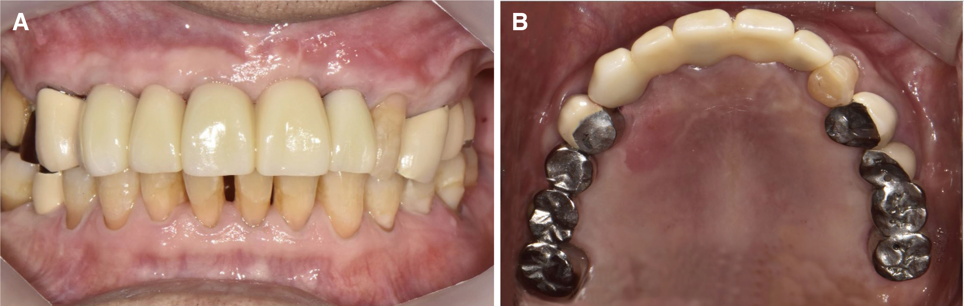
Fig. 9 Final restoration. (A) Intraoral photo: Front view, (B) Intraoral photo: Occlusal view.
Reference
-
References
1. Levin EI. 1978; Dental esthetics and the golden proportion. J Prosthet Dent. 40:244–52. DOI: 10.1016/0022-3913(78)90028-8. PMID: 279670. PMID: https://www.scopus.com/inward/record.uri?partnerID=HzOxMe3b&scp=0018013322&origin=inward.
Article2. Vercruyssen M, Laleman I, Jacobs R, Quirynen M. 2015; Computer-supported implant planning and guided surgery: a narrative review. Clin Oral Implants Res. 26 Suppl 11:69–76. DOI: 10.1111/clr.12638. PMID: 26385623. PMID: https://www.scopus.com/inward/record.uri?partnerID=HzOxMe3b&scp=84941887677&origin=inward.3. Jung RE, Schneider D, Ganeles J, Wismeijer D, Zwahlen M, Hämmerle CH, Tahmaseb A. 2009; Computer technology applications in surgical implant dentistry: a systematic review. Int J Oral Maxillofac Implants. 24 Suppl:92–109. DOI: 10.11607/jomi.2014suppl.g1.2. PMID: 24660188. PMID: https://www.scopus.com/inward/record.uri?partnerID=HzOxMe3b&scp=84901928997&origin=inward.
Article4. Sesemann MR. 2017; Understanding and providing - Appropriate line angles to optimize smile design restorations. J Cosmet Dent. 33:66–75. PMID: https://www.scopus.com/inward/record.uri?partnerID=HzOxMe3b&scp=84901928997&origin=inward.5. Chiche G, Pinault A. 1994. Esthetics of anterior fixed prosthodontics. 1st ed. Quitessence Pub.;Hanover Park: p. 25–31. PMID: https://www.scopus.com/inward/record.uri?partnerID=HzOxMe3b&scp=84901928997&origin=inward.6. Bhuvaneswaran M. 2010; Principles of smile design. J Conser Dent. 13:225–32. DOI: 10.4103/0972-0707.73387. PMID: 21217950. PMCID: PMC3010027.
Article7. Stiesch-Scholz M, Demling A, Rossbach A. 2006; Reproducibility of jaw movements in patients with craniomandibular disorders. J Oral Rehabil. 33:807–12. DOI: 10.1111/j.1365-2842.2006.01636.x. PMID: 17002739. PMID: https://www.scopus.com/inward/record.uri?partnerID=HzOxMe3b&scp=33749317209&origin=inward.
Article8. Becker CM, Kaiser DA. 1993; Evolution of occlusion and occlusal instruments. J Prosthodont. 2:33–43. DOI: 10.1111/j.1532-849X.1993.tb00379.x. PMID: 8374710. PMID: https://www.scopus.com/inward/record.uri?partnerID=HzOxMe3b&scp=0027568118&origin=inward.
Article9. Leung CC, Palomo L, Griffith R, Hans MG. 2010; Accuracy and reliability of cone-beam computed tomography for measuring alveolar bone height and detecting bony dehiscences and fenestrations. Am J Orthod Dentofacial Orthop. 137 Suppl 4:S109–19. DOI: 10.1016/j.ajodo.2009.07.013. PMID: 20381751. PMID: https://www.scopus.com/inward/record.uri?partnerID=HzOxMe3b&scp=77950360946&origin=inward.
Article10. Li J, Chen Z, Decker AM, Wang HL, Joda T, Mendonca G, Lepidi L. 2022; Trueness and Precision of Economical Smartphone-Based Virtual Facebow Records. J Prosthodont. 31:22–9. DOI: 10.1111/jopr.13366. PMID: 33876857. PMCID: PMC8526632. PMID: https://www.scopus.com/inward/record.uri?partnerID=HzOxMe3b&scp=85104964130&origin=inward.
Article
- Full Text Links
-
- Actions
-
Cited
- CITED
-
- Close
- Share
-
- Similar articles
-
- Digital intraoral impression for immediate provisional restoration of maxillary single implant: A case report
- Immediate restoration through gingiva conditioning of maxillary anterior implant installed labially: A case report
- Usage of digital technique to facilitate communication between dentist, dental lab technician, and patients in diagnosis and restoration for maxillary anterior implant: a case report
- Aesthetic prosthetic restoration through immediate implant placement and provisional restoration in the maxillary anterior region using a digital guide
- Decoronation and implant restoration of ankylosed tooth resulted from anterior avulsion: A case report
