Aesthetic prosthetic restoration through immediate implant placement and provisional restoration in the maxillary anterior region using a digital guide
- Affiliations
-
- 1Department of Prosthodontics, College of Dentistry, Dankook University, Cheonan, Republic of Korea
- KMID: 2562773
- DOI: http://doi.org/10.14368/jdras.2024.40.4.291
Abstract
- Immediate implant placement and immediate loading in the anterior maxilla is an effective approach to rapidly address aesthetic demands. To achieve successful outcomes, bone quality, soft tissue condition, and accurate implant positioning are essential factors. For optimal results, procedures such as bone augmentation, precise implant placement, and, when necessary, soft tissue grafting should be considered. Furthermore, provisional restoration play a crucial role in achieving the desired appearance of prosthetic restorations and improving the aesthetics of the soft tissue. By performing soft tissue molding through provisional restoration, an ideal emergence profile can be established, which can be subsequently transferred to the final prosthesis, leading to a functional and aesthetically pleasing restoration. This approach aims to optimize the aesthetic outcomes in the anterior region while preserving the natural contours of the peri-implant soft tissue. In this case, a patient requiring extraction of maxillary anterior tooth underwent immediate implantation and alveolar bone grafting using a guide fabricated in advance from CT data. The patient received a provisional restoration on the same day. Subsequent steps included transitioning from the provisional prosthesis to the definitive prosthesis, ultimately achieving an aesthetically pleasing and functional implant restoration. We report this case to highlight the successful approach to maxillary anterior implant rehabilitation.
Keyword
Figure
-

Fig. 1 Initial panoramic radiographic image. (A) Periapical radiograph, (B) Panoramic radiograph.

Fig. 2 Intraoral photograph before treatment. (A) Occlusal view, (B) Frontal view, (C) Lateral view.
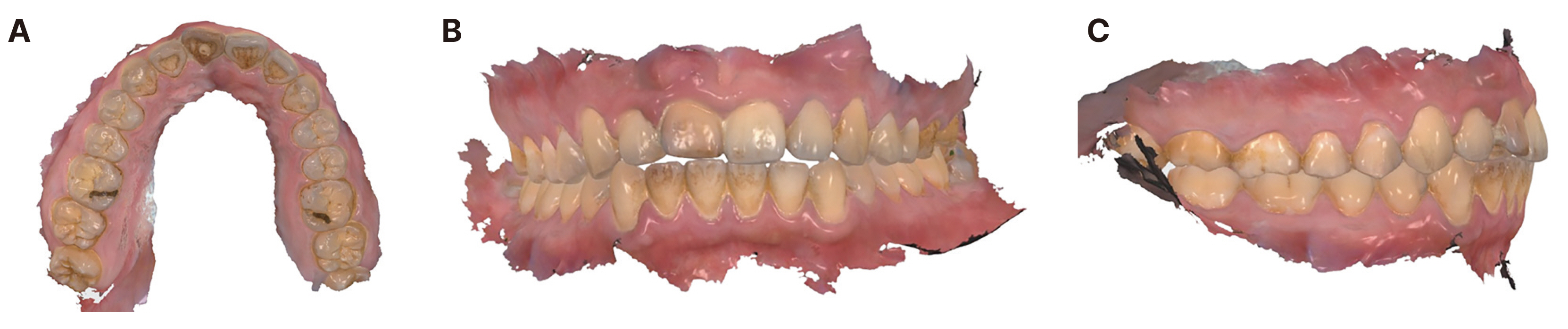
Fig. 3 Digital impression taking for definitive restoration by intraoral scanner. (A) Screenshot of the digital impression of the occlusal side, (B) Screenshot of the digital impression of frontal side, (C) Screenshot of digital impression of lateral side.
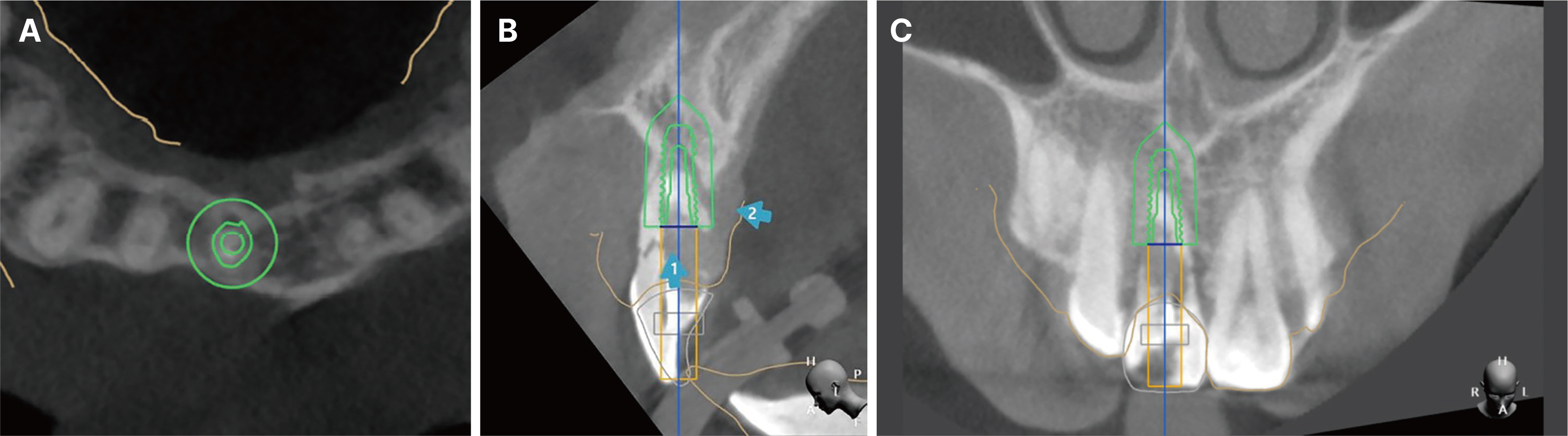
Fig. 4 3D digital implant planning with software. (A) Implant position on occlusal view, (B) Implant position on sagittal view, (C) Implant position on frontal view.

Fig. 5 Provisional abutment & crown design with software. (A) Design of provisional titanium abutment, (B) Design of provisional crown.

Fig. 6 Immediate implant placement with guide. (A) Extraction of #11, (B) Surgical guide try-in.
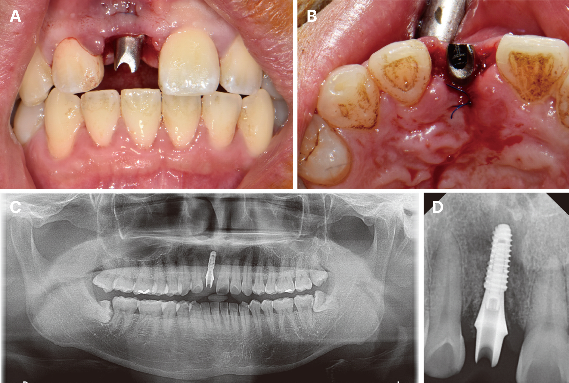
Fig. 7 Delivered provisional titanium abutment. (A) Intraoral photograph of provisional titanium abutment placement (Left: Frontal view, Right: Occlusal view.), (B) Panoramic radiograph after #11 abutment placement, (C) Periapical radiograph after #11 abutment placement.

Fig. 8 Contouring of provisional crown.

Fig. 9 Immediate provisional restoration. (A) Occlusal view, (B) Frontal view.

Fig. 10 Stable soft tissues after 8 weeks with the provisional crown insertion. (A) Frontal view, (B) Occlusal view after the removal of provisional restoration.
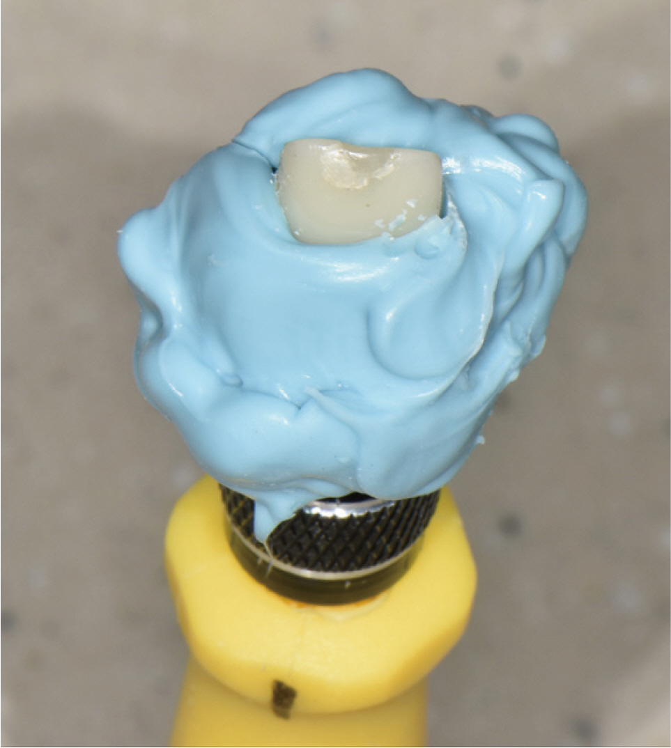
Fig. 11 Fabrication of customized impression coping.
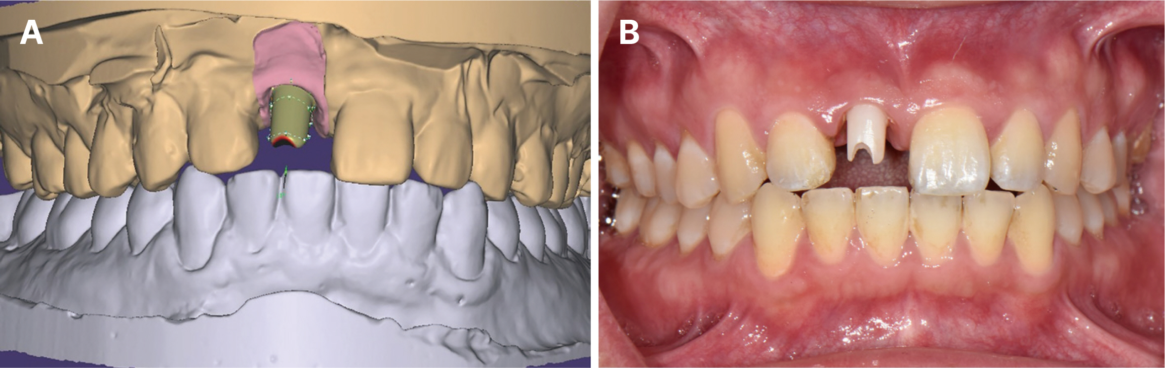
Fig. 12 Procedure of designing zirconia abutments using software. (A) Computer aided design for zirconia abutment, (B) Intraoral insertion of zirconia abutment.
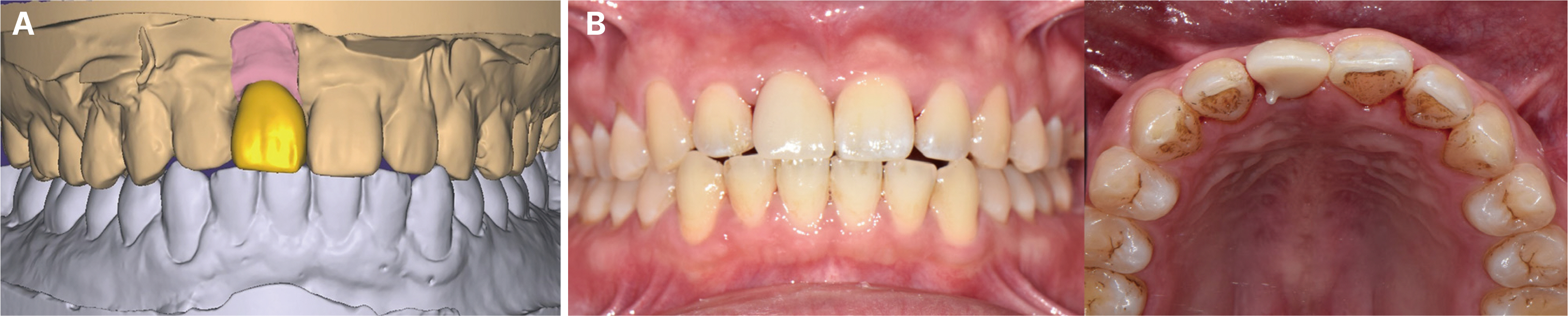
Fig. 13 Definitive prosthesis. (A) Computer aided design for definitive porcelain fused zirconia, (B) Intraoral insertion of definitive prosthesis (Left: Frontal view, Right: Occlusal view).
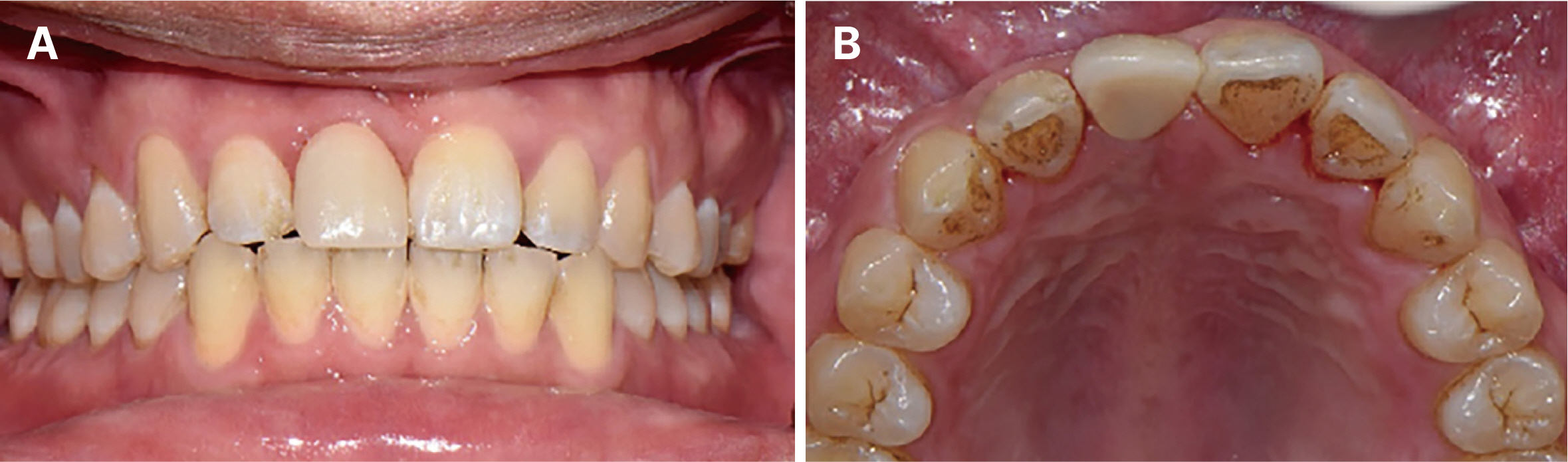
Fig. 14 Clinical photo of definitive restoration after 6 months. (A) Frontal view, (B) Occlusal view.
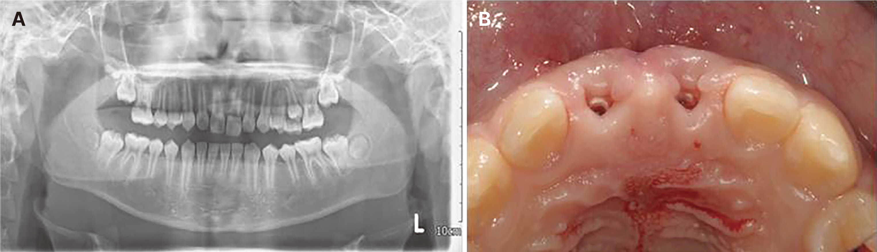
Fig. 15 Initial image. (A) Panoramic radiograph, (B) Clinical occlusal view.
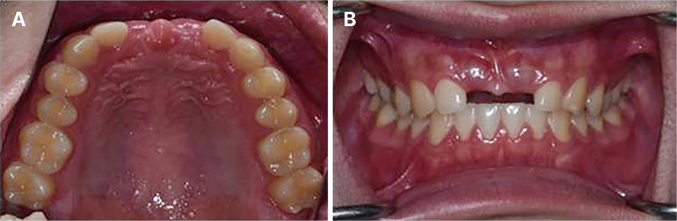
Fig. 16 Intraoral photograph before treatment. (A) Occlusal view, (B) Frontal view.

Fig. 17 Periapical radiograph.
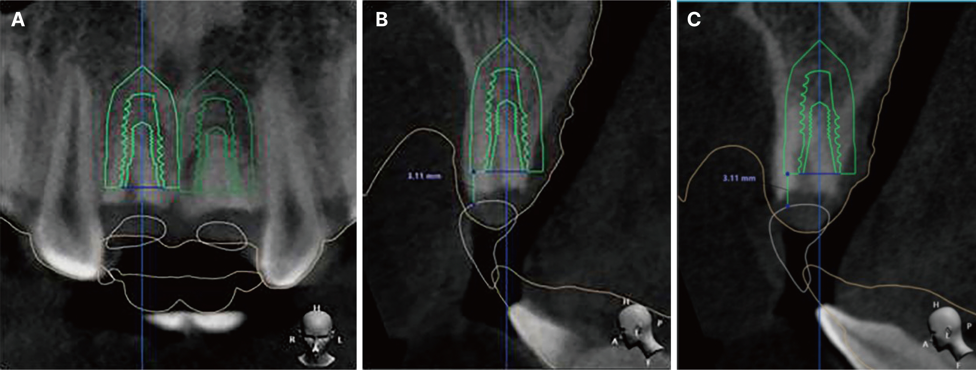
Fig. 18 3D digital implant planning with software. (A) Implant position on frontal view, (B) Implant position on sagittal view (#11), (C) Implant position on sagittal view (#21).
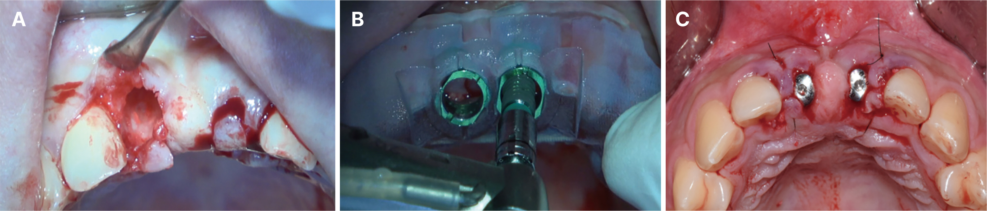
Fig. 19 Immediate implant placement with guide. (A) Extraction of #11, 21, (B) Surgical guide for #11, 21 implantation, (C) Occlusal view after implant surgery.
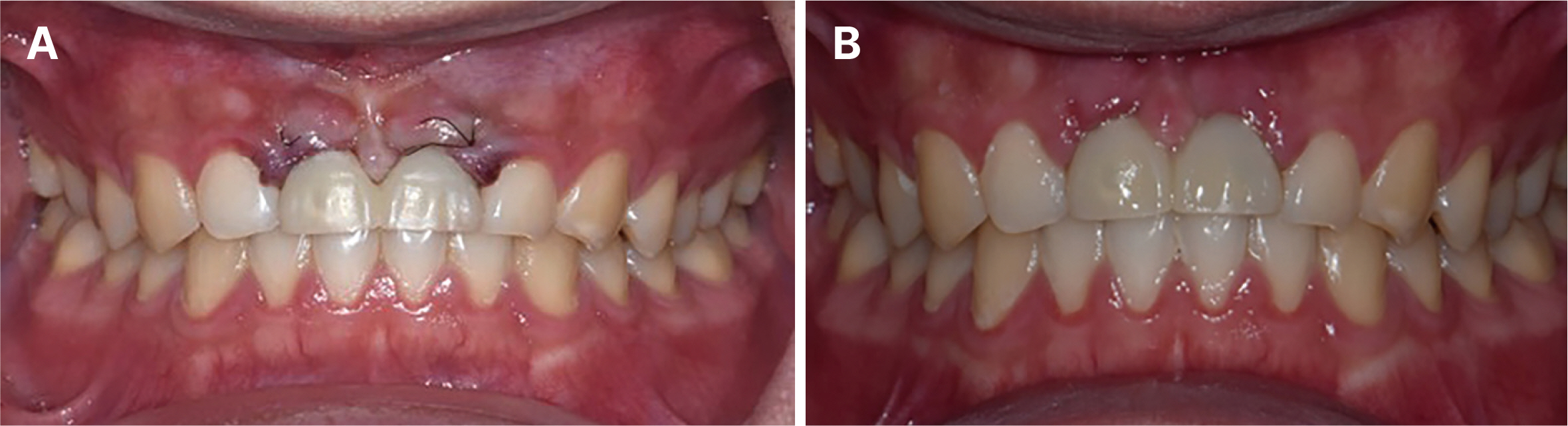
Fig. 20 Immediate provisional restoration. (A) Frontal view: Immediate provisional restoration, (B) Frontal view: Stable soft tissues after 2 weeks with the provisional crown insertion.
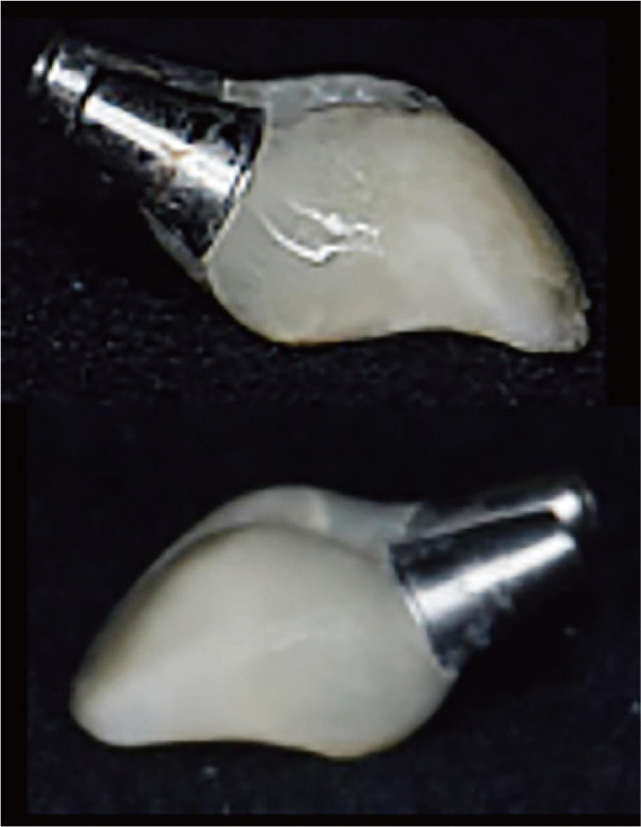
Fig. 21 Contouring of provisional crown.

Fig. 22 Digital custom impression to record emergence profile. (A) Intraoral scan with provisional in place, (B) Intraoral scan of soft tissue after removing provisional restoration, (C) Digitally locking of the soft tissue profile, (D) Digital scan tissue surface of provisional prosthesis.

Fig. 23 Frontal view of Intraoral insertion of zirconia abutment.

Fig. 24 Definitive prosthesis. (A) Frontal view, (B) Occlusal view.

Fig. 25 Check occlusal relationship with T-scan Novus. (A) Protrusion, (B) Maximum intercuspation, (C) Right lateroguidance, (D) Left lateroguidance.
Reference
-
References
1. Arora H, Ivanovski S. 2021; Ten Year Clinical and Aesthetic Outcomes of an Immediately Placed and Restored Implant in the Anterior Maxilla: A Case Report. Prosthesis. 3:129–36. DOI: 10.3390/prosthesis3020014.2. Hämmerle CH, Chen ST, Wilson TG Jr. Consensus statements and recommended clinical procedures regarding the placement of implants in extraction sockets. Int J Oral Maxillofac Implants. 2004; 19 Suppl:26–8.3. Esposito M, Grusovin MG, Coulthard P, Worthington HV. 2006; The efficacy of various bone augmentation procedures for dental implants: a Cochrane systematic review of randomized controlled clinical trials. Int J Oral Maxillofac Implants. 21:696–710.4. Gallucci GO, Benic GI, Eckert SE, Papaspyridakos P, Schimmel M, Schrott A, Weber HP. Consensus Statements and Clinical Recommendations for Implant Loading Protocols. Int J Oral Maxillofac Implants. 2014; 29 Suppl:287–90. DOI: 10.11607/jomi.2013.g4. PMID: 24660204.5. Spray JR, Black CG, Morris HF, Ochi S. 2000; The influence of bone thickness on facial marginal bone response: stage 1 placement through stage 2 uncovering. Ann Periodontol. 5:119–28. DOI: 10.1902/annals.2000.5.1.119. PMID: 11885170.6. Buser D, Martin W, Belser UC. Optimizing esthetics for implant restorations in the anterior maxilla: anatomic and surgical considerations. Int J Oral Maxillofac Implants. 2004; 19 Suppl:43–61.7. Tarnow D, Elian N, Fletcher P, Froum S, Magner A, Cho SC, Salama M, Salama H, Garber DA. 2003; Vertical distance from the crest of bone to the height of the interproximal papilla between adjacent implants. J Periodontol. 74:1785–8. DOI: 10.1902/jop.2003.74.12.1785. PMID: 14974820.8. Choquet V, Hermans M, Adriaenssens P, Daelemans P, Tarnow DP, Malevez C. 2001; Clinical and radiographic evaluation of the papilla level adjacent to single-tooth dental implants. A retrospective study in the maxillary anterior region. J Periodontol. 72:1364–71. DOI: 10.1902/jop.2001.72.10.1364. PMID: 11699478.9. Grütter L, Belser UC. Implant loading protocols for the partially edentulous esthetic zone. Int J Oral Maxillofac Implants. 2009; 24 Suppl:169–79.10. Todescan S, Lavigne S, Kelekis-Cholakis A. 2012; Guidance for the Maintenance Care of Dental Implants: Clinical Review. J Can Dent Assoc. 78:c107.11. Roccuzzo M, Roccuzzo A, Ramanuskaite A. 2018; Papilla height in relation to the distance between bone crest and interproximal contact point at single-tooth implants: a systematic review. Clin Oral Implants Res. 29 Suppl 15:50–61. DOI: 10.1111/clr.13116. PMID: 29498124.12. Morton D, Chen ST, Martin WC, Levine RA, Buser D. Consensus statements and recommended clinical procedures regarding optimizing esthetic outcomes in implant dentistry. Int J Oral Maxillofac Implants. 2014; 29 Suppl:216–20. DOI: 10.11607/jomi.2013.g3. PMID: 24660199.13. Van Steenberghe D, Callens A, Geers L, Jacobs R. 2000; The clinical use of deproteinized bovine bone mineral on bone regeneration in conjunction with immediate implant installation. Clin Oral Implants Res. 11:210–6. DOI: 10.1034/j.1600-0501.2000.011003210.x. PMID: 11168212.14. Funato A, Salama MA, Ishikawa T, Garber DA, Salama H. 2007; Timing, positioning, and sequential staging in esthetic implant therapy: a four-dimensional perspective. Int J Periodontics Restorative Dent. 27:313–23.15. Wilson TG Jr, Schenk R, Buser D, Cochran D. 1998; Implants placed in immediate extraction sites: a report of histologic and histometric analyses of human biopsies. Int J Oral Maxillofac Implants. 13:333–41.16. Felice P, Marchetti C, Iezzi G, Piattelli A, Worthington H, Pellegrino G, Esposito M. 2009; Vertical ridge augmentation of the atrophic posterior mandible with interpositional bloc grafts: bone from the iliac crest vs. bovine anorganic bone. Clinical and histological results up to one year after loading from a randomized-controlled clinical trial. Clin Oral Implants Res. 20:1386–93. DOI: 10.1111/j.1600-0501.2009.01765.x. PMID: 19681966.17. Guichet D. 2015; Digitally enhanced dentistry: the power of digital design. J Calif Dent Assoc. 43:135–41. DOI: 10.1080/19424396.2015.12222824. PMID: 25864301.18. Greenberg AM. 2015; Digital technologies for dental implant treatment planning and guided surgery. Oral Maxillofac Surg Clin North Am. 27:319–40. DOI: 10.1016/j.coms.2015.01.010. PMID: 25951962.19. Colombo M, Mangano C, Mijiritsky E, Krebs M, Hauschild U, Fortin T. 2017; Clinical applications and effectiveness of guided implant surgery: a critical review based on randomized controlled trials. BMC Oral Health. 17:150. DOI: 10.1186/s12903-017-0441-y. PMID: 29237427. PMCID: PMC5729259.20. Su H, Gonzalez-Martin O, Weisgold A, Lee E. 2010; Considerations of implant abutment and crown contour: critical contour and subcritical contour. Int J Periodontics Restorative Dent. 30:335–43.21. Grütter L, Belser UC. Implant loading protocols for the partially edentulous esthetic zone. Int J Oral Maxillofac Implants. 2009; 24 Suppl:169–79.22. Chen ST, Buser D. Esthetic outcomes following immediate and early implant placement in the anterior maxilla - a systematic review. Int J Oral Maxillofac Implants. 2014; 29 Suppl:186–215. DOI: 10.11607/jomi.2014suppl.g3.3. PMID: 24660198.23. Yildirim M, Edelhoff D, Hanisch O, Spiekermann H. 2000; Ceramic abutments - a new era in achieving optimal esthetics in implant dentistry. Int J Periodontics Restorative Dent. 20:81–91.24. Foong JK, Judge RB, Palamara JE, Swain MV. 2013; Fracture resistance of titanium and zirconia abutments: an in vitro study. J Prosthet Dent. 109:304–12. DOI: 10.1016/S0022-3913(13)60306-6. PMID: 23684280.25. Sailer I, Sailer T, Stawarczyk B, Jung RE, Hämmerle CH. 2009; In vitro study of the influence of the type of connection on the fracture load of zirconia abutments with internal and external implant-abutment connections. Int J Oral Maxillofac Implants. 24:850–8.26. Leutert CR, Stawarczyk B, Truninger TC, Hämmerle CH, Sailer I. 2012; Bending moments and types of failure of zirconia and titanium abutments with internal implant-abutment connections: a laboratory study. Int J Oral Maxillofac Implants. 27:505–12.
- Full Text Links
-
- Actions
-
Cited
- CITED
-
- Close
- Share
-
- Similar articles
-
- Immediate restoration through gingiva conditioning of maxillary anterior implant installed labially: A case report
- Complete mouth rehabilitation with fixed implant-supported prosthesis in an edentulous maxilla using dental CAD-CAM technology
- Decoronation and implant restoration of ankylosed tooth resulted from anterior avulsion: A case report
- Digital immediate implantation and aesthetic immediate loading on maxillary incisor displaced due to root fracture: a case report
- Digital intraoral impression for immediate provisional restoration of maxillary single implant: A case report
