Assessing Rectocele Depth and Its Association with Symptoms of Pelvic Floor Disorders Using 2D Transperineal Ultraound
- Affiliations
-
- 1Department of Surgery, Hallym Hospital, Incheon, Korea
- 2Department of Surgery, Seoul Song Do Hospital, Seoul, Korea
- KMID: 2538386
- DOI: http://doi.org/10.46268/jsu.2022.9.2.42
Abstract
- We investigated the clinical features of symptomatic rectoceles, as measured by transperineal ultrasound (TPUS), and evaluated the association between rectocele size and the clinical symptoms of pelvic floor disorders. This was a retrospective study using data obtained at a pelvic floor center between August 2020 and January 2021. A total of 125 patients with defecation disorders, such as constipation and fecal incontinence, were included. The preoperative questionnaire included the Cleveland Clinic Constipation Scoring System (CCCS, Wexner constipation score), Cleveland Clinic Incontinence Score (CCIS, Wexner incontinence score), fecal incontinence severity index (FISI), and fecal incontinence quality of life (FIQOL) scale. The size of the rectocele was measured on 2D-TPUSimages. Patients were assigned to three groups based on rectocele size: no rectocele (<10 mm), ≥10 mm rectocele, and ≥15 mm rectocele. In the study population, 43 participants (34.4%) had no rectocele, 50 (40.0%) had ≥10 mm rectocele, and 32 (25.6%) had ≥15 mm rectocele. With the increase in the size of the rectocele from the no rectocele to ≥15 mm rectocelegroup, the scores for the symptoms of incontinence and constipation increased, and the quality of life worsened. The increase in the scores for the three groups was as follows: CCIS (6.00 ± 4.95 vs. 8.62 ± 5.77 vs. 11.08 ± 5.63, P = 0.004), FIQOL (13.72 ± 4.19 vs. 13.42 ± 4.35 vs. 10.38 ± 3.88, P = 0.006), FISI (18.83 ± 17.67 vs. 25.15 ± 17.34 vs. 33.42 ± 15.49, P = 0.010), and CCCS (7.50 ± 6.26 vs. 8.65 ± 5.31 vs. 13.11 ± 5.90, P = 0.006), respectively. TPUS is a valuable method for the anatomical evaluation of symptomatic rectocele. The larger the size of the symptomatic rectocele measured using TPUS, the more severe were the clinical symptoms.
Figure
-

Fig. 1 Rectoceles on transperineal ultrasound. Transperineal 2D imaging with rest (A and C) and Valsalva maneuver (B and D) in two patients demonstrates a ≥10 mm rectocele (B) and a ≥20 mm rectocele (D) B = bladder; PR = puborectalis muscle; PS = symphysis pubis; R = rectum. The white line represents the horizon-tal line from the lower margin of the symphysis pubis. The green line represents the extended ventral line of the internal sphincter. (B) and (D) show a rectocele with the measure-ment of its caudate extent (white dotted line) and depth (red dotted line).
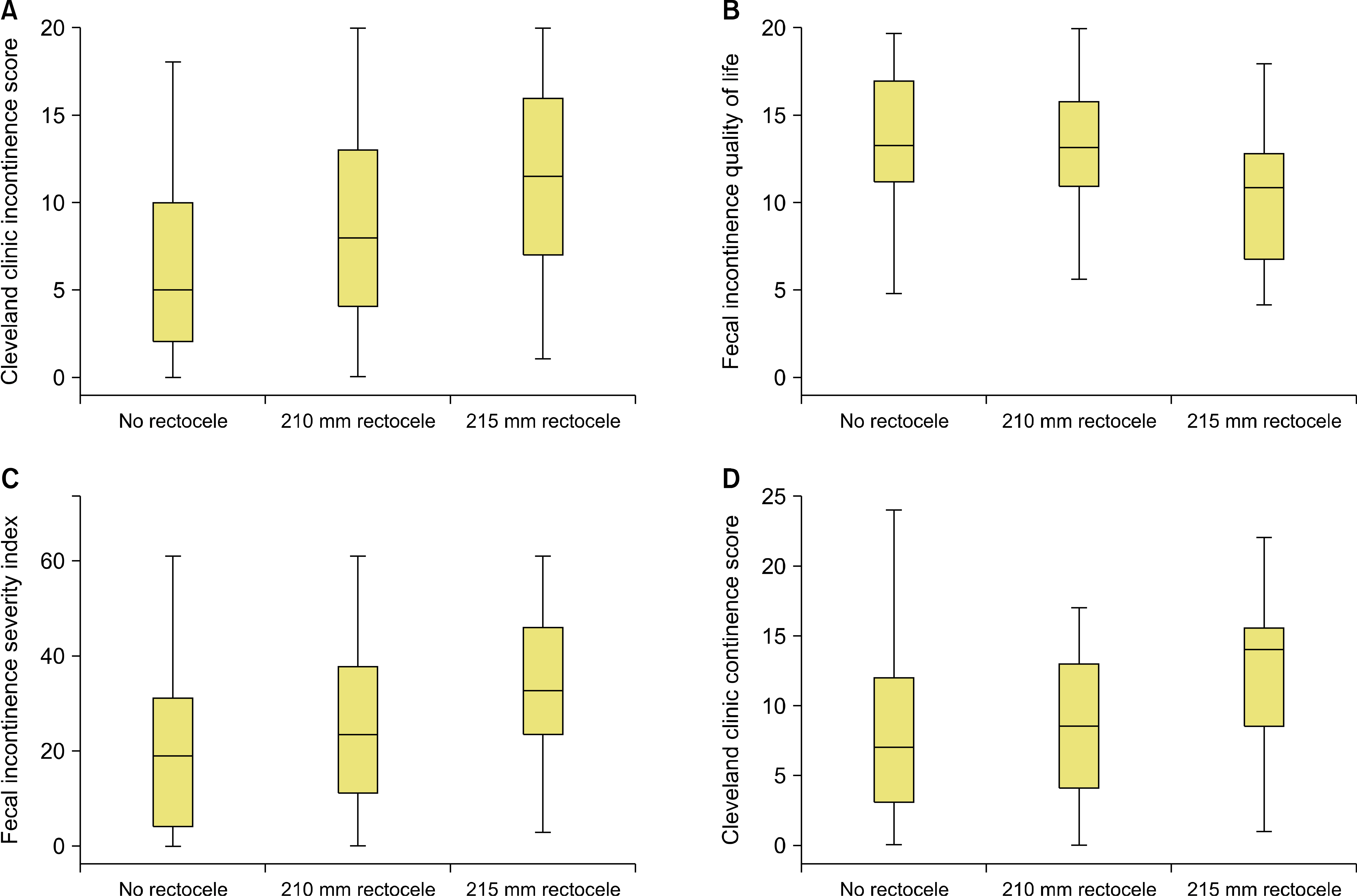
Fig. 2 Incontinence and constipation scores according to the rectocele size. (A–D) The x-axis denotes the groups according to the size of the rectocele. (A) The y-axis denotes the cleveland clinic incontinence score, (B) the y-axis denotes the fecal incontinence quality of life score, (C) the y-axis denotes the fecal incontinence severity index, (D) the y-axis denotes the cleveland clinic constipation score.
Reference
-
1. Dietz HP, Beer-Gabel M. 2012; Ultrasound in the investigation of posterior compartment vaginal prolapse and obstructed defecation. Ultrasound Obstet Gynecol. 40:14–27. DOI: 10.1002/uog.10131. PMID: 22045564.
Article2. Porter WE, Steele A, Walsh P, Kohli N, Karram MM. 1999; The anatomic and functional outcomes of defect-specific rectocele repairs. Am J Obstet Gynecol. 181:1353–8. discussion 1358-9. DOI: 10.1016/S0002-9378(99)70376-5. PMID: 10601912.
Article3. Burrows LJ, Meyn LA, Walters MD, Weber AM. 2004; Pelvic symptoms in women with pelvic organ prolapse. Obstet Gynecol. 104(5 Pt 1):982–8. DOI: 10.1097/01.AOG.0000142708.61298.be. PMID: 15516388.
Article4. Andromanakos N, Skandalakis P, Troupis T, Filippou D. 2006; Constipation of anorectal outlet obstruction: pathophysiology, evaluation and management. J Gastroenterol Hepatol. 21:638–46. DOI: 10.1111/j.1440-1746.2006.04333.x. PMID: 16677147.
Article5. Zbar AP, Lienemann A, Fritsch H, Beer-Gabel M, Pescatori M. 2003; Rectocele: pathogenesis and surgical management. Int J Colorectal Dis. 18:369–84. DOI: 10.1007/s00384-003-0478-z. PMID: 12665990.
Article6. Dietz HP, Korda A. 2005; Which bowel symptoms are most strongly associated with a true rectocele? Aust N Z J Obstet Gynaecol. 45:505–8. DOI: 10.1111/j.1479-828X.2005.00486.x. PMID: 16401217.
Article7. Pitchford CA. 1967; Rectocele: a cause of anorectal pathologic changes in women. Dis Colon Rectum. 10:464–6. DOI: 10.1007/BF02616820. PMID: 6066371.8. Bartolo DC, Roe AM, Virjee J, Mortensen NJ, Locke-Edmunds JC. 1988; An analysis of rectal morphology in obstructed defae-cation. Int J Colorectal Dis. 17–22. DOI: 10.1007/BF01649677. PMID: 3361219.
Article9. Ayabaca SM, Zbar AP, Pescatori M. 2002; Anal continence after rectocele repair. Dis Colon Rectum. 45:63–9. DOI: 10.1007/s10350-004-6115-2. PMID: 11786766.
Article10. Marti. Roche. Déléaval. 1999; Rectoceles: value of videodefaeco-graphy in selection of treatment policy. Colorectal Dis. 1:324–9. DOI: 10.1046/j.1463-1318.1999.00075.x. PMID: 23574594.11. Harvey CJ, Halligan S, Bartram CI, Hollings N, Sahdev A, Kingston K. 1999; Evacuation proctography:a prospective study of diagnostic and therapeutic effects. Radiology. 211:223–7. DOI: 10.1148/radiology.211.1.r99mr16223. PMID: 10189475.
Article12. Beer-Gabel M, Carter D. 2015; Comparison of dynamic transperineal ultrasound and defecography for the evaluation of pelvic floor disorders. Int J Colorectal Dis. 30:835–41. DOI: 10.1007/s00384-015-2195-9. PMID: 25820786.
Article13. Perniola G, Shek C, Chong CC, Chew S, Cartmill J, Dietz HP. 2008; Defecation proctography and translabial ultrasound in the investigation of defecatory disorders. Ultrasound Obstet Gynecol. 31:567–71. DOI: 10.1002/uog.5337. PMID: 18409183.
Article14. Kenton K, Shott S, Brubaker L. 1999; The anatomic and functional variability of rectoceles in women. Int Urogynecol J Pelvic Floor Dysfunct. 10:96–9. DOI: 10.1007/PL00004019. PMID: 10384970.
Article15. Agachan F, Chen T, Pfeifer J, Reissman P, Wexner SD. 1996; A constipation scoring system to simplify evaluation and management of constipated patients. Dis Colon Rectum. 39:681–5. DOI: 10.1007/BF02056950. PMID: 8646957.
Article16. Rockwood TH, Church JM, Fleshman JW, Kane RL, Mavrantonis C, Thorson AG, et al. 2000; Fecal Incontinence Quality of Life Scale: quality of life instrument for patients with fecal incontinence. Dis Colon Rectum. 43:9–16. discussion 16-7. DOI: 10.1007/BF02237236. PMID: 10813117.17. Shobeiri SA, LeClaire E, Nihira MA, Quiroz LH, O'Donoghue D. 2009; Appearance of the levator ani muscle subdivisions in endovaginal three-dimensional ultrasonography. Obstet Gynecol. 114:66–72. DOI: 10.1097/AOG.0b013e3181aa2c89. PMID: 19546760.
Article18. Morgan DM, Cardoza P, Guire K, Fenner DE, DeLancey JO. 2010; Levator ani defect status and lower urinary tract symptoms in women with pelvic organ prolapse. Int Urogynecol J. 21:47–52. DOI: 10.1007/s00192-009-0970-2. PMID: 19885634. PMCID: PMC2866151.
Article19. Shobeiri SA, Rostaminia G, White D, Quiroz LH. 2013; The determinants of minimal levator hiatus and their relationship to the puborectalis muscle and the levator plate. BJOG. 120:205–11. Erratum in: BJOG 2013120:655. DOI: 10.1111/1471-0528.12055. PMID: 23157458.
Article20. Bordeianou LG, Carmichael JC, Paquette IM, Wexner S, Hull TL, Bernstein M, et al. 2018; Consensus statement of definitions for anorectal physiology testing and pelvic floor terminology (Revised). Dis Colon Rectum. 61:421–7. DOI: 10.1097/DCR.0000000000001070. PMID: 29521821.
Article21. Dietz HP, Haylen BT, Broome J. 2001; Ultrasound in the quantification of female pelvic organ prolapse. Ultrasound Obstet Gynecol. 18:511–4. DOI: 10.1046/j.0960-7692.2001.00494.x. PMID: 11844174.
Article22. Dietz HP, Steensma AB. 2005; Posterior compartment prolapse on two-dimensional and three-dimensional pelvic floor ultrasound: the distinction between true rectocele, perineal hypermobility and enterocele. Ultrasound Obstet Gynecol. 26:73–7. DOI: 10.1002/uog.1930. PMID: 15973648.
Article23. Dietz HP. 2013; Pelvic floor ultrasound. Curr Surg Rep. 167–81. DOI: 10.1007/s40137-013-0026-x. PMID: 31529202.
Article24. Dietz HP, Zhang X, Shek KL, Guzman RR. 2015; How large does a rectocele have to be to cause symptoms? A 3D/4D ultrasound study. Int Urogynecol J. 26:1355–9. DOI: 10.1007/s00192-015-2709-6. PMID: 25944658.
Article25. Guzman Rojas R, Kamisan Atan I, Shek KL, Dietz HP. 2016; The prevalence of abnormal posterior compartment anatomy and its association with obstructed defecation symptoms in urogynecological patients. Int Urogynecol J. 27:939–44. DOI: 10.1007/s00192-015-2914-3. PMID: 26670577.
Article26. Altman D, López A, Kierkegaard J, Zetterström J, Falconer C, Pollack J, et al. 2005; Assessment of posterior vaginal wall prolapse: comparison of physical findings to cystodefecoperitoneography. Int Urogynecol J Pelvic Floor Dysfunct. 16:96–103. discussion 103. DOI: 10.1007/s00192-004-1220-2. PMID: 15372142.
Article27. Turnbull GK, Bartram CI, Lennard-Jones JE. 1988; Radiologic studies of rectal evacuation in adults with idiopathic constipation. Dis Colon Rectum. 31:190–7. DOI: 10.1007/BF02552545. PMID: 3349875.
Article28. Yoshioka K, Matsui Y, Yamada O, Sakaguchi M, Takada H, Hioki K, et al. 1991; Physiologic and anatomic assessment of patients with rectocele. Dis Colon Rectum. 34:704–8. DOI: 10.1007/BF02050355. PMID: 1855428.
Article29. Kelvin FM, Maglinte DD, Hornback JA, Benson JT. 1992; Pelvic prolapse: assessment withevacuation proctography (defecography). Radiology. 184:547–51. DOI: 10.1148/radiology.184.2.1620863. PMID: 1620863.
Article30. Capps WF Jr. 1975; Rectoplasty and perineoplasty for the symptomatic rectocele: a report of fifty cases. Dis Colon Rectum. 18:237–43. DOI: 10.1007/BF02587281. PMID: 1140053.31. Yang A, Mostwin JL, Rosenshein NB, Zerhouni EA. 1991; Pelvic floor descent in women: dynamic evaluation with fast MR imaging and cinematic display. Radiology. 179:25–33. DOI: 10.1148/radiology.179.1.2006286. PMID: 2006286.
Article32. Tan C, Geng J, Tang J, Yang X. 2020; The relationship between obstructed defecation and true rectocele in patients with pelvic organ prolapse. Sci Rep. 5599. DOI: 10.1038/s41598-020-62376-2. PMID: 32221359. PMCID: PMC7101397.
Article33. Rotholtz NA, Efron JE, Weiss EG, Nogueras JJ, Wexner SD. 2002; Anal manometric predictors of significant rectocele in constipated patients. Tech Coloproctol. 6:73–6. discussion 76-7. DOI: 10.1007/s101510200016. PMID: 12402049.
Article34. Dietz HP, Steensma AB. 2006; The role of childbirth in the aetiology of rectocele. BJOG. 113:264–7. DOI: 10.1111/j.1471-0528.2006.00860.x. PMID: 16487196.
Article35. Richardson AC. 1993; The rectovaginal septum revisited: its relationship to rectocele and its importance in rectocele repair. Clin Obstet Gynecol. 36:976–83. DOI: 10.1097/00003081-199312000-00022. PMID: 8293598.
Article36. Hendrix SL, Clark A, Nygaard I, Aragaki A, Barnabei V, McTiernan A. 2002; Pelvic organ prolapse in the Women's Health Initiative: gravity and gravidity. Am J Obstet Gynecol. 186:1160–6. DOI: 10.1067/mob.2002.123819. PMID: 12066091.
Article37. Shek KL, Dietz HP. 2010; Can levator avulsion be predicted antenatally? Am J Obstet Gynecol. 202:586.e1–6. DOI: 10.1016/j.ajog.2009.11.038. PMID: 20079479.
Article
- Full Text Links
-
- Actions
-
Cited
- CITED
-
- Close
- Share
-
- Similar articles
-
- Comparison of 3-Dimensional Pelvic Floor Ultrasonography and Defecography for Assessment of Posterior Pelvic Floor Disorders
- Anal canal coronal-sagittal ratio: a novel parameter for diagnosing pelvic floor injury in 2-dimensional transanal ultrasound
- Characteristics of the pelvic floor during pregnancy by 2D and 3D ultrasound
- Mesh-Based Transperineal Repair of a Perineal Hernia After a Laparoscopic Abdominoperineal Resection
- Transperineal rectocele repair is ideal for patients presenting with fecal incontinence
