Prog Med Phys.
2022 Dec;33(4):114-120. 10.14316/pmp.2022.33.4.114.
Monte Carlo Investigation of Dose Enhancement due to Gold Nanoparticle in Carbon-12, Helium-4, and Proton Beam Therapy
- Affiliations
-
- 1Department of Radiation Oncology, Samsung Medical Center, Sungkyunkwan University School of Medicine, Seoul, Korea
- KMID: 2537879
- DOI: http://doi.org/10.14316/pmp.2022.33.4.114
Abstract
- Purpose
Particle beam therapy is advantageous over photon therapy. However, adequately delivering therapeutic doses to tumors near critical organs is difficult. Nanoparticle-aided radiation therapy can be used to alleviate this problem, wherein nanoparticles can passively accumulate at higher concentrations in the tumor tissue compared to the surrounding normal tissue. In this study, we investigate the dose enhancement effect due to gold nanoparticle (GNP) when Carbon-12, He-4, and proton beams are irradiated on GNP.
Methods
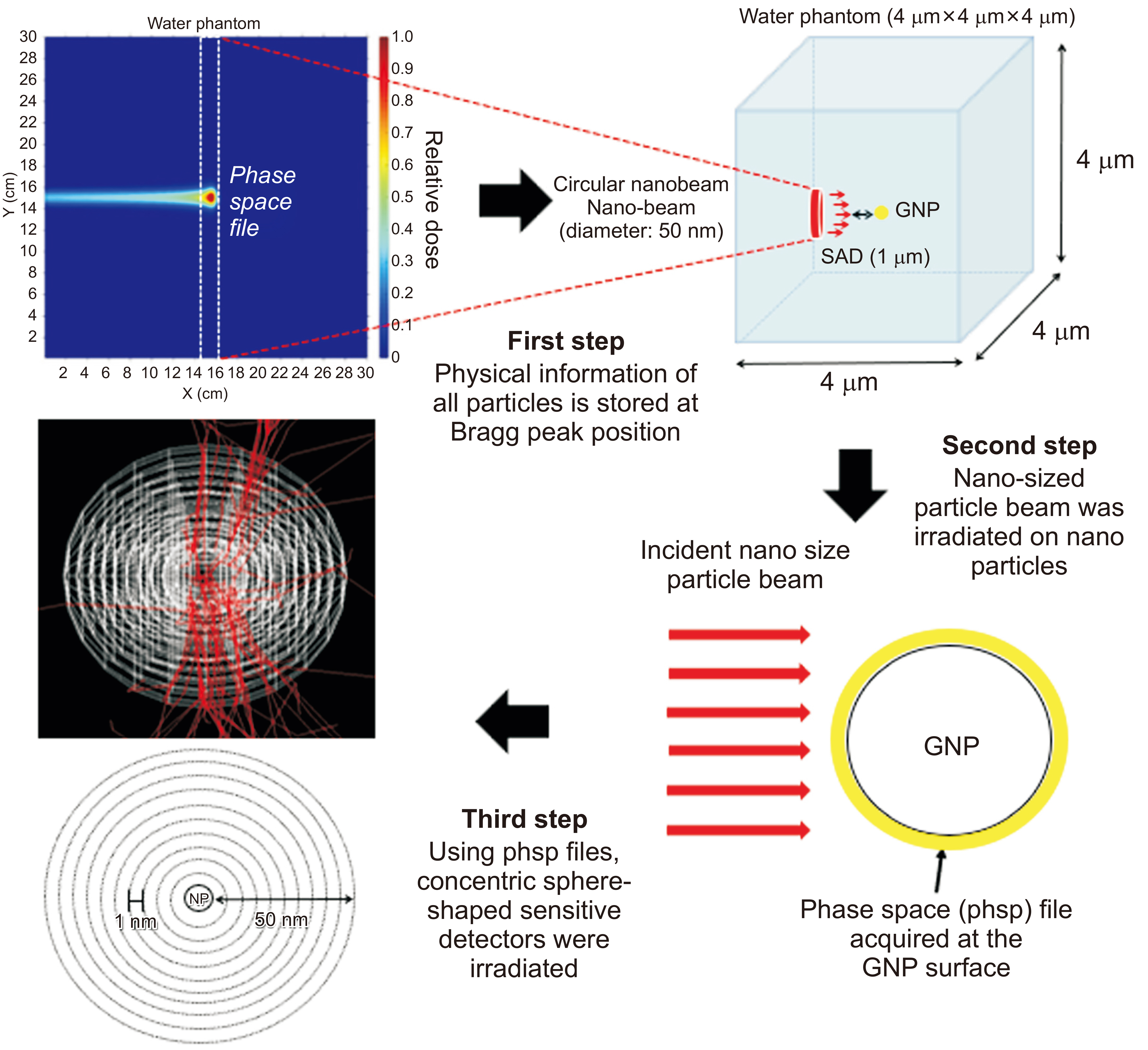
First, monoenergetic Carbon-12 and He-4 ion beams of energy of 283.33 MeV/u and 150 MeV/u, respectively, and a proton beam of energy of 150 MeV were irradiated on a water phantom of dimensions 30 cm×30 cm×30 cm. Subsequently, the secondary-particle information generated near the Bragg peak was recorded in a phase-space (phsp) file. Second, the obtained phsp file was scaled down to a nanometer scale to irradiate GNP of diameter 50 nm located at the center of a 4 µm×4 µm×4 µm water phantom. The dose enhancement ratio (DER) was calculated in intervals of 1 nm from the GNP surface.
Results
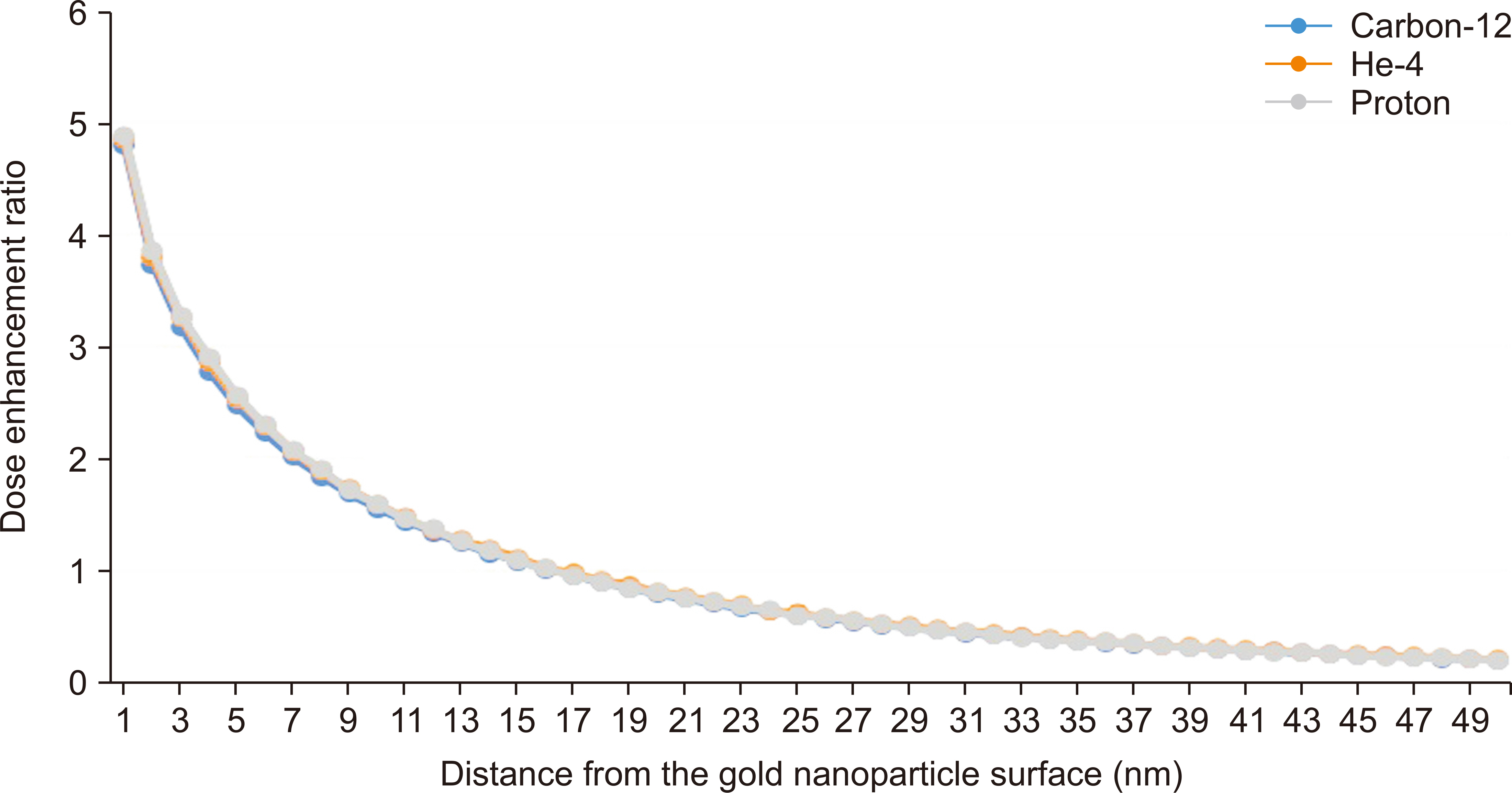
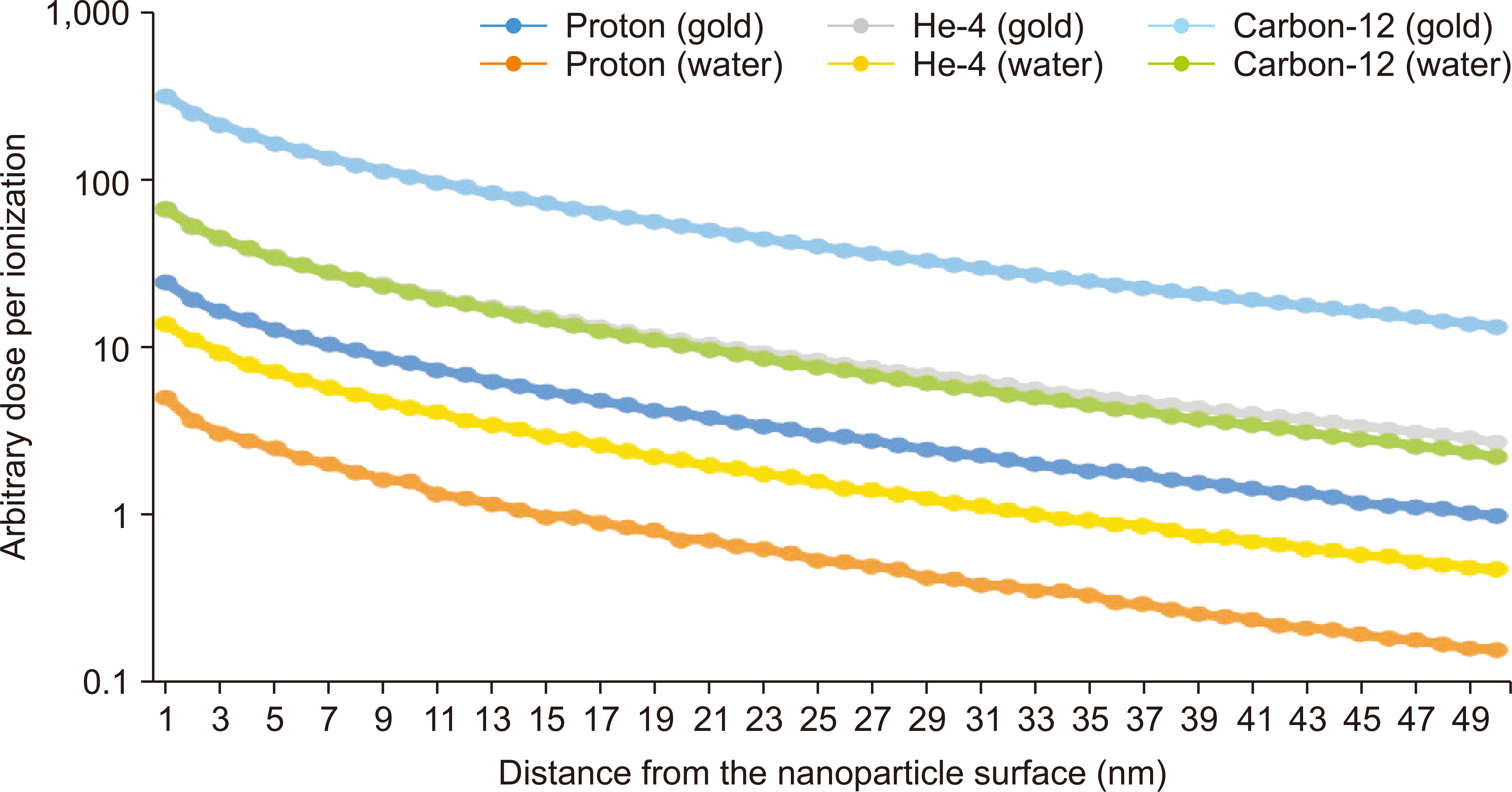
The DER of GNP computed at 1 nm from the GNP surface was 4.70, 4.86, and 4.89 for Carbon-12, He-4, and proton beams, respectively; the DER decreased rapidly with increasing distance from the GNP surface.
Conclusions
The results indicated that GNP can be used as radiosensitizers in particle beam therapy. Furthermore, the dose enhancement effect of the GNP absorbed by tumor cells can aid in delivering higher therapeutic doses.
Keyword
Figure
Reference
-
References
1. Doyen J, Falk AT, Floquet V, Hérault J, Hannoun-Lévi JM. 2016; Proton beams in cancer treatments: clinical outcomes and dosimetric comparisons with photon therapy. Cancer Treat Rev. 43:104–112. DOI: 10.1016/j.ctrv.2015.12.007. PMID: 26827698.
Article2. Jakobi A, Stützer K, Bandurska-Luque A, Löck S, Haase R, Wack LJ, et al. 2015; NTCP reduction for advanced head and neck cancer patients using proton therapy for complete or sequential boost treatment versus photon therapy. Acta Oncol. 54:1658–1664. DOI: 10.3109/0284186X.2015.1071920. PMID: 26340301.
Article3. Hainfeld JF, Slatkin DN, Smilowitz HM. 2004; The use of gold nanoparticles to enhance radiotherapy in mice. Phys Med Biol. 49:N309–N315. DOI: 10.1088/0031-9155/49/18/N03. PMID: 15509078.
Article4. Fang J, Nakamura H, Maeda H. 2011; The EPR effect: unique features of tumor blood vessels for drug delivery, factors involved, and limitations and augmentation of the effect. Adv Drug Deliv Rev. 63:136–151. DOI: 10.1016/j.addr.2010.04.009. PMID: 20441782.
Article5. Knäusl B, Fuchs H, Dieckmann K, Georg D. 2016; Can particle beam therapy be improved using helium ions? - a planning study focusing on pediatric patients. Acta Oncol. 55:751–759. DOI: 10.3109/0284186X.2015.1125016. PMID: 26750803.
Article6. Perl J, Shin J, Schumann J, Faddegon B, Paganetti H. 2012; TOPAS: an innovative proton Monte Carlo platform for research and clinical applications. Med Phys. 39:6818–6837. DOI: 10.1118/1.4758060. PMID: 23127075. PMCID: PMC3493036.
Article7. Ahn SH, Lee N, Choi C, Shin SW, Han Y, Park HC. 2018; Feasibility study of Fe3O4/TaOx nanoparticles as a radiosensitizer for proton therapy. Phys Med Biol. 63:114001. DOI: 10.1088/1361-6560/aac27b. PMID: 29726404.8. Martínez-Rovira I, Prezado Y. 2015; Evaluation of the local dose enhancement in the combination of proton therapy and nanoparticles. Med Phys. 42:6703–6710. DOI: 10.1118/1.4934370. PMID: 26520760.
Article9. Kyriakou I, Incerti S, Francis Z. 2015; Technical Note: Improvements in geant4 energy-loss model and the effect on low-energy electron transport in liquid water. Med Phys. 42:3870–3876. DOI: 10.1118/1.4921613. PMID: 26133588.
Article10. Bernal MA, Bordage MC, Brown JMC, Davídková M, Delage E, El Bitar Z, et al. 2015; Track structure modeling in liquid water: a review of the Geant4-DNA very low energy extension of the Geant4 Monte Carlo simulation toolkit. Phys Med. 31:861–874. DOI: 10.1016/j.ejmp.2015.10.087. PMID: 26653251.
Article11. Nikjoo H, Emfietzoglou D, Liamsuwan T, Taleei R, Liljequist D, Uehara S. 2016; Radiation track, DNA damage and response-a review. Rep Prog Phys. 79:116601. DOI: 10.1088/0034-4885/79/11/116601. PMID: 27652826.
Article12. Egorov V, Egorov E. 2018. Ion beams for materials analysis: conventional and advanced approaches. Ion beam applications. IntechOpen;London: p. 37–71. DOI: 10.5772/intechopen.76297.
Article13. Rudek B, McNamara A, Ramos-Méndez J, Byrne H, Kuncic Z, Schuemann J. 2019; Radio-enhancement by gold nanoparticles and their impact on water radiolysis for x-ray, proton and carbon-ion beams. Phys Med Biol. 64:175005. DOI: 10.1088/1361-6560/ab314c. PMID: 31295730.
Article14. Peukert D, Kempson I, Douglass M, Bezak E. 2020; Gold nanoparticle enhanced proton therapy: a Monte Carlo simulation of the effects of proton energy, nanoparticle size, coating material, and coating thickness on dose and radiolysis yield. Med Phys. 47:651–661. DOI: 10.1002/mp.13923. PMID: 31725910.
Article
- Full Text Links
- Actions
-
Cited
- CITED
-
- Close
- Share
- Similar articles
-
- A Monte Carlo Simulation Study of a Therapeutic Proton Beam Delivery System Using the Geant4 Code
- Monte Carlo Simulation of the Carbon Beam Nozzle for the Biomedical Research Facility in RAON
- Study on Optimization of Detection System of Prompt Gamma Distribution for Proton Dose Verification
- Dosimetric Influence of Implanted Gold Markers in Proton Therapy for Prostate Cancer
- Measurement of Proton Beam Dose-Averaged Linear Energy Transfer Using a Radiochromic Film


