Nasal septal anatomical variations among Saudi population and their possible coincidence with sinusitis: a computed tomography scan study
- Affiliations
-
- 1Department of Anatomy, Faculty of Medicine, The National University, Khartoum, Sudan
- 2Department of Anatomy, College of Medicine and Medical Sciences, Arabian Gulf University, Manama, Bahrain
- 3Department of Human Anatomy and Embryology, Faculty of Medicine, Suez Canal University, Ismailia, Egypt
- 4Department of Human Anatomy and Embryology, Faculty of Medicine, Menoufia University, Menoufia, Egypt
- 5Department of Family and Community Medicine, Ibn Sina National College for Medical Studies, Jeddah, Saudi Arabia
- KMID: 2537461
- DOI: http://doi.org/10.5115/acb.22.110
Abstract
- The nasal septum is a crucial supporting factor for the nasal cavity and may develop several anatomical variants including septal deviation, spur and pneumatization. These variants could be associated with a higher incidence of sinusitis due to structural and functional alterations. So, the aim of this study was to assess the prevalence of nasal septal deviation (NSD), nasal septal spur (NSS) and nasal septal pneumatization (NSP) among the Saudi adult population and their links with the incidence of sinusitis by using computed tomography (CT). A retrospective study was achieved over a twenty-two months period on 681 adult Saudi subjects (420 males and 261 females) aged 20 years or older, referred for coronal CT evaluation of the paranasal sinuses. NSD and NSS were significantly more prevalent in males than females (80.0% vs. 67.4% respectively for NSD, and 34.5% vs. 24.9% respectively for NSS), while there was no statistical difference in frequency of NSP regarding gender (P=0.670). The incidence of sinusitis was significantly higher in presence of NSD and/or NSS (P<0.001 for both). On the contrary, NSP was not associated with a significant increase in the prevalence of sinusitis (P=0.131). In conclusion, NSD and NSS are more prevalent in males than females among the Saudi population with no statistical difference between both genders regarding the presence of septal pneumatization. Furthermore, sinusitis is more prevalent with the occurrence of NSD and NSS, and not related to the incidence of NSP.
Figure
-
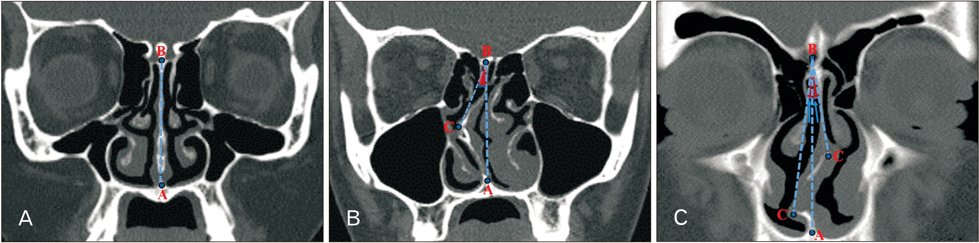
Fig. 1 Coronal CT images demonstrating measurement procedures of nasal septal angle showing point A is the maxillary crest, point B is the junction point of the perpendicular and the cribriform plate of ethmoid bone, and point C is the most prominent point of the deviated septum. (A) Normal nasal septum. (B) Right-sided NSD. (C) S-shaped NSD. CT, computed tomography; NSD, nasal septal deviation.
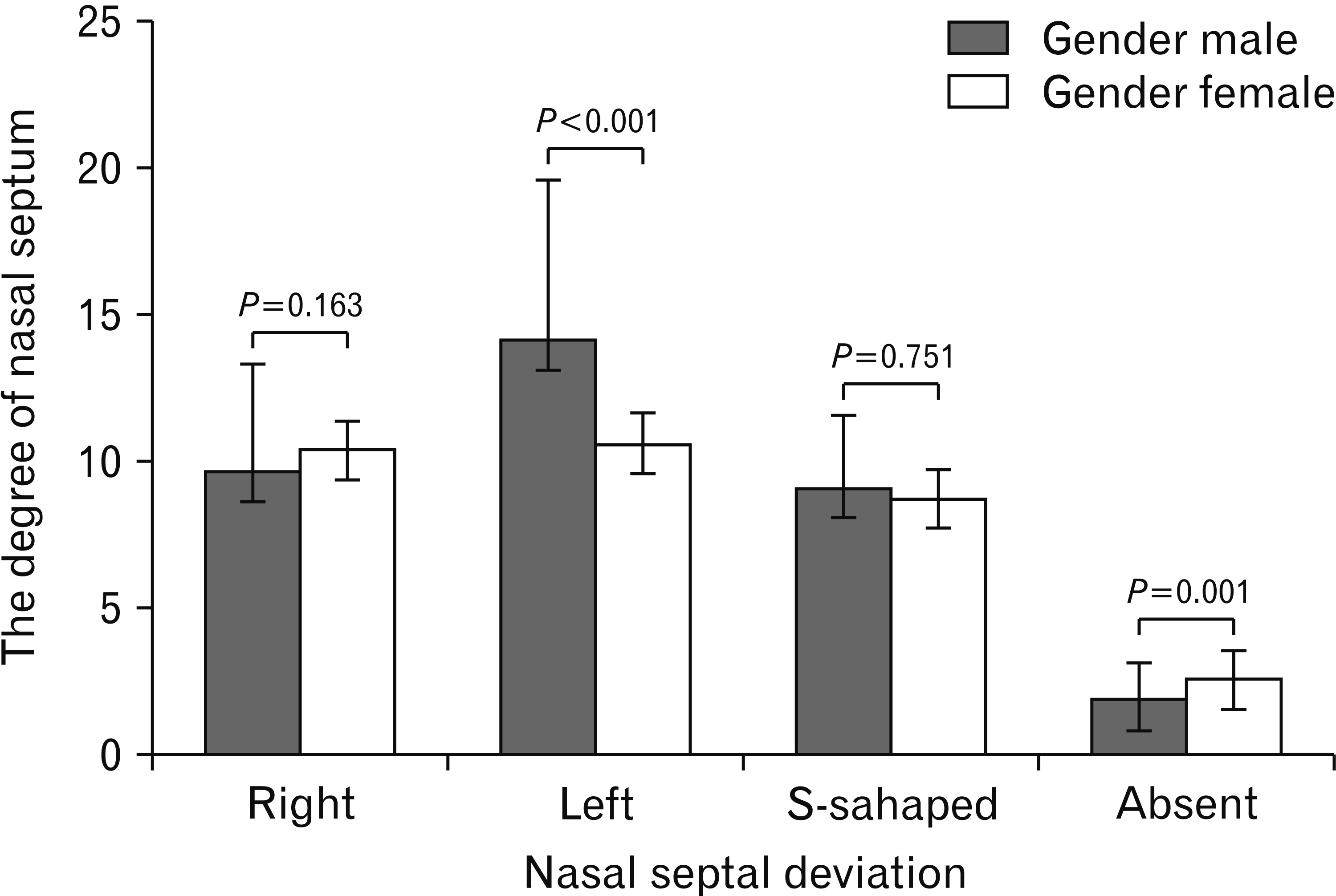
Fig. 2 The degree of NSD in male and female subjects. Values are presented as means with the minimal and maximal values. Statistical analysis was performed by unpaired Student’s t-test. NSD, nasal septal deviation.
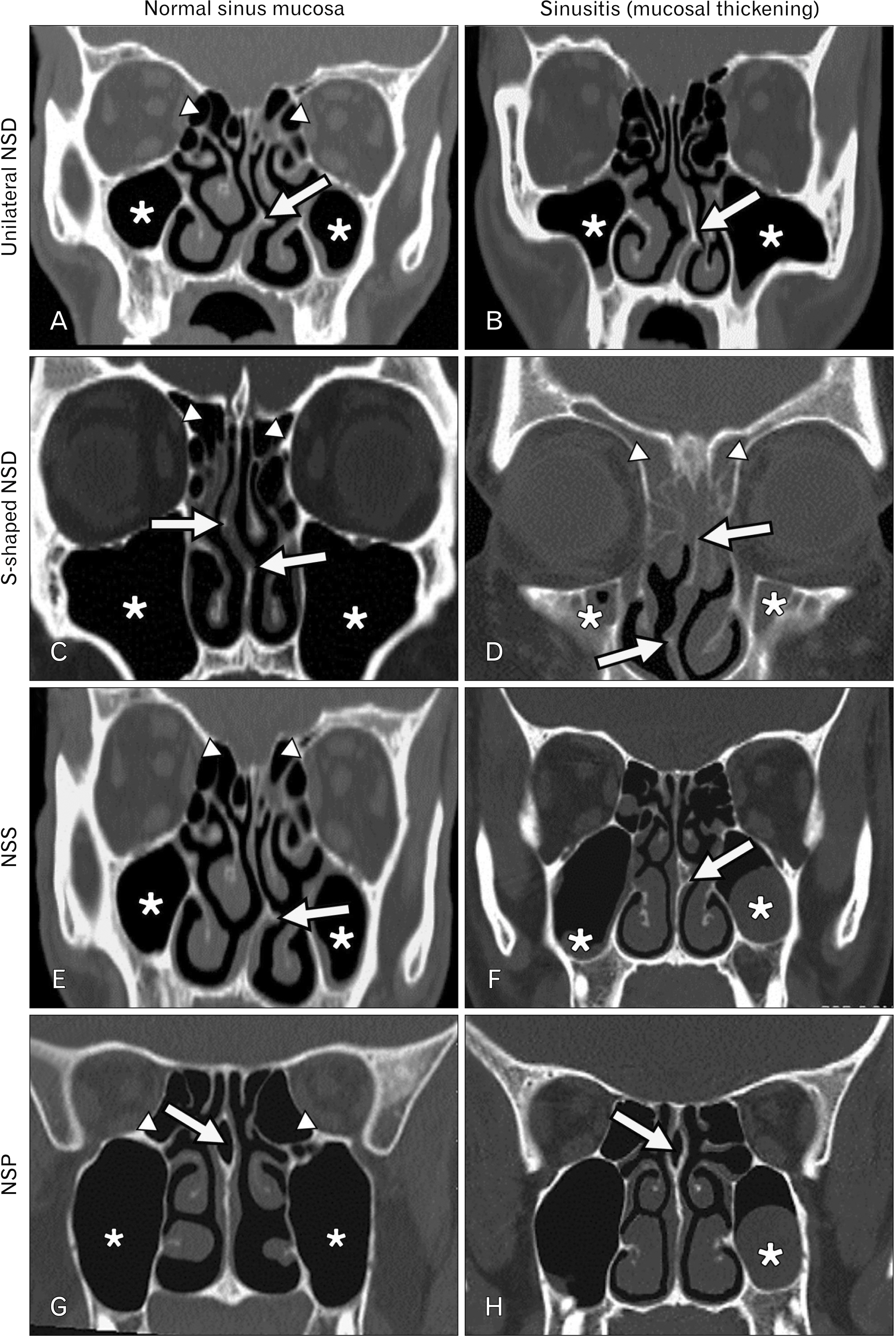
Fig. 3 Coronal CT images. (A) Left-sided NSD (arrow) with bilateral normal mucosa of both maxillary (asterisks) and ethmoid (arrowheads) sinuses. (B) Left-sided NSD (arrow) with mild sinusitis of both maxillary sinuses (asterisks). (C) S-shaped NSD (arrows) with bilateral normal mucosa of both maxillary (asterisks) and ethmoid (arrowheads) sinuses. (D) S-shaped NSD (arrows) with bilateral maxillary (asterisks) and ethmoid (arrowheads) sinusitis. (E) Left-sided NSD with ipsilateral NSS (arrow), and normal mucosa of both maxillary (asterisks) and ethmoid (arrowheads) sinuses. (F) Centralized nasal septum with left NSS (arrow) and mucosal thickening of both maxillary sinuses (asterisks). (G) Pneumatization (arrow) of a centralized nasal septum with normal mucosa of both maxillary (asterisks) and ethmoid (arrowheads) sinuses. (H) Pneumatization (arrow) of a centralized nasal septum with left maxillary sinusitis (asterisk). CT, computed tomography; NSD, nasal septal deviation; NSS, nasal septal spur.
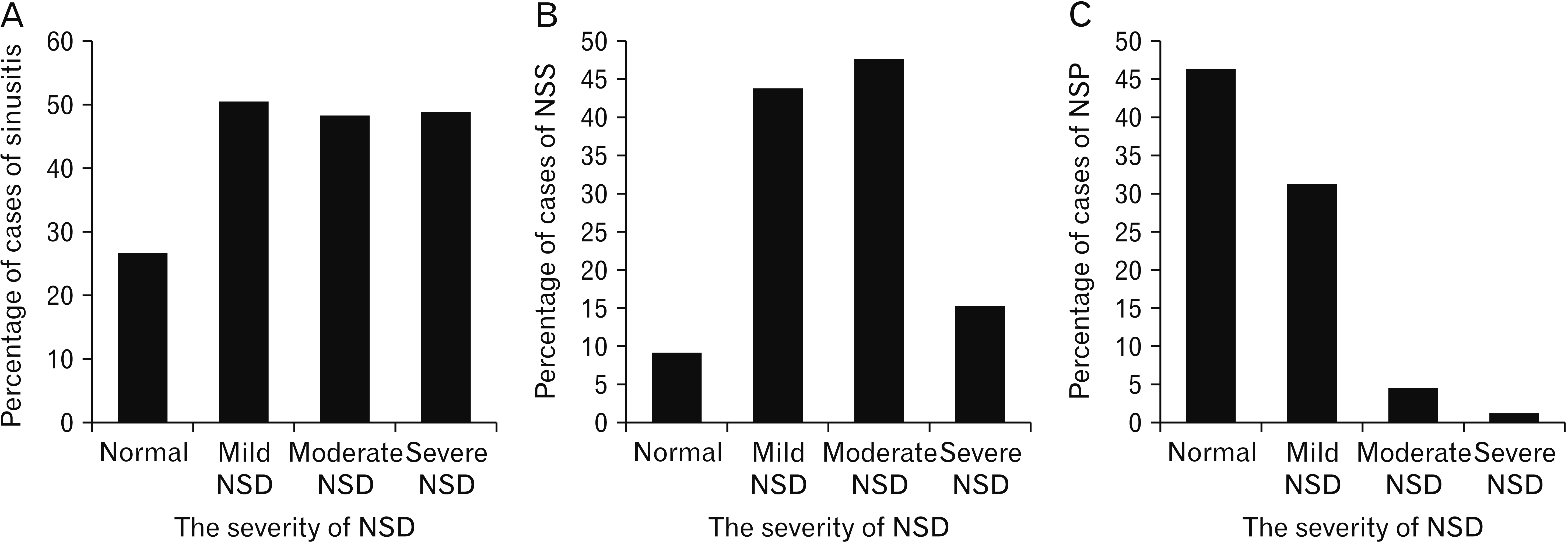
Fig. 4 Percentages of incidence of sinusitis according to the severity of NSD. (B) Percentages of incidence of NSS according to the severity of NSD. (C) Percentages of incidence of NSP according to the severity of NSD. NSD, nasal septal deviation; NSS, nasal septal spur.
Reference
-
References
1. Daghighi M, Daryani A, Nejad KC. 2006; Evaluation of anatomic variations of paranasal sinuses. Int J Otorhinolaryngol. 7:1–5. DOI: 10.5580/1a5d.2. Chandel NS, Gupta R, Vyas MM. 2015; Computerized tomographic evaluation of anatomical variations of paranasal sinus and nose. Natl J Med Dent Res. 4:48–52.3. iprakash V Sr. 2017; Prevalence and clinical features of nasal septum deviation: a study in an urban centre. Int J Otorhinolaryngol Head Neck Surg. 3:842–4. DOI: 10.18203/issn.2454-5929.ijohns20173670.4. Wang IJ, Lin SL, Tsou KI, Hsu MC, Chiu WT, Tsai SH, Lee LM, Lin TJ. 2010; Congenital midline nasal mass: cases series and review of the literature. Turk J Pediatr. 52:520–4. PMID: 21434538.5. Gray LP. 1978; Deviated nasal septum. Incidence and etiology. Ann Otol Rhinol Laryngol Suppl. 87(3 Pt 3 Suppl 50):3–20. DOI: 10.1177/00034894780873S201. PMID: 99070.6. Hsia JC, Camacho M, Capasso R. 2014; Snoring exclusively during nasal breathing: a newly described respiratory pattern during sleep. Sleep Breath. 18:159–64. DOI: 10.1007/s11325-013-0864-x. PMID: 23716022.7. Cellina M, Gibelli D, Cappella A, Martinenghi C, Belloni E, Oliva G. 2020; Nasal cavities and the nasal septum: anatomical variants and assessment of features with computed tomography. Neuroradiol J. 33:340–7. DOI: 10.1177/1971400920913763. PMID: 32193968. PMCID: PMC7416352.8. Beale TJ, Madani G, Morley SJ. 2009; Imaging of the paranasal sinuses and nasal cavity: normal anatomy and clinically relevant anatomical variants. Semin Ultrasound CT MR. 30:2–16. DOI: 10.1053/j.sult.2008.10.011. PMID: 19388234.9. Biswas J, Patil CY, Deshmukh PT, Kharat R, Nahata V. 2013; Tomographic evaluation of structural variations of nasal cavity in various nasal pathologies. Int J Otolaryngol Head Neck Surg. 2:129–34. DOI: 10.4236/ijohns.2013.24028.10. Mostafa SY, Abd-Elgaber FM, Mohamed BA, Hammad AS. 2021; Impact of septoplasty alone or with endoscopic sinus surgery for treatment of chronic rhinosinusitis with deviated septum. Egypt J Otolaryngol. 37:2. DOI: 10.1186/s43163-020-00066-6. PMID: 5a88664dc37e4d09a94f4a81f0853187.11. Janovic N, Janovic A, Milicic B, Djuric M. 2020; Oct. 10. Relationship between nasal septum morphology and nasal obstruction symptom severity: computed tomography study. Braz J Otorhinolaryngol. [Epub]. https://doi.org/10.1016/j.bjorl.2020.09.004. DOI: 10.1016/j.bjorl.2020.09.004. PMID: 33132090. PMCID: PMC9483930.12. Periyasamy V, Bhat S, ee Ram MN Sr. 2019; Classification of naso septal deviation angle and its clinical implications: a CT scan imaging study of Palakkad population, India. Indian J Otolaryngol Head Neck Surg. 71(Suppl 3):2004–10. DOI: 10.1007/s12070-018-1425-1. PMID: 31763284. PMCID: PMC6848586.13. Smith KD, Edwards PC, Saini TS, Norton NS. 2010; The prevalence of concha bullosa and nasal septal deviation and their relationship to maxillary sinusitis by volumetric tomography. Int J Dent. 2010:404982. DOI: 10.1155/2010/404982. PMID: 20862205. PMCID: PMC2938434.14. Bora A, Koç M, Durmuş K, Altuntas EE. 2021; Evaluating the frequency of anatomical variations of the sinonasal region in pediatric and adult age groups according to gender: computed tomography findings of 1532 cases. Egypt J Otolaryngol. 37:58. DOI: 10.1186/s43163-021-00122-9. PMID: e317ecc747c84039b8d29a52b8ac9cd3.15. Shrestha KK, Acharya K, Joshi R, Maharjan S, Adhikari D. 2019; Anatomical variations of the paranasal sinuses and the nasal cavity. Nepal Med Coll J. 21:7–11. DOI: 10.3126/nmcj.v21i1.24837.16. Badia L, Lund VJ, Wei W, Ho WK. 2005; Ethnic variation in sinonasal anatomy on CT-scanning. Rhinology. 43:210–4. PMID: 16218515.17. Sazgar AA, Massah J, Sadeghi M, Bagheri A, Rasool E. 2008; The incidence of concha bullosa and the correlation with nasal septal deviation. B-ENT. 4:87–91. PMID: 18681204.18. Devareddy MM, Devakar S. 2019; Evaluation of anatomical variations in nose and paranasal sinuses by using multidetector computed tomography. Int J Contemp Med Surg Radiol. 4:C146–51. DOI: 10.21276/ijcmsr.2019.4.3.32.19. Turna Ö, Aybar MD, Karagöz Y, Tuzcu G. 2014; Anatomic variations of the paranasal sinus region: evaluation with multidetector CT. Istanbul Med J. 15:104–9. DOI: 10.5152/imj.2013.74429.20. Qureshi MF, Usmani A. 2021; A CT-Scan review of anatomical variants of sinonasal region and its correlation with symptoms of sinusitis (nasal obstruction, facial pain and rhinorrhea). Pak J Med Sci. 37:195–200. DOI: 10.12669/pjms.37.1.3260. PMID: 33437276. PMCID: PMC7794148.21. Adeel M, Rajput MS, Akhter S, Ikram M, Arain A, Khattak YJ. 2013; Anatomical variations of nose and para-nasal sinuses; CT scan review. J Pak Med Assoc. 63:317–9. PMID: 23914628.22. Onwuchekwa RC, Alazigha N. 2017; Computed tomography anatomy of the paranasal sinuses and anatomical variants of clinical relevants in Nigerian adults. Egypt J Ear Nose Throat Allied Sci. 18:31–8. DOI: 10.1016/j.ejenta.2016.11.001.23. Espinosa W, Genito R, Ramos RZ. 2018; Anatomic variations of the nasal cavity and paranasal sinus and their correlation with chronic rhinosinusitis using Harvard staging system. J Otolaryngol ENT Res. 10:190–3. DOI: 10.15406/joentr.2018.10.00343.24. Pérez-Piñas , Sabaté J, Carmona A, Catalina-Herrera CJ, Jiménez-Castellanos J. 2000; Anatomical variations in the human paranasal sinus region studied by CT. J Anat. 197(Pt 2):221–7. DOI: 10.1017/S0021878299006500. PMID: 11005714. PMCID: PMC1468121.25. Clark DW, Del Signore AG, Raithatha R, Senior BA. 2018; Nasal airway obstruction: prevalence and anatomic contributors. Ear Nose Throat J. 97:173–6. DOI: 10.1177/014556131809700615. PMID: 30036414.26. Bagri N, Kavirajan K, Chandra R, Agarwal Y, Gupta N, Mandal S. 2019; Nasal septal angle deviation: effect on lateral wall in nasal obstruction. Int J Res Med Sci. 7:90–5. DOI: 10.18203/2320-6012.ijrms20185095.27. Madani SA, Hashemi SA, Modanluo M. 2015; The incidence of nasal septal deviation and its relation with chronic rhinosinusitis in patients undergoing functional endoscopic sinus surgery. J Pak Med Assoc. 65:612–4. PMID: 26060156.28. Poorey VK, Gupta N. 2014; Endoscopic and computed tomographic evaluation of influence of nasal septal deviation on lateral wall of nose and its relation to sinus diseases. Indian J Otolaryngol Head Neck Surg. 66:330–5. DOI: 10.1007/s12070-014-0726-2. PMID: 25032124. PMCID: PMC4071434.29. Stallman JS, Lobo JN, Som PM. 2004; The incidence of concha bullosa and its relationship to nasal septal deviation and paranasal sinus disease. AJNR Am J Neuroradiol. 25:1613–8. PMID: 15502150. PMCID: PMC7976404.30. Earwaker J. 1993; Anatomic variants in sinonasal CT. Radiographics. 13:381–415. DOI: 10.1148/radiographics.13.2.8460226. PMID: 8460226.31. Elahi MM, Frenkiel S, Fageeh N. 1997; Paraseptal structural changes and chronic sinus disease in relation to the deviated septum. J Otolaryngol. 26:236–40. PMID: 9263892.32. Hatipoglu HG, Cetin MA, Yuksel E. 2008; Nasal septal deviation and concha bullosa coexistence: CT evaluation. B-ENT. 4:227–32. PMID: 19227028.33. Mohebbi A, Ahmadi A, Etemadi M, Safdarian M, Ghourchian S. 2012; An epidemiologic study of factors associated with nasal septum deviation by computed tomography scan: a cross sectional study. BMC Ear Nose Throat Disord. 12:15. DOI: 10.1186/1472-6815-12-15. PMID: 23244707. PMCID: PMC3541255.34. Alshaikh N, Aldhurais A. 2018; Anatomic variations of the nose and paranasal sinuses in Saudi population: computed tomography scan analysis. Egypt J Otolaryngol. 34:234–41. DOI: 10.4103/1012-5574.244904. PMID: 990fcb38955d45b6b3bd9d0be9c00ef3.35. Alsubael MO, Hegazy AA. 2009; Anatomical variations of the human nasal osteomeatal complex, studied by CT. Zagazig Univ Med J. 16:72–83.36. Dua K, Chopra H, Khurana A, Munjal M. 2005; CT scan variations in chronic sinusitis. Indian J Radiol Imaging. 15:315–20. DOI: 10.4103/0971-3026.29144.37. Azila A, Irfan M, Rohaizan Y, Shamim AK. 2011; The prevalence of anatomical variations in osteomeatal unit in patients with chronic rhinosinusitis. Med J Malaysia. 66:191–4. PMID: 22111438.38. Zinreich S, Albayram S, Benson M, Oliverio P. Som PM, Curtin HD, editors. 2003. The ostiomeatal complex and functional endoscopic surgery. Head and Neck Imaging. 4th ed. Mosby;St. Louis: p. 149–73.39. Al- Qudah MA. 2010; Anatomical variations in sino-nasal region: a computer tomography (CT) study. J Med J. 44:290–7.
- Full Text Links
-
- Actions
-
Cited
- CITED
-
- Close
- Share
-
- Similar articles
-
- Prevalence of the anatomical variations of concha bullosa and its relation with sinusitis among Saudi population: a computed tomography scan study
- The Relationship between Anatomic Variation of Nasal Cavity and Paranasal Sinusitis
- Anatomical Factors Influencing Unilateral Chronic Sinusitis
- The Relationship between Clinical Patterns of Sinusitis and Anatomical Variation
- Correlations between anatomical variations of the nasal cavity and ethmoidal sinuses on cone-beam computed tomography scans
