Prevalence of the anatomical variations of concha bullosa and its relation with sinusitis among Saudi population: a computed tomography scan study
- Affiliations
-
- 1Department of Anatomy, College of Medicine and Medical Sciences, Arabian Gulf University, Manama, Bahrain, Egypt
- 2Department of Human Anatomy and Embryology, Faculty of Medicine, Suez Canal University, Ismailia, Egypt
- 3Department of Anatomy, Faculty of Medicine, The National University, Khartoum, Sudan
- 4Department of Radiology, National Cancer Institute (NCI), Cairo University, Cairo, Egypt
- 5Department of Medical Imaging, Saudi German Hospitals Group, Jeddah, Saudi Arabia
- 6Department of Human Anatomy and Embryology, Faculty of Medicine, Menoufia University, Menoufia, Egypt
- 7Department of Anatomy, Ibn Sina National College for Medical Studies, Jeddah, Saudi Arabia
- KMID: 2516903
- DOI: http://doi.org/10.5115/acb.20.247
Abstract
- Concha bullosa (CB) is a pneumatic cavitation inside a concha in the nasal cavity. It is one of the most widely recognized nasal variations and is mostly found in the middle concha. CB is divided according to its site into three types; lamellar, bulbous and extensive. The goal of our study was to estimate the prevalence of CB among Saudi adult population and its association with sinusitis by using multidetector computed tomography (MDCT). This was a retrospective study carried out over a three-year period on 879 adult Saudi patients aged 18 years or older, referred for MDCT assessment of paranasal sinuses. Males were 540 and females were 339. Patients with facial congenital anomalies or nasal trauma were excluded from our study. CB was prevalent in both males and females among Saudi population (55.4%, 55.7%) respectively. Bilateral CB (55.5%) was more frequent than unilateral (44.5%). Extensive CB (44.0%) was the most frequent type. Sinusitis was associated more in patients with CB (48.0%) versus those who have no CB (5.9%). In conclusion, CB was prevalent among Saudi population and the most frequently recorded is the extensive type. Furthermore, the most common type associated with sinusitis was extensive CB (49.6%).
Figure
-
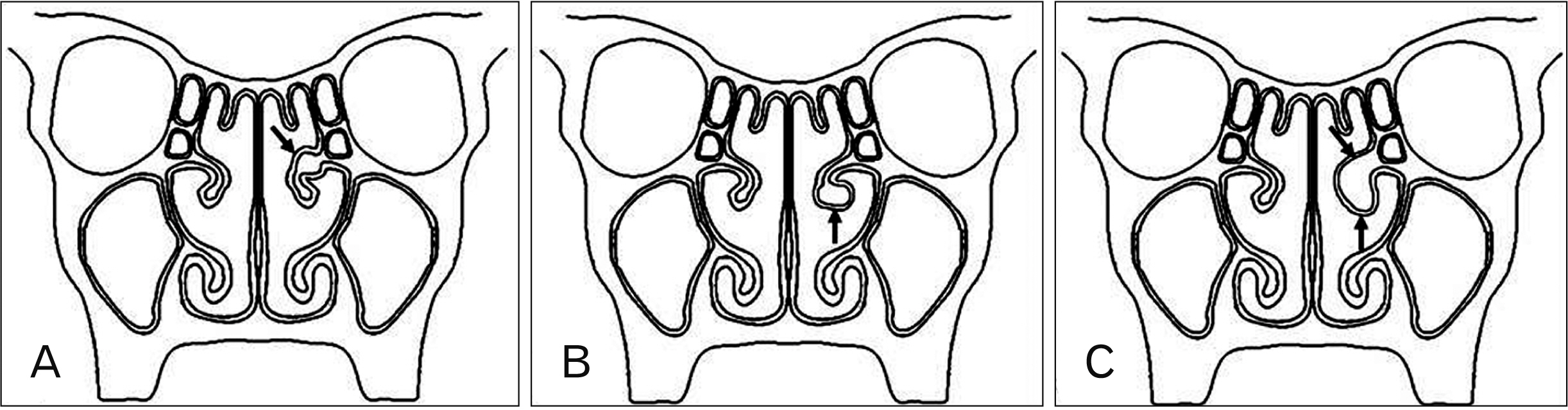
Fig. 1 Types of concha bullosa (arrows): (A) lamellar type, (B) bulbous type, and (C) extensive type. (Illustrated by Nasr El-Din et al.).
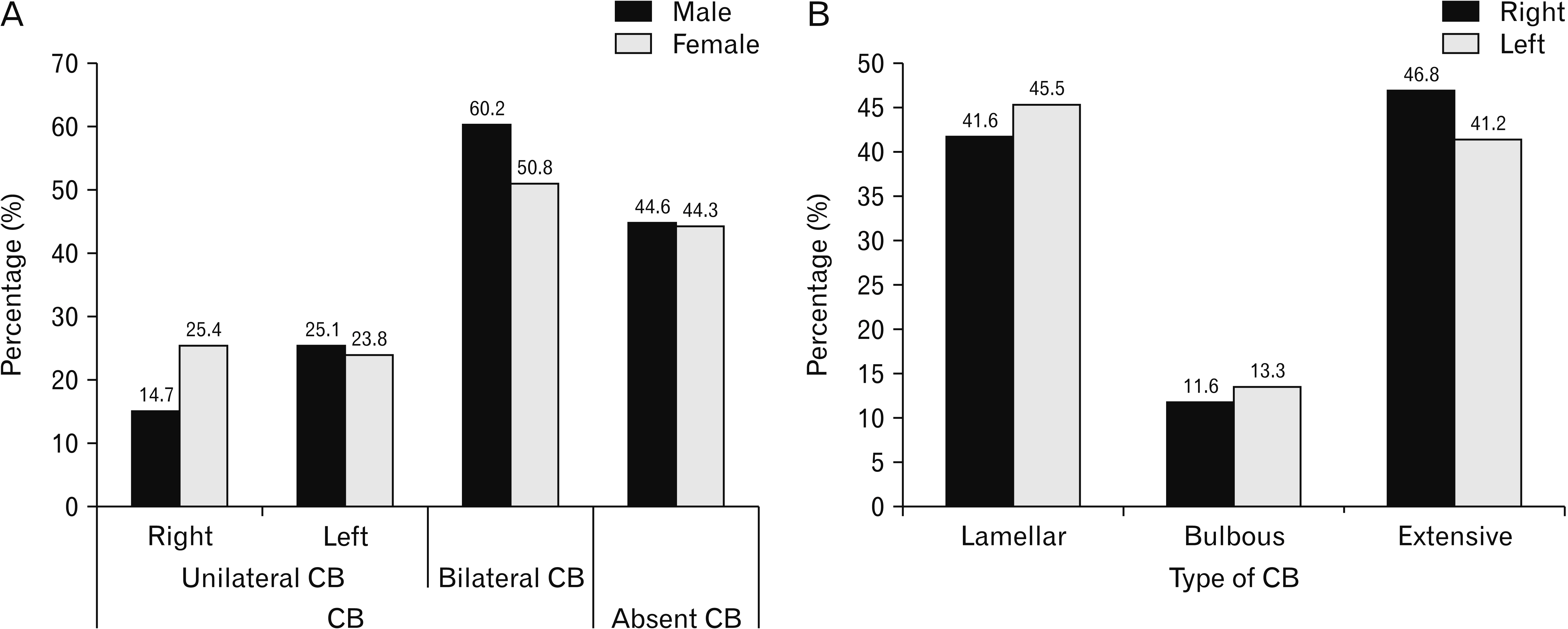
Fig. 2 Relations between CB, side and sex. Values are presented as percentages. (A) Relation between the incidence of CB, side and sex. CB vs. absent CB according to sex chi-square P-value=0.912; unilateral CB vs. bilateral CB according to sex chi-square P-value=0.041, right CB vs. left CB according to sex chi-square P-value=0.033, right CB vs. bilateral CB according to sex chi-square P-value=0.003 and left CB vs. bilateral CB according to sex chi-square P-value=0.604. (B) Relation between the different types of CB and the side. Types of CB according to the side chi-square P-value=0.453. CB, concha bullosa.
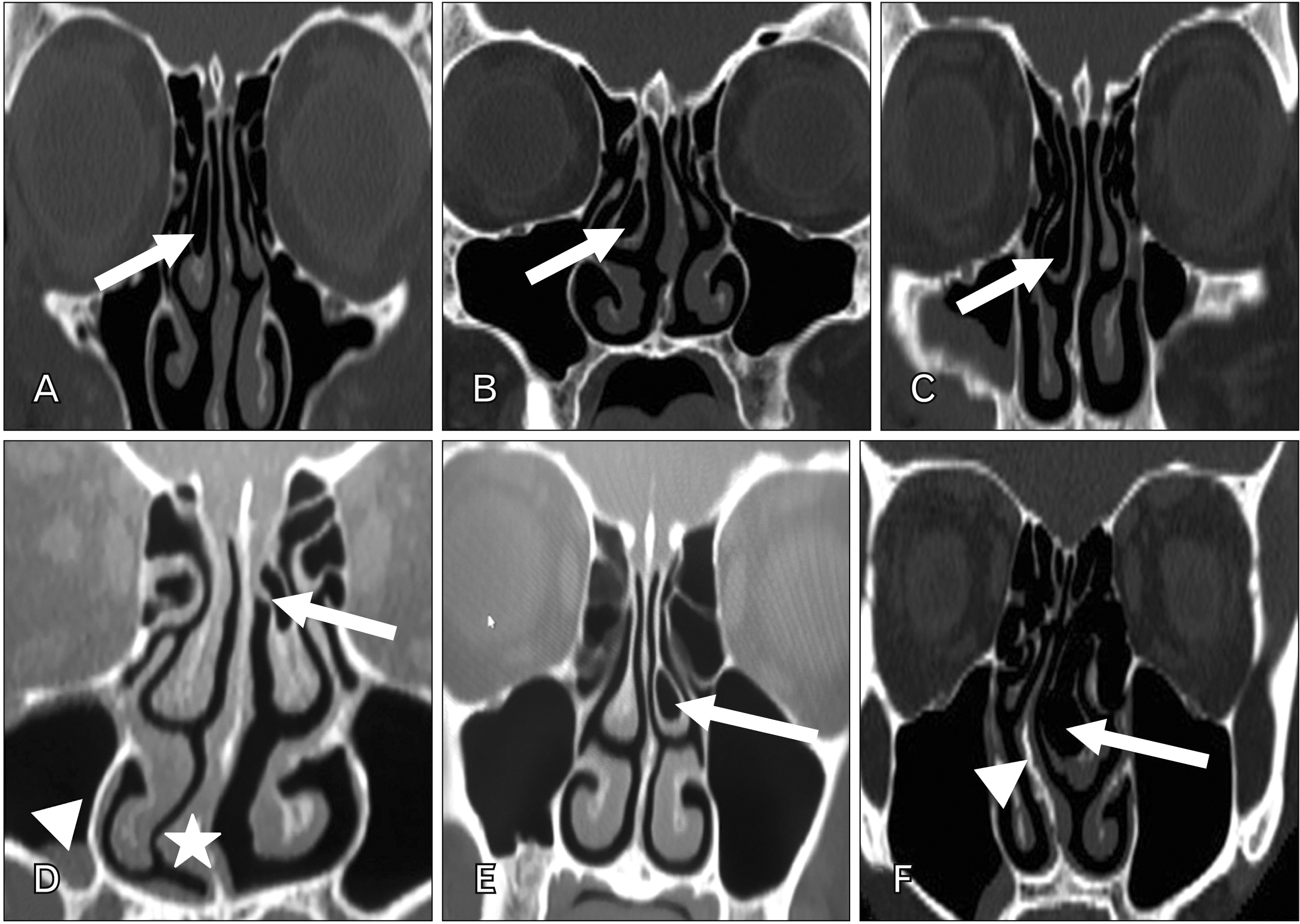
Fig. 3 Different types of unilateral concha bullosa (CB) shown by coronal computed tomography images. (A) Right lamellar type CB (arrow). (B) Right bulbous CB (arrow). (C) Right extensive CB (arrow). (D) Left lamellar CB (arrow) with right maxillary sinusitis (arrowhead) and deviated nasal septum to right (asterisk). (E) Left bulbous CB (arrow). (F) Left extensive CB (arrow), with nasal septum deviation having a convexity to the right side (arrowhead).
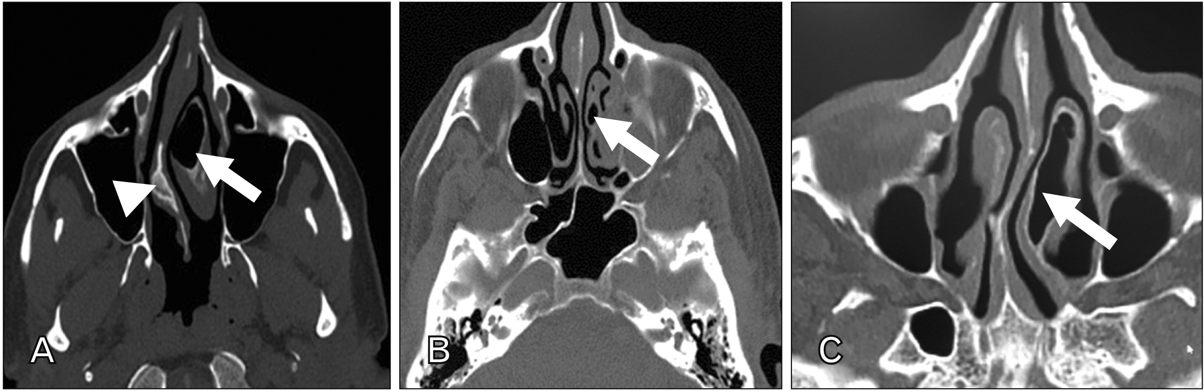
Fig. 4 Different types of unilateral concha bullosa (CB) shown by axial computed tomography images. (A) Left large bulbous CB (arrow) with severe deviation of the nasal septum convexity to the right side (arrowhead). (B) Left lamellar CB (arrow). (C) Left extensive CB (arrow).
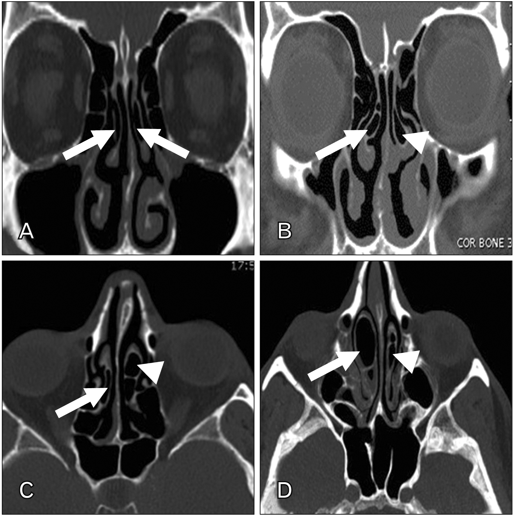
Fig. 5 Different types of bilateral concha bullosa (CB) shown by coronal (A, B) and axial (C, D) computed tomography images. (A) Bilateral lamellar type of CB (arrows). (B) Right lamellar (arrow) and left bulbous CB (arrowhead). (C) Right lamellar (arrow) and left extensive CB (arrowhead). (D) Right extensive CB (arrow) and left bulbous type (arrowhead).
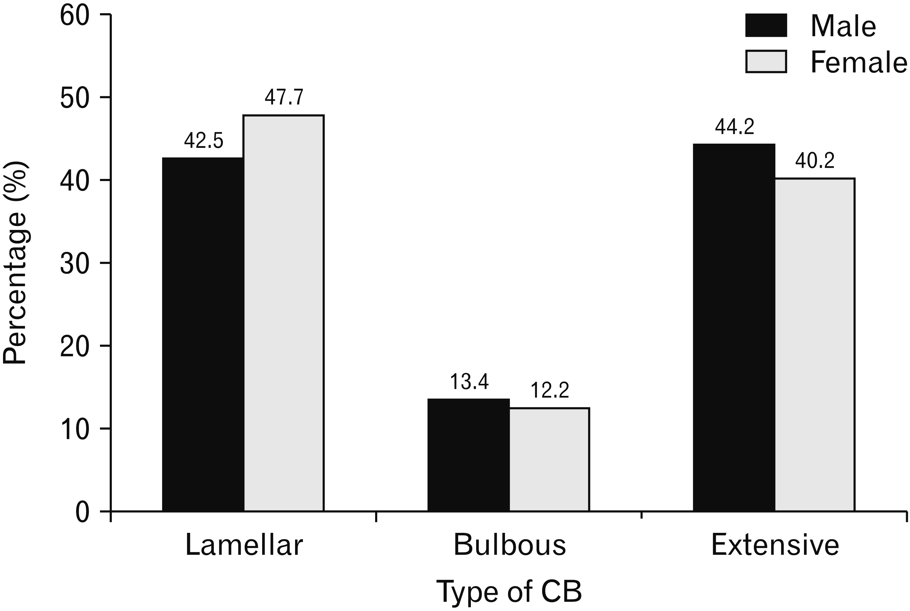
Fig. 6 Relation between different types of CB and sex. Values are presented as number (percentage). Types of CB according to the sex chi-square P-value=0.693. CB, concha bullosa.

Fig. 7 Different types of concha bullosa (CB) associated with different types of sinusitis shown by coronal (A, C) and axial (D) computed tomography images. (A) Left bulbous CB (thin arrow), with deviated nasal septum (asterisk) to the right and solitary right ethmoidal sinusitis (thick arrow). (B) Moderate sized right extensive CB (arrow) with ipsilateral mucosal thickening in the maxillary sinus (MS). (C) Left extensive CB (thin arrow) with mild left frontal mucosal thickening (sinusitis) (arrowhead) associated with nasal septum deviation to right side (asterisk). Right bulbous CB is also noted (thick arrow). (D) Left bulbous CB (arrow) associated with sphenoid sinusitis (arrowhead) and deviated nasal septum to right side (asterisk).
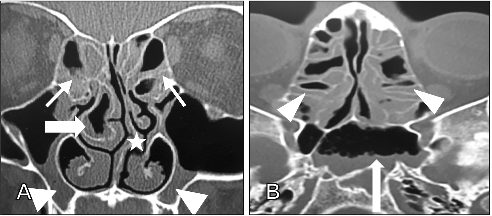
Fig. 8 Pansinusitis in the same subject having concha bullosa (CB) shown by coronal (A) and axial (B) computed tomography images. (A) Right bulbous CB (thick arrow) with partial opacification denoting infected CB, bilateral ethmoidal (thin arrows) and bilateral maxillary sinusitis (arrowheads). Deviated nasal septum to left is also noted (asterisk). (B) Sphenoid (arrow) and bilateral ethmoidal (arrowheads) sinusitis.
Reference
-
References
1. Bolger WE, Butzin CA, Parsons DS. 1991; Paranasal sinus bony anatomic variations and mucosal abnormalities: CT analysis for endoscopic sinus surgery. Laryngoscope. 101(1 Pt 1):56–64. DOI: 10.1288/00005537-199101000-00010. PMID: 1984551.2. Subramanian S, Lekhraj Rampal GR, Wong EF, Mastura S, Razi A. 2005; Concha bullosa in chronic sinusitis. Med J Malaysia. 60:535–9. PMID: 16515102.3. Hatipoğlu HG, Cetin MA, Yüksel E. 2005; Concha bullosa types: their relationship with sinusitis, ostiomeatal and frontal recess disease. Diagn Interv Radiol. 11:145–9. PMID: 16206055.4. Flint P, Haughey B, Lund V, Niparko J, Robbins K, Thomas J, Lesperance M. 2015. Cummings otolaryngology: head & neck surgery. 6th ed. Elsevier/Saunders;Philadelphia: p. 658–78.5. Tonai A, Baba S. 1996; Anatomic variations of the bone in sinonasal CT. Acta Otolaryngol Suppl. 525:9–13. PMID: 8908262.6. Al-Qudah MA. 2010; Anatomical variations in sino-nasal region: a computer tomography (CT) study. J Med J. 44:290–7.7. Stammberger H, Hawke M. 1993. Essentials of endoscopic sinus surgery. 2nd ed. Mosby;St. Louis: p. 1–108.8. Mamatha H, Shamasundar NM, Bharathi MB, Prasanna LC. 2010; Variations of ostiomeatal complex and its applied anatomy: a CT scan study. Indian J Sci Technol. 3:904–7. DOI: 10.17485/ijst/2010/v3i8.17.
Article9. Cukurova I, Yaz A, Gumussoy M, Yigitbasi OG, Karaman Y. 2012; A patient presenting with concha bullosa in another concha bullosa: a case report. J Med Case Rep. 6:87. DOI: 10.1186/1752-1947-6-87. PMID: 22448660. PMCID: PMC3338398.
Article10. Ahmed EA, Hanci D, Üstün O, Aydogdu I, Özdemir E, Karaketir S, Uyar Y, Kumral TL. 2018; Surgıcal techniques for the treatment of concha bullosa: a systematic review. Otolaryngol Open J. 4:9–14. DOI: 10.17140/OTLOJ-4-146.
Article11. Cohen SD, Matthews BL. 2008; Large concha bullosa mucopyocele replacing the anterior ethmoid sinuses and contiguous with the frontal sinus. Ann Otol Rhinol Laryngol. 117:15–7. DOI: 10.1177/000348940811700104. PMID: 18254365.
Article12. Okuyucu S, Akoğlu E, Dağli AS. 2008; Concha bullosa pyocele. Eur Arch Otorhinolaryngol. 265:373–5. DOI: 10.1007/s00405-007-0448-0. PMID: 17882444.
Article13. Al-Sebeih KH, Bu-Abbas MH. 2014; Concha bullosa mucocele and mucopyocele: a series of 4 cases. Ear Nose Throat J. 93:28–31. DOI: 10.1177/014556131409300107. PMID: 24452890.14. Pérez-Piñas , Sabaté J, Carmona A, Catalina-Herrera CJ, Jiménez-Castellanos J. 2000; Anatomical variations in the human paranasal sinus region studied by CT. J Anat. 197(Pt 2):221–7. DOI: 10.1017/S0021878299006500. PMID: 11005714. PMCID: PMC1468121.15. Smith KD, Edwards PC, Saini TS, Norton NS. 2010; The prevalence of concha bullosa and nasal septal deviation and their relationship to maxillary sinusitis by volumetric tomography. Int J Dent. 2010:404982. DOI: 10.1155/2010/404982. PMID: 20862205. PMCID: PMC2938434.
Article16. Anbiaee N, Khodabakhsh R, Bagherpour A. 2019; Relationship between anatomical variations of sinonasal area and maxillary sinus pneumatization. Iran J Otorhinolaryngol. 31:229–34. PMID: 31384589. PMCID: PMC6666940.17. Kalaiarasi R, Ramakrishnan V, Poyyamoli S. 2018; Anatomical variations of the middle turbinate concha bullosa and its relationship with chronic sinusitis: a prospective radiologic study. Int Arch Otorhinolaryngol. 22:297–302. DOI: 10.1055/s-0038-1625978. PMID: 29983772. PMCID: PMC6033609.
Article18. Bahemmat N, Hadian H. 2016; The frequency of nasal septal deviation and concha bullosa and their relationship with maxillary sinusitis based on CBCT finding. Int J Med Res Health Sci. 5:152–6.19. Stallman JS, Lobo JN, Som PM. 2004; The incidence of concha bullosa and its relationship to nasal septal deviation and paranasal sinus disease. AJNR Am J Neuroradiol. 25:1613–8. PMID: 15502150. PMCID: PMC7976404.20. Maru YK, Gupta V. 2001; Anatomic variations of the bone in sinonasal C.T. Indian J Otolaryngol Head Neck Surg. 53:123–8. DOI: 10.1007/BF02991504. PMID: 23119772. PMCID: PMC3450832.
Article21. Gupta AK, Gupta B, Gupta N, Tripathi N. 2012; Computerized tomography of paranasal sinuses: a roadmap to endoscopic surgery. Clin Rhinol Int J. 5:1–10. DOI: 10.5005/jp-journals-10013-1106.
Article22. Al-Rawi NH, Uthman AT, Abdulhameed E, Al Nuaimi AS, Seraj Z. 2019; Concha bullosa, nasal septal deviation, and their impacts on maxillary sinus volume among Emirati people: a cone-beam computed tomography study. Imaging Sci Dent. 49:45–51. DOI: 10.5624/isd.2019.49.1.45. PMID: 30941287. PMCID: PMC6444003.
Article23. Tiwari R, Goyal R. 2019; Role of concha bullosa in chronic rhinosinusitis. Indian J Otolaryngol Head Neck Surg. 71:128–31. DOI: 10.1007/s12070-018-1497-y. PMID: 30906729. PMCID: PMC6401031.
Article24. Ahmed DF. 2020; Prevalence and correlation of nasal septum deviation, concha bullosa and maxillary sinusitis in a group of adult Egyptian population: a retrospective CBCT study. Egypt Dent J. 66:143–52. DOI: 10.21608/edj.2020.77528.
Article25. Kinsui MM, Guilherme A, Yamashita HK. 2002; Anatomical variations and sinusitis: a computed tomographic study. Rev Bras Otorrinolaringol. 68:645–52. DOI: 10.1590/S0034-72992002000500008.26. Bagri N, Kavirajan K, Chandra R, Agarwal Y, Gupta N, Mandal S. 2019; Nasal septal angle deviation: effect on lateral wall in nasal obstruction. Int J Res Med Sci. 7:90–5. DOI: 10.18203/2320-6012.ijrms20185095.
Article27. Van der Veken P, Clement PA, Buisseret T, Desprechins B, Kaufman L, Derde MP. 1989; [CAT-scan study of the prevalence of sinus disorders and anatomical variations in 196 children]. Acta Otorhinolaryngol Belg. 43:51–8. Dutch. PMID: 2801096.28. Javadrashid R, Naderpour M, Asghari S, Fouladi DF, Ghojazadeh M. 2014; Concha bullosa, nasal septal deviation and paranasal sinusitis; a computed tomographic evaluation. B-ENT. 10:291–8. PMID: 25654953.29. Rajashree , Ali FA, Deepthi P, Viswanatha B. 2018; Impact of concha bullosa on osteomeatal complex drainage and septal deviation. Res Otolaryngol. 7:1–4.30. de Araújo Neto SA, de Sá Leite Martins P, Souza AS, Baracat ECE, Nanni L. 2006; The role of osteomeatal complex anatomical variations in chronic rhinosinusitis. Radiol Bras. 39:227–32. DOI: 10.1590/S0100-39842006000300014.31. Tunçyürek Ö, Eyigör H, Songu M. 2013; The relationship among concha bullosa, septal deviation and chronic rhinosinusitis. J Med Updates. 3:1–7. DOI: 10.2399/jmu.2013001002.
Article32. Unlü HH, Akyar S, Caylan R, Nalça Y. 1994; Concha bullosa. J Otolaryngol. 23:23–7. PMID: 8170015.33. Scribano E, Ascenti G, Loria G, Cascio F, Gaeta M. 1997; The role of the ostiomeatal unit anatomic variations in inflammatory disease of the maxillary sinuses. Eur J Radiol. 24:172–4. DOI: 10.1016/S0720-048X(96)01073-X. PMID: 9232387.
Article34. Mokhasanavisu VJP, Singh R, Balakrishnan R, Kadavigere R. 2019; Ethnic variation of sinonasal anatomy on CT scan and volumetric analysis. Indian J Otolaryngol Head Neck Surg. 71(Suppl 3):2157–64. DOI: 10.1007/s12070-019-01600-6. PMID: 31763314. PMCID: PMC6848680.
Article35. Alshaikh N, Aldhurais A. 2018; Anatomic variations of the nose and paranasal sinuses in Saudi population: computed tomography scan analysis. Egypt J Otolaryngol. 34:234–41. DOI: 10.4103/1012-5574.244904.
Article36. Gouripur K, Udaya Kumar M, Janagond AB, Elangovan S, inivasa V Sr. 2017; Incidence of sinonasal anatomical variations associated with chronic sinusitis by CT scan in Karaikal, South India. Int J Otorhinolaryngol Head Neck Surg. 3:576–80. DOI: 10.18203/issn.2454-5929.ijohns20172291.
Article37. Calvo-Henriquez C, Ruano-Ravina A, Martinez-Capoccioni G, Martins-Neves S, Mayo-Yáñez M, Alosilla-Sandoval S, Junquera-Olay S, Varela E. 2020; Is an increase in the depth of the olfactory fossa caused by excessive vertical facial growth? A radiological study. B-ENT. 16:25–30. DOI: 10.5152/B-ENT.2020.19013.
Article38. Sirik M, Inan I. 2019; Evaluation of the relationship between concha bullosa and nasal septum deviation with diameter of nasolacrimal duct. Ann Med Res. 26:209–12.
Article39. Fatterpekar GM, Delman BN, Som PM. 2008; Imaging the paranasal sinuses: where we are and where we are going. Anat Rec (Hoboken). 291:1564–72. DOI: 10.1002/ar.20773. PMID: 18951498.
Article40. Orhan K, Aksoy S, Oz U. Gendeh BS, editor. 2017. CBCT imaging of paranasal sinuses and variations. Paranasal Sinuses. IntechOpen;London: p. 57–77. DOI: 10.5772/intechopen.69090.
Article41. Wadhwa S, Sharma N, Garg U, Dutta P. 2017; Concha bullosa: types and relationship with chronic sinusitis. Int J Otorhinolaryngol Head Neck Surg. 3:482–5. DOI: 10.18203/issn.2454-5929.ijohns20172651.
Article
- Full Text Links
-
- Actions
-
Cited
- CITED
-
- Close
- Share
-
- Similar articles
-
- Concha Bullosa: Incidence and Relationship with Chronic Sinusitis on OMU CT
- The Contents of Concha Bullosa and Its Origin Site
- Massive Concha Bullosa with Secondary Maxillary Sinusitis
- The Relationship between Clinical Patterns of Sinusitis and Anatomical Variation
- Nasal septal anatomical variations among Saudi population and their possible coincidence with sinusitis: a computed tomography scan study
