Comparison of the bite force and occlusal contact area of the deviated and non-deviated sides after intraoral vertical ramus osteotomy in skeletal Class III patients with mandibular asymmetry: Two-year follow-up
- Affiliations
-
- 1Department of Orthodontics, College of Dentistry, Yonsei University, Seoul, Korea
- 2Department of Orthodontics, The Institute of Craniofacial Deformity, College of Dentistry, Yonsei University, Seoul, Korea
- 3Department of Preventive Dentistry and Public Oral Health, College of Dentistry, Yonsei University, Seoul, Korea
- 4Department of Orthodontics, The Institute of Craniofacial Deformity, Gangnam Severance Dental Hospital, College of Dentistry, Yonsei University, Seoul, Korea
- KMID: 2529941
- DOI: http://doi.org/10.4041/kjod21.236
Abstract
Objective
The objectives of this study were to compare the time-dependent changes in occlusal contact area (OCA) and bite force (BF) of the deviated and non-deviated sides in mandibular prognathic patients with mandibular asymmetry before and after orthognathic surgery and investigate the factors associated with the changes in OCA and BF on each side.
Methods
The sample consisted of 67 patients (33 men and 34 women; age range 15-36 years) with facial asymmetry who underwent 2-jaw orthognathic surgery. OCA and BF were taken before presurgical orthodontic treatment, within 1 month before surgery, and 1 month, 3 months, 6 months, 1 year, and 2 years after surgery. OCA and BF were measured using the Dental Prescale System.
Results
The OCA and BF decreased gradually before surgery and increased after surgery on both sides. The OCA and BF were significantly greater on the deviated side than on the non-deviated side before surgery, and there was no difference after surgery. According to the linear mixed-effect model, only the changes in the mandibular plane angle had a significant effect on BF (p < 0.05).
Conclusions
There was a difference in the amount of the OCA and BF between the deviated and non-deviated sides before surgery. The change in mandibular plane angle affects the change, especially on the non-deviated side, during the observation period.
Figure
-

Figure 1 The Dental Prescale System (FujiFilm Corp., Tokyo, Japan). A, Pressure-sensitive sheet (50H, type R-L). B, The image scanner (Occluzer FPD-707). C, D, An example of the results of the bite force and occlusal contact area. The results are presented on the screen comparing the left and the right sides.
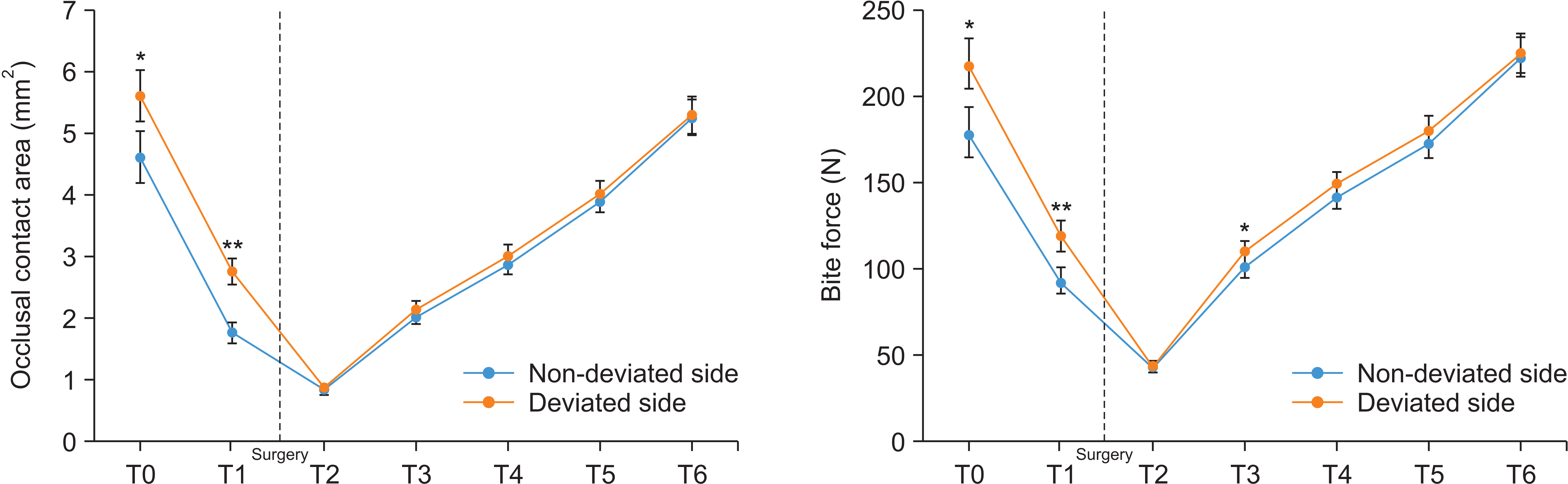
Figure 2 Comparison of the occlusal contact area and bite force of the deviated side and non-deviated sides at each time point. T0, before treatment; T1, 1 month before surgery; T2, T3, T4, T5, and T6 indicate 1, 3, 6, 12, and 24 months after surgery, respectively. The dashed line indicates the time when surgery was performed. The vertical bars indicate the 95% confidence intervals. *p < 0.05; **p < 0.01.
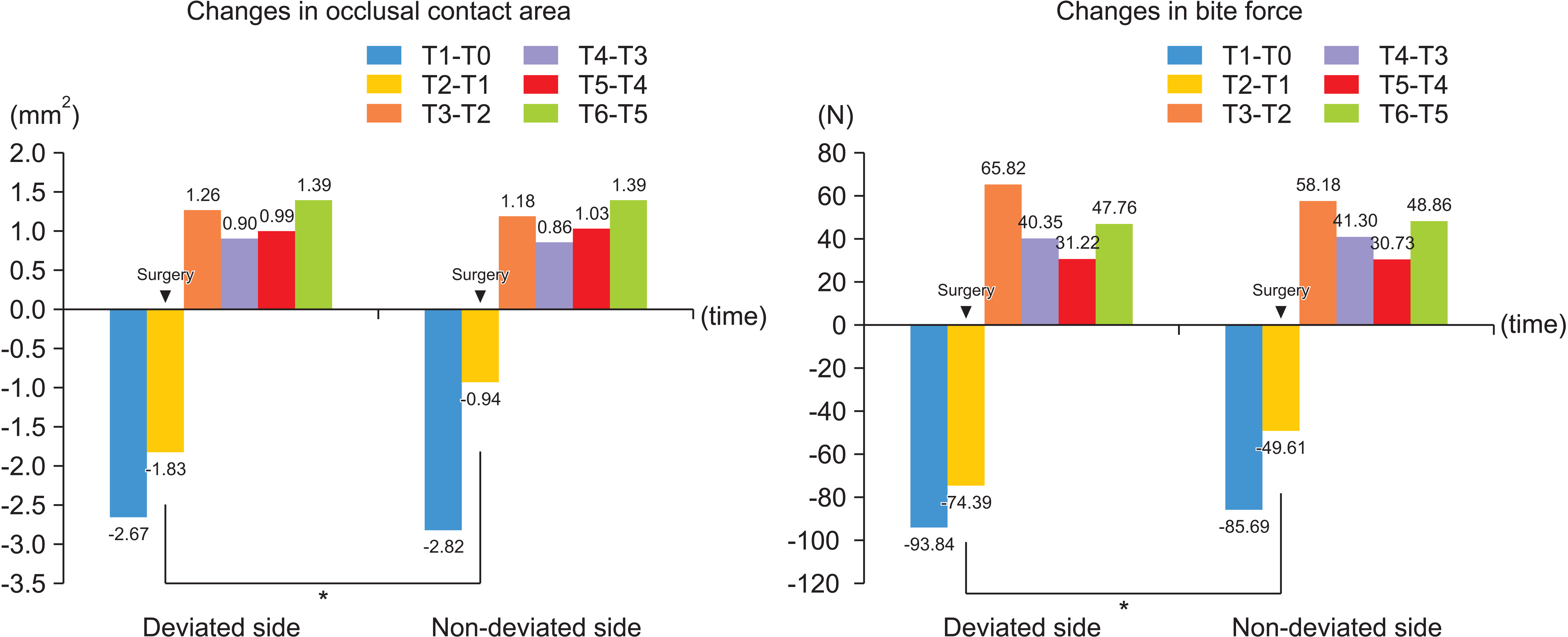
Figure 3 Comparison of the magnitudes of change in occlusal contact area and bite force of the deviated and non-deviated sides at each time point. T0, before treatment; T1, 1 month before surgery; T2, T3, T4, T5, and T6 indicate 1, 3, 6, 12, and 24 months after surgery, respectively. The time indicated by an inverted triangle is when surgery was performed. *p < 0.05.
Reference
-
1. Bailey LJ, Haltiwanger LH, Blakey GH, Proffit WR. 2001; Who seeks surgical-orthodontic treatment: a current review. Int J Adult Orthodon Orthognath Surg. 16:280–92.2. Severt TR, Proffit WR. 1997; The prevalence of facial asymmetry in the dentofacial deformities population at the University of North Carolina. Int J Adult Orthodon Orthognath Surg. 12:171–6.3. Anistoroaei D, Golovcencu L, Saveanu I, Zegan G. 2014; The prevalence of facial asymmetry in preorthodontic treatment. Int J Med Dent. 4:210–5.4. Kobayashi T, Honma K, Nakajima T, Hanada K. 1993; Masticatory function in patients with mandibular prognathism before and after orthognathic surgery. J Oral Maxillofac Surg. 51:997–1001. discussion 1002–3. DOI: 10.1016/S0278-2391(10)80043-6. PMID: 8355107.
Article5. Ellis E 3rd, Throckmorton GS, Sinn DP. 1996; Bite forces before and after surgical correction of mandibular prognathism. J Oral Maxillofac Surg. 54:176–81. DOI: 10.1016/S0278-2391(96)90443-7. PMID: 8604066.
Article6. Kim YG, Oh SH. 1997; Effect of mandibular setback surgery on occlusal force. J Oral Maxillofac Surg. 55:121–6. discussion 126–8. DOI: 10.1016/S0278-2391(97)90224-X. PMID: 9024347.
Article7. Iwase M, Sugimori M, Kurachi Y, Nagumo M. 1998; Changes in bite force and occlusal contacts in patients treated for mandibular prognathism by orthognathic surgery. J Oral Maxillofac Surg. 56:850–5. discussion 855–6. DOI: 10.1016/S0278-2391(98)90013-1. PMID: 9663576.
Article8. Harada K, Watanabe M, Ohkura K, Enomoto S. 2000; Measure of bite force and occlusal contact area before and after bilateral sagittal split ramus osteotomy of the mandible using a new pressure-sensitive device: a preliminary report. J Oral Maxillofac Surg. 58:370–3. discussion 373–4. DOI: 10.1016/S0278-2391(00)90913-3. PMID: 10759115.
Article9. Kobayashi T, Honma K, Shingaki S, Nakajima T. 2001; Changes in masticatory function after orthognathic treatment in patients with mandibular prognathism. Br J Oral Maxillofac Surg. 39:260–5. DOI: 10.1054/bjom.2000.0576. PMID: 11437420.
Article10. Nagai I, Tanaka N, Noguchi M, Suda Y, Sonoda T, Kohama G. 2001; Changes in occlusal state of patients with mandibular prognathism after orthognathic surgery: a pilot study. Br J Oral Maxillofac Surg. 39:429–33. DOI: 10.1054/bjom.2001.0681. PMID: 11735137.11. Ohkura K, Harada K, Morishima S, Enomoto S. 2001; Changes in bite force and occlusal contact area after orthognathic surgery for correction of mandibular prognathism. Oral Surg Oral Med Oral Pathol Oral Radiol Endod. 91:141–5. DOI: 10.1067/moe.2001.112334. PMID: 11174588.
Article12. Sultana MH, Yamada K, Hanada K. 2002; Changes in occlusal force and occlusal contact area after active orthodontic treatment: a pilot study using pressure-sensitive sheets. J Oral Rehabil. 29:484–91. DOI: 10.1046/j.1365-2842.2002.00849.x. PMID: 12028498.
Article13. Iwase M, Ohashi M, Tachibana H, Toyoshima T, Nagumo M. 2006; Bite force, occlusal contact area and masticatory efficiency before and after orthognathic surgical correction of mandibular prognathism. Int J Oral Maxillofac Surg. 35:1102–7. DOI: 10.1016/j.ijom.2006.08.014. PMID: 17097270.
Article14. Ueki K, Marukawa K, Shimada M, Nakagawa K, Yamamoto E. 2007; Changes in occlusal force after mandibular ramus osteotomy with and without Le Fort I osteotomy. Int J Oral Maxillofac Surg. 36:301–4. DOI: 10.1016/j.ijom.2006.09.022. PMID: 17239561.
Article15. Goto TK, Yamada T, Yoshiura K. 2008; Occlusal pressure, contact area, force and the correlation with the morphology of the jaw-closing muscles in patients with skeletal mandibular asymmetry. J Oral Rehabil. 35:594–603. DOI: 10.1111/j.1365-2842.2007.01837.x. PMID: 18482354.
Article16. Choi YJ, Chung C, Kim KH. 2010; Changes in occlusal force and occlusal contact area after orthodontic treatment. Korean J Orthod. 40:176–83. DOI: 10.4041/kjod.2010.40.3.176.
Article17. Choi YJ, Lim H, Chung CJ, Park KH, Kim KH. 2014; Two-year follow-up of changes in bite force and occlusal contact area after intraoral vertical ramus osteotomy with and without Le Fort I osteotomy. Int J Oral Maxillofac Surg. 43:742–7. DOI: 10.1016/j.ijom.2014.02.005. PMID: 24630069.
Article18. Islam I, Lim AAT, Wong RCW. 2017; Changes in bite force after orthognathic surgical correction of mandibular prognathism: a systematic review. Int J Oral Maxillofac Surg. 46:746–55. DOI: 10.1016/j.ijom.2017.01.012. PMID: 28209396.
Article19. Moroi A, Ishihara Y, Sotobori M, Iguchi R, Kosaka A, Ikawa H, et al. 2015; Changes in occlusal function after orthognathic surgery in mandibular prognathism with and without asymmetry. Int J Oral Maxillofac Surg. 44:971–6. DOI: 10.1016/j.ijom.2015.03.015. PMID: 25864000.
Article20. Yoon HR, Choi YJ, Kim KH, Chung C. 2010; Comparisons of occlusal force according to occlusal relationship, skeletal pattern, age and gender in Koreans. Korean J Orthod. 40:304–13. DOI: 10.4041/kjod.2010.40.5.304.
Article21. Goto TK, Nishida S, Yahagi M, Langenbach GE, Nakamura Y, Tokumori K, et al. 2006; Size and orientation of masticatory muscles in patients with mandibular laterognathism. J Dent Res. 85:552–6. DOI: 10.1177/154405910608500614. PMID: 16723654.
Article22. Lee DH, Yu HS. 2012; Masseter muscle changes following orthognathic surgery: a long-term three-dimensional computed tomography follow-up. Angle Orthod. 82:792–8. DOI: 10.2319/111911-717.1. PMID: 22390633. PMCID: PMC8823124.23. Iodice G, Danzi G, Cimino R, Paduano S, Michelotti A. 2016; Association between posterior crossbite, skeletal, and muscle asymmetry: a systematic review. Eur J Orthod. 38:638–51. DOI: 10.1093/ejo/cjw003. PMID: 26823371.
Article24. Kim HJ, Hong M, Park HS. 2019; Analysis of dental compensation in patients with facial asymmetry using cone-beam computed tomography. Am J Orthod Dentofacial Orthop. 156:493–501. DOI: 10.1016/j.ajodo.2018.10.025. PMID: 31582121.
Article25. Kim KA, Park HS, Lee SY, Kim SJ, Baek SH, Ahn HW. 2019; Short-term changes in muscle activity and jaw movement patterns after orthognathic surgery in skeletal Class III patients with facial asymmetry. Korean J Orthod. 49:254–64. DOI: 10.4041/kjod.2019.49.4.254. PMID: 31367580. PMCID: PMC6658898.
Article26. Verna C. 2016; Regional acceleratory phenomenon. Front Oral Biol. 18:28–35. DOI: 10.1159/000351897. PMID: 26599115.
Article27. Raadsheer MC, van Eijden TM, van Ginkel FC, Prahl-Andersen B. 1999; Contribution of jaw muscle size and craniofacial morphology to human bite force magnitude. J Dent Res. 78:31–42. DOI: 10.1177/00220345990780010301. PMID: 10065943.
Article28. Proffit WR, Fields HW, Nixon WL. 1983; Occlusal forces in normal- and long-face adults. J Dent Res. 62:566–70. DOI: 10.1177/00220345830620051201. PMID: 6573373.
Article29. Braun S, Bantleon HP, Hnat WP, Freudenthaler JW, Marcotte MR, Johnson BE. 1995; A study of bite force, part 2: relationship to various cephalometric measurements. Angle Orthod. 65:373–7. DOI: 10.1043/0003-3219(1995)065<0373:ASOBFP>2.0.CO;2. PMID: 8526297.30. Alkayyal MA, Turkistani KA, Al-Dharrab AA, Abbassy MA, Melis M, Zawawi KH. 2020; Occlusion time, occlusal balance and lateral occlusal scheme in subjects with various dental and skeletal characteristics: a prospective clinical study. J Oral Rehabil. 47:1503–10. DOI: 10.1111/joor.13095. PMID: 32966657.
Article
- Full Text Links
-
- Actions
-
Cited
- CITED
-
- Close
- Share
-
- Similar articles
-
- Unilateral intraoral vertical ramus osteotomy and sagittal split ramus osteotomy for the treatment of asymmetric mandibles
- A study on the vertical dysplasia in the skeletal class iii malocclusion
- Unilateral intraoral vertical ramus osteotomy based on preoperative three-dimensional simulation surgery in a patient with facial asymmetry
- Positional changes of the mandibular condyle in unilateral sagittal split ramus osteotomy combined with intraoral vertical ramus osteotomy for asymmetric class III malocclusion
- Postoperative stability and occlusal plane alternation by orthgnathic surgery of skeletal Class III malocclusion with anterior open bite
