Ultrasonographic Findings of Post-Operative Changes after Breast Cancer Surgery
- Affiliations
-
- 1Department of Surgery, Kyungpook National University Chilgok Hospital, School of Medicine, Kyungpook National University, Daegu, Korea
- KMID: 2525878
- DOI: http://doi.org/10.46268/jsu.2021.8.2.32
Abstract
- An accurate understanding of ultrasound findings of post-operative changes in breast cancer would be one of the most important areas of breast cancer surveillance. This is especially true when the ultrasound is performed by breast surgeons who have in-depth knowledge of oncoplastic breast surgery as it would be easier for them to distinguish between true recurrence of breast cancer and post-operative changes. In this article, the various but typical post-operative changes in breast cancer are described. These findings would be truly helpful to breast surgeons for the detection of early recurrence and treatment of post-operative complications.
Figure
-

Fig. 1 Ultrasonographicfindings after breast-conserving surgery. (A, B) A vertical shape of postoperative scar is observed from the skin to the pectoralis major muscle, which will be remained almost permanently. If any nodule is found around the scar or there is an increasing sign of blood flow, it should be confirmed by tissue biopsy due to possibility of tumor recurrence.
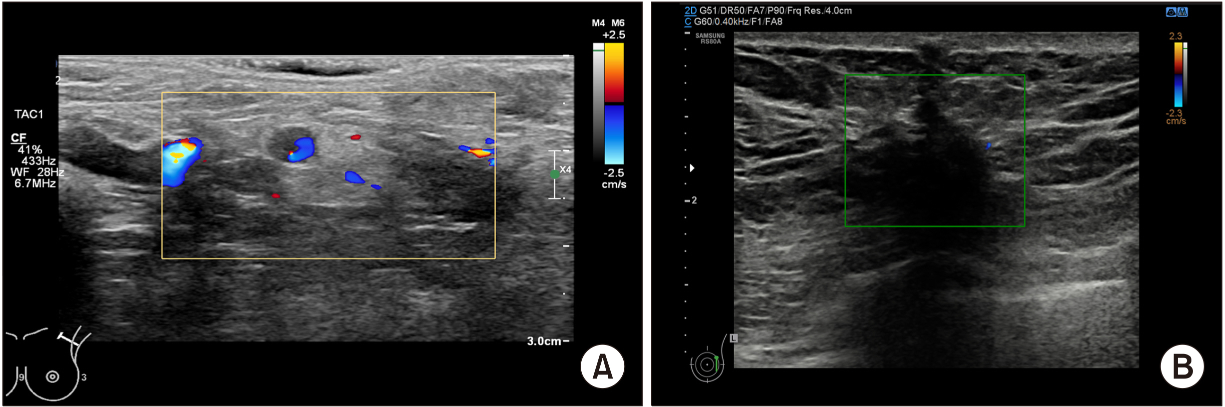
Fig. 2 Doppler imaging of breast ultrasonography in suspected nodule of local recurrence. (A) A 5 mm-sized hypoechoic nodule (yellow square) occurred near the axillary region, and showed increased blood flow on the doppler mode which indicating a proliferative lesion (confirmed as recurrent invasive cancer on histological examination). (B) A 2 cm-sized hypoechoic nodule (green squares) was identified around the postoperative scar. However, there is no evidence of increased blood flow in the doppler mode, suggesting a high possibility of non-proliferative lesions (fat necrosis was confirmed in biopsy).
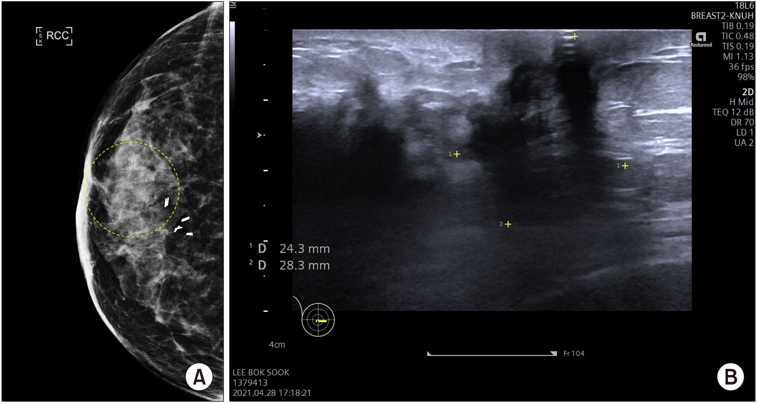
Fig. 3 Local recurrence of breast cancer. (A) Malignant microcalcifi-cations (yellow dotted circle) on subareolar region were identified on mammography. (B) In ultrasound finding, a hypoechoic nodule with irregular margins were found at the same location. The lesion was dia-gnosed as a local recurrence of breast cancer in core needle biopsy.

Fig. 4 Ultrasound findings of a patient who were received left side of mastectomy for breast cancer. (A) The right side of contralateral breast is a normal breast. All of the breast structures are observed. (B) The left side of ipsilateral breast, which was removed as total mastectomy. Only the skin is remained overlying the pectoralis major muscle.
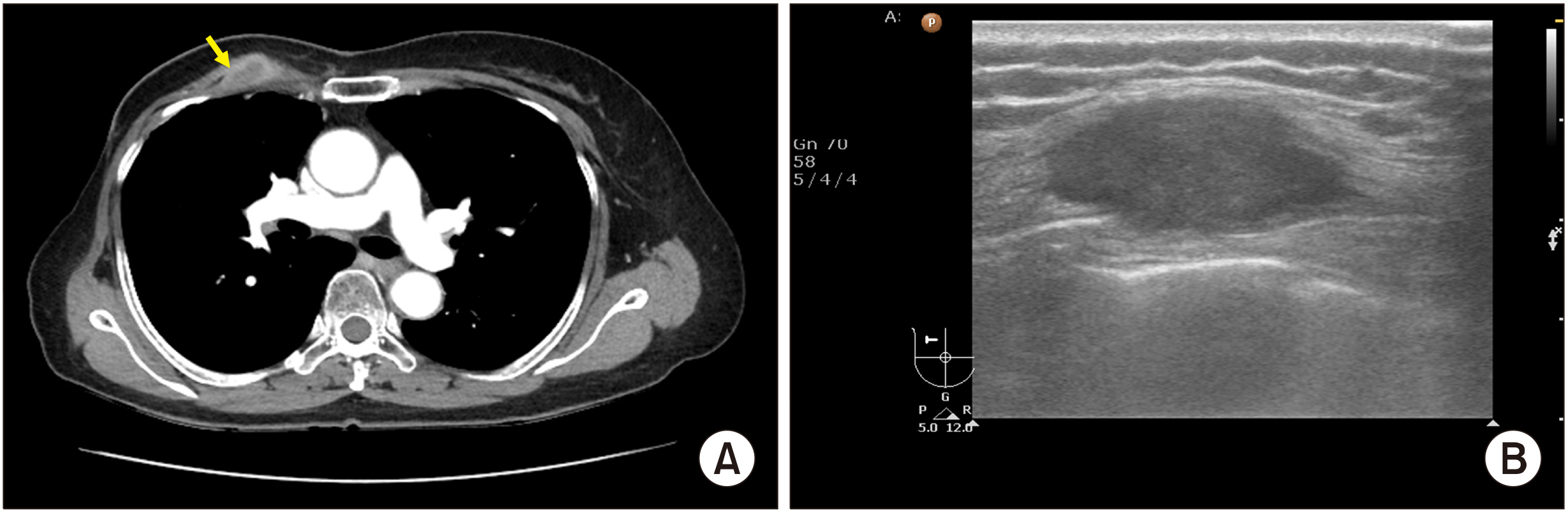
Fig. 5 Ultrasonographic finding of locally recurred nodule on ipsilateral chest wall. (A) On chest CT, an enhancing nodule was developed under the pectoralis major muscle, suggesting that blood flow was slightly increased. (B) Ultrasound finding of the same nodule. A 3.1 cm-sized nodule was identified under the pectoralis major muscle, showing irregular margins and hypoechoic material inside, which is strongly suspected of local recurrence (it was confirmed as invasive ductal carcinoma, recurred.).
Reference
-
1. Chansakul T, Lai KC, Slanetz PJ. 2012; The postconservation breast: part 1, expected imaging findings. AJR Am J Roentgenol. 198:321–30. DOI: 10.2214/AJR.10.7298. PMID: 22268174.
Article2. Duijm LE, Guit GL, Zaat JO, Koomen AR, Willebrand D. 1997; Sensitivity, specificity and predictive values of breast imaging in the detection of cancer. Br J Cancer. 76:377–81. DOI: 10.1038/bjc.1997.393. PMID: 9252206. PMCID: PMC2224070.
Article3. Yoon JH, Kim MJ, Kim EK, Moon HJ. 2015; Imaging surveillance of patients with breast cancer after primary treatment: current recommendations. Korean J Radiol. 16:219–28. DOI: 10.3348/kjr.2015.16.2.219. PMID: 25741186. PMCID: PMC4347260.
Article4. Chang JF, Huang CS, Chang RF. 2020; Automated whole breast segmentation for hand-held ultrasound with position information: application to breast density estimation. Comput Methods Programs Biomed. 197:105727. DOI: 10.1016/j.cmpb.2020.105727. PMID: 32916544.
Article5. Houssami N, Sainsbury R. 2006; Breast cancer: multidisciplinary care and clinical outcomes. Eur J Cancer. 2480–91. DOI: 10.1016/j.ejca.2006.05.023. PMID: 16904313.
Article6. Fragomeni SM, Sciallis A, Jeruss JS. 2018; Molecular subtypes and local-regional control of breast cancer. SurgOncol Clin N Am. 27:95–120. DOI: 10.1016/j.soc.2017.08.005. PMID: 29132568. PMCID: PMC5715810.
Article7. Chen JH, Nie K, Bahri S, Hsu CC, Hsu FT, Shih HN, et al. 2010; Decrease in breast density in the contralateral normal breast of patients receiving neoadjuvant chemotherapy:MR imaging evaluation. Radiology. 255:44–52. DOI: 10.1148/radiol.09091090. PMID: 20308443. PMCID: PMC2843830.
Article8. Yi A, Kim HH, Shin HJ, Huh MO, Ahn SD, Seo BK. 2009; Radiation-induced complications after breast cancer radiation therapy: a pictorial review of multimodality imaging findings. Korean J Radiol. 10:496–507. DOI: 10.3348/kjr.2009.10.5.496. PMID: 19721835. PMCID: PMC2731868.
Article9. Yilmaz ZN, Neal CH, Noroozian M, Klein KA, Sundaram B, Kazerooni EA, et al. 2014; Imaging of breast cancer-related changes after nonsurgical therapy. AJR Am J Roentgenol. 202:675–83. DOI: 10.2214/AJR.13.11518. PMID: 24555607.
Article10. Margolis NE, Morley C, Lotfi P, Shaylor SD, Palestrant S, Moy L, et al. 2014; Update on imaging of the postsurgical breast. Radiographics. 34:642–60. DOI: 10.1148/rg.343135059. PMID: 24819786.
Article11. Stavros AT. 2004. Breast ultrasound. Lippincott Williams & Wilkins;Philadelphia:12. Gutierrez R, Horst KC, Dirbas FM, Ikeda DM. Dirbas F, Scott-Conner C, editors. 2011. Breast imaging following breast conservation therapy. Breast Surgical Techniques and Interdisciplinary Management. Springer;New York: p. 975–95. DOI: 10.1007/978-1-4419-6076-4_81.
Article13. Destounis SV. 2005; Imaging of the post-surgical breast. Contemp Diagn Radiol. 1–6. DOI: 10.1097/00219246-200512310-00001.
Article14. Stuhrmann M, Aronius R, Schietzel M. 2000; Tumor vascularity of breast lesions: potentials and limits of contrast-enhanced Doppler sonography. AJR Am J Roentgenol. 175:1585–9. DOI: 10.2214/ajr.175.6.1751585. PMID: 11090380.
- Full Text Links
-
- Actions
-
Cited
- CITED
-
- Close
- Share
-
- Similar articles
-
- Ultrasonographic Findings of Postoperative Change after Breast Reconstruction
- The analysis of ultrasonographic findings in breast carcinoma
- Ultrasonographic Findings of Breast Cancer
- Ultrasonographic Findings of Breast Diseases During Pregnancy and Lactating Period
- Radiologic findings of male breast cancer: two cases report
