Microsurgical Reconstruction of Lower Limb Using Thoracodorsal Artery Perforator Chimeric Free Flap after Popliteal Artery Revascularization: A Case Report
- Affiliations
-
- 1Department of Plastic and Reconstructive Surgery, Hanyang University College of Medicine, Seoul, Korea
- KMID: 2522630
- DOI: http://doi.org/10.12790/ahm.21.0130
Abstract
- Although traumatic popliteal artery injury is uncommon, it can significantly increase the risk of limb amputation because of the anatomical complexity and delayed diagnosis and treatment. Various tools are available for treatment. Recently, an endovascular approach has been attempted for such injuries; however, open surgical repair remains the standard treatment. An integrated and stepwise procedure involving multidisciplinary specialists, including emergency department personnel for initial evaluation, orthopedic surgeons for treating accompanying fractures or dislocations, vascular and plastic surgeons for vessel repair, and interventional radiologists for immediate diagnosis and implementation of the endovascular approach, is needed. Covering wound defects due to skin and soft tissue necrosis and irreversible ischemic damage remains difficult despite successfully revascularizing the injured vessels. Here, we describe a case of revascularization after popliteal artery injury along with successful reconstruction of a complex defect with a thoracodorsal artery perforator chimeric free flap when recipient vessel selection was limited.
Keyword
Figure
-
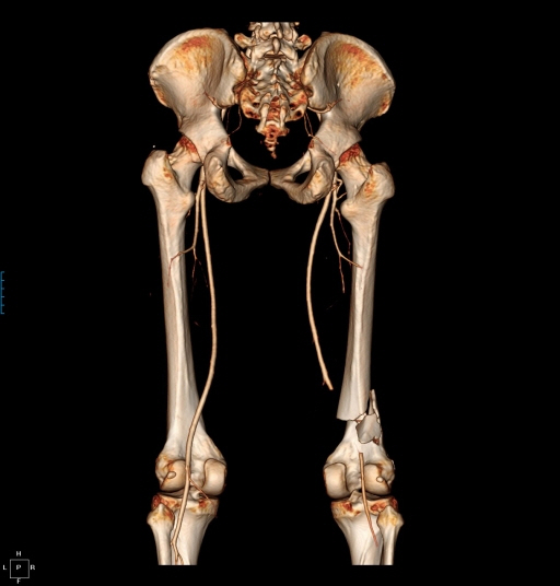
Fig. 1. Posterior view of computed tomography angiography showing long segmental obstruction about 8 cm in length above the tibial plateau.
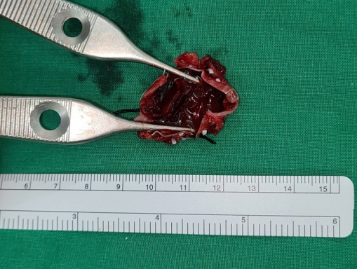
Fig. 2. The excised section of the allograft with an intravascular thrombus can be observed.
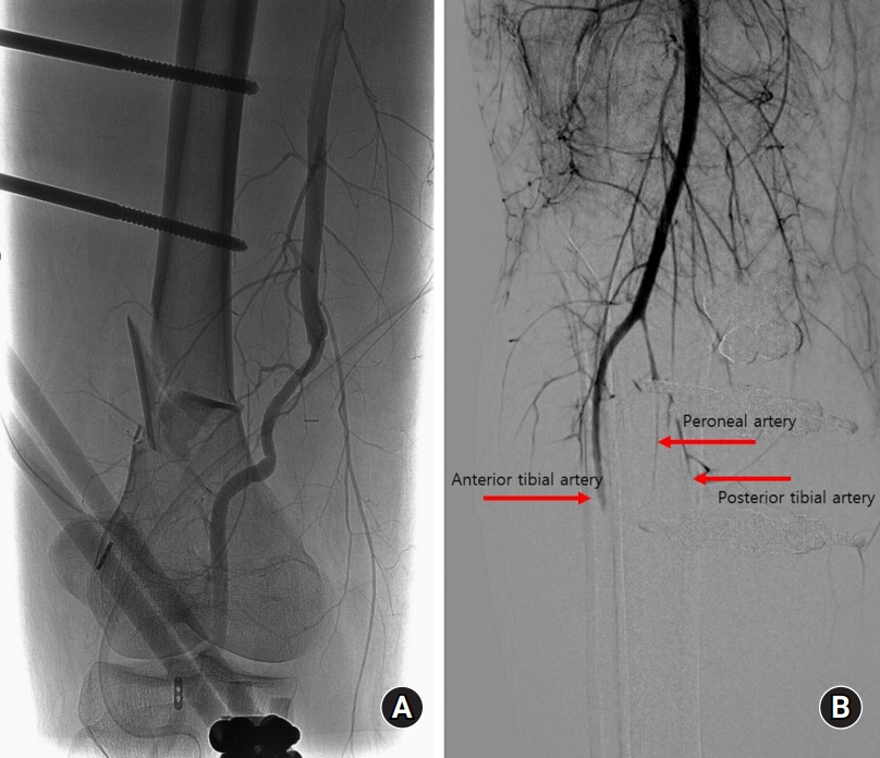
Fig. 3. Angiography was performed by a radiologist. (A) No occlusions were observed at the anastomosis site. (B) Diffuse vasospasm was observed at the proximal level of the lower extremity in all vessels, including the anterior tibial artery, peroneal artery, and posterior tibial artery.
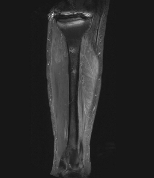
Fig. 4. Magnetic resonance imaging revealed diffuse muscle necrosis and fatty degeneration below the knee level, especially involving the gastrocnemius and soleus muscles.
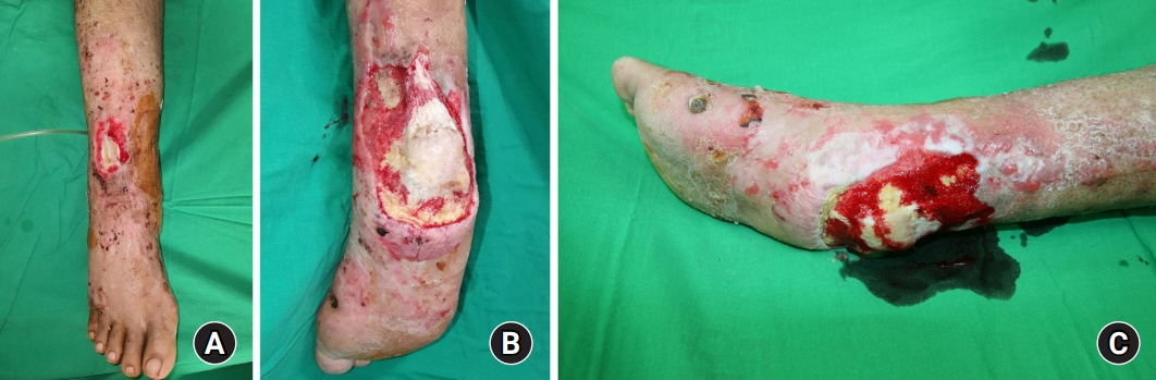
Fig. 5. (A) Image showing the dorsal ankle defect with exposure of the extensor tendon. (B) Posterior view of the heel defect with exposure of the calcaneus and Achilles tendon. (C) Lateral view of the heel defect. This heel defect was managed with periodic negative pressure wound therapy, and the fresh granulation tissue had grown into the wound.
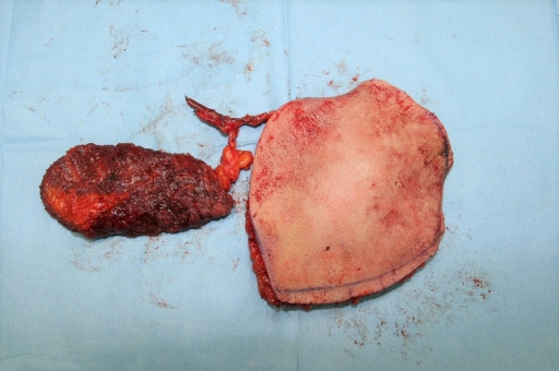
Fig. 6. The harvested thoracodorsal artery perforator chimeric flap consisting of 6 × 7 cm-sized latissimus dorsi muscle and 16 × 12 cm-sized skin perforator flap.
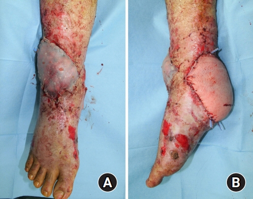
Fig. 7. Immediate postoperative view. (A) A muscle flap was inserted into the dorsal ankle defect with a split-thickness skin graft. (B) Skin flap of the thoracodorsal artery perforator chimeric flap was incorporated into the heel defect area.
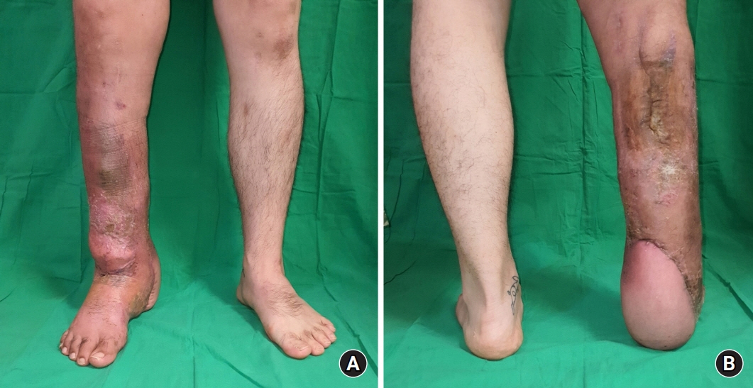
Fig. 8. (A) Anterior view, 6 months after the trauma. (B) Posterior view, 6 months after the trauma. No wound problems were observed, and independent walking was possible.
Reference
-
1. Makaloski V, Stellmes A, Wyss D, et al. Posterior approach for revascularization in blunt popliteal vessel injury. Ann Vasc Surg. 2018; 48:89–96.
Article2. Mullenix PS, Steele SR, Andersen CA, Starnes BW, Salim A, Martin MJ. Limb salvage and outcomes among patients with traumatic popliteal vascular injury: an analysis of the National Trauma Data Bank. J Vasc Surg. 2006; 44:94–100.
Article3. Asensio JA, Dabestani PJ, Miljkovic SS, et al. Popliteal artery injuries. Less ischemic time may lead to improved outcomes. Injury. 2020; 51:2524–31.
Article4. Butler WJ, Calvo RY, Sise MJ, et al. Outcomes for popliteal artery injury repair after discharge: a large-scale population-based analysis. J Trauma Acute Care Surg. 2019; 86:173–80.
Article5. Cooper N, Roshdy M, Sciarretta JD, et al. Multidisciplinary team approach in the management of popliteal artery injury. J Multidiscip Healthc. 2018; 11:399–403.
Article6. Sriussadaporn S, Pak-art R. Temporary intravascular shunt in complex extremity vascular injuries. J Trauma. 2002; 52:1129–33.
Article7. Rehman ZU. Outcomes of popliteal artery injuries repair: autologous vein versus prosthetic interposition grafts. Ann Vasc Surg. 2020; 69:141–5.
Article8. Sharrock M, Antoniou SA, Antoniou GA. Vein versus prosthetic graft for femoropopliteal bypass above the knee: a systematic review and meta-analysis of randomized controlled trials. Angiology. 2019; 70:649–61.
Article9. Horstmann R, Daum H, Heller M, Schröder A. Critical ischemia of the extremities caused by ergotism. Treatment with intra-arterial prostaglandin E1 infusion. Dtsch Med Wochenschr. 1993; 118:1067–71.10. Lee KT, Wiraatmadja ES, Mun GH. Free latissimus dorsi muscle-chimeric thoracodorsal artery perforator flaps for reconstruction of complicated defects: does muscle still have a place in the domain of perforator flaps? Ann Plast Surg. 2015; 74:565–72.
- Full Text Links
-
- Actions
-
Cited
- CITED
-
- Close
- Share
-
- Similar articles
-
- Reconstruction of a Mangled Hand with a Thoracodorsal Artery Perforator Free Flap: A Report of Two Cases
- Reconstruction of the Soft Tissue Defect Using Thoracodorsal Artery Perforator Skin Flap
- Reconstruction of Greater Trochanteric defect using Lumbar Artery Perforator Free Flap: A Case Report
- Resurfacing the defect from wide excision of a malignant peripheral nerve sheath tumor based on a thoracodorsal artery perforator free flap: a case report
- The Retrograde Limb of the Internal Mammary Artery: An Alternative Inflow Option for Free Flap Breast Reconstruction
