Rosette-forming epithelioid osteosarcoma in the rib: a rare case of location and morphology
- Affiliations
-
- 1Department of Pathology, Kosin University College of Medicine, Busan, Korea
- KMID: 2522442
- DOI: http://doi.org/10.4132/jptm.2021.06.22
Abstract
- The rib is an unusual location for osteosarcoma and is reported in only 2% of all cases. The major histological variants of osteosarcoma are osteoblastic, chondroblastic, and fibroblastic, with a few rare variants including one epithelioid type. This report describes a 44-year-old male with an osteolytic mass in the right seventh rib. Histological examination revealed osteosarcoma with unique features of epithelioid appearance and rosette structures. To the best of our knowledge, this is the first reported case of a rosette-forming osteosarcoma of the rib that showed epithelioid morphology. Despite successful surgery, the patient’s prognosis was poor because this malignancy had an unusual location within the axial skeleton and was a rare histological variant.
Keyword
Figure
-
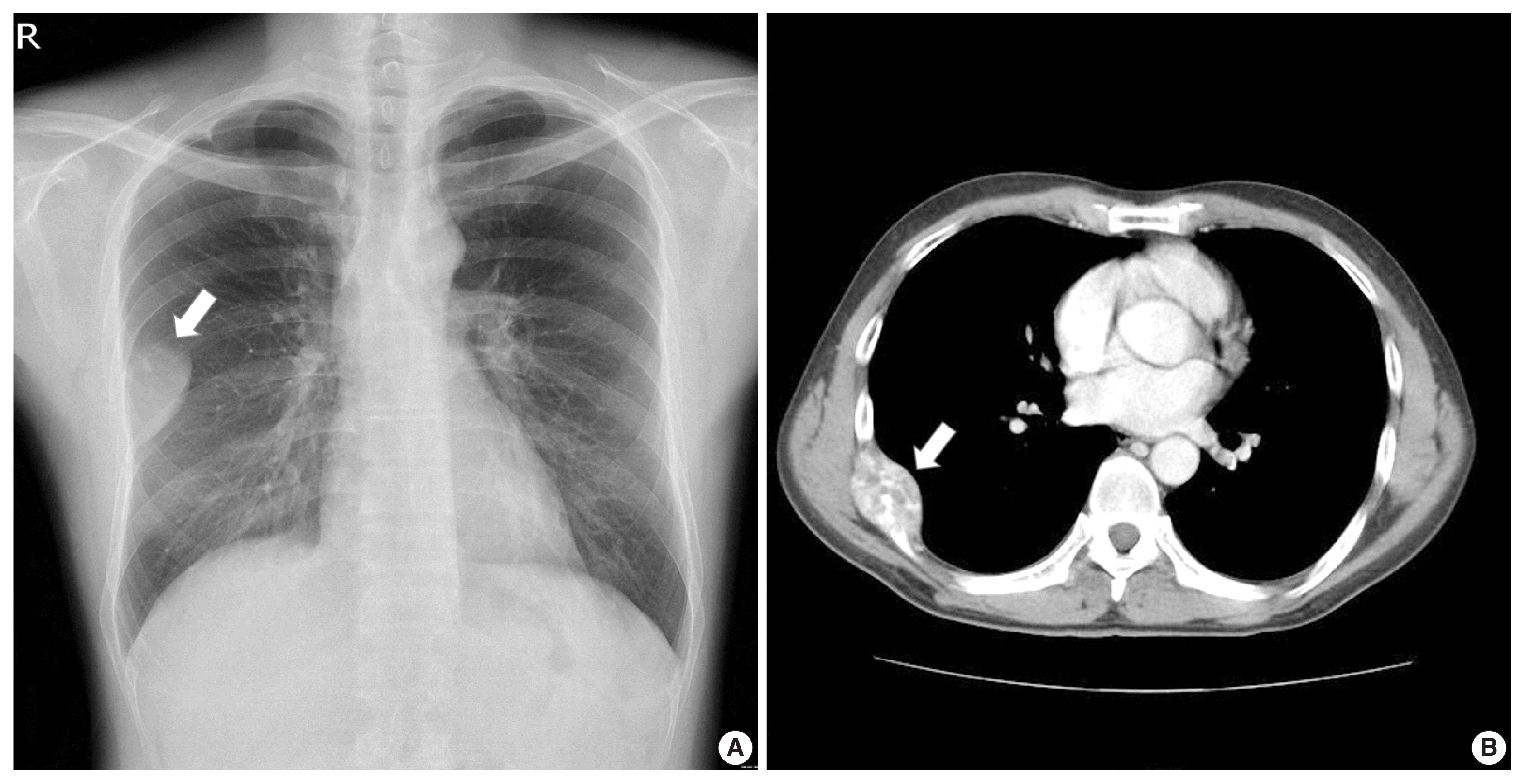
Fig. 1 Radiologic images. (A) Chest radiograph showing an oval mass (arrow) in the right thorax. (B) Computed tomography scan of the chest, revealing a large, heterogeneously enhanced mass (arrow) that originated from the right seventh rib, with lytic destruction of the bone and coarse calcification.

Fig. 2 Epithelioid osteosarcoma of the rib. (A) Macroscopic picture of an ill-defined lytic mass of the rib with hemorrhagic cystic degeneration. (B) Microscopic view of the trabecular and rosette-forming pattern that contained a fibrillar matrix in the center. (C) Rosette-like structures of plasmacyoid tumor cells with eccentrically located nuclei and abundant pale cytoplasm. (D) Mixed epithelioid and plasmacytoid tumor cells. (E) Blood-filled cystic spaces showing telangiectatic features. (F) Neoplastic bones produced by malignant tumor cells are found throughout the lesion.
Reference
-
References
1. Unni KK, Inwards CY. Dahlin’s bone tumors. 6th ed.Philadelphia: Lippincott Williams & Wilkins;2010. p. 122–57.2. Chattopadhyay A, Nagendhar Y, Kumar V. Osteosarcoma of the rib. Indian J Pediatr. 2004; 71:543–4.
Article3. Ikeda H, Takeo M, Kayata H, Mikami R, Nakamoto Y, Yamamoto M. A case of rapidly growing osteosarcoma of the rib. Ann Thorac Cardiovasc Surg. 2014; 20(Suppl):521–4.
Article4. Deitch J, Crawford AH, Choudhury S. Osteogenic sarcoma of the rib: a case presentation and literature review. Spine (Phila Pa 1976). 2003; 28:E74–7.5. Mohanty S, Inchara YK, Crasta JA, Ananthamurthy A. An unusual case of primary osteosarcoma of the rib in an adult. Indian J Med Paediatr Oncol. 2010; 31:18–20.
Article6. Scranton PE Jr, DeCicco FA, Totten RS, Yunis EJ. Prognostic factors in osteosarcoma: a review of 20 year’s experience at the University of Pittsburgh Health Center Hospitals. Cancer. 1975; 36:2179–91.
Article7. Kramer K, Hicks DG, Palis J, et al. Epithelioid osteosarcoma of bone: immunocytochemical evidence suggesting divergent epithelial and mesenchymal differentiation in a primary osseous neoplasm. Cancer. 1993; 71:2977–82.
Article8. Carlos-Bregni R, Contreras E, Hiraki KR, Vargas PA, Leon JE, de Almeida OP. Epithelioid osteosarcoma of the mandible: a rare case with unusual immunoprofile. Oral Surg Oral Med Oral Pathol Oral Radiol Endod. 2008; 105:e47–52.
Article9. Okada K, Hasegawa T, Yokoyama R. Rosette-forming epithelioid osteosarcoma: a histologic subtype with highly aggressive clinical behavior. Hum Pathol. 2001; 32:726–33.
Article10. Kaveri H, Rekha K, Punnya VA. Epithelioid osteosarcoma of the maxilla: report of an unusual case. Br J Oral Maxillofac Surg. 2009; 47:143–5.
Article11. Kuwabara H, Fujita K, Yuki M, Goto I, Hanafusa T, Shibayama Y. Cytokeratin-positive rib osteosarcoma metastasizing to the small intestine. Indian J Pathol Microbiol. 2014; 57:109–12.
- Full Text Links
-
- Actions
-
Cited
- CITED
-
- Close
- Share
-
- Similar articles
-
- A rare histopathological variant of Schwannoma with rosette-like arrangements and epithelioid cells: a case report from a histopathologist’s perspective
- Well-differentiated osteosarcoma of the rib.
- A study of Corelation between Rosette Positivity and Plasma Level of Immunoglobulins in the Pulmonary Tuberculosis Patients
- Metachronous osteosarcoma
- Primary Osteoblastic Osteosarcoma of the Rib in an Adult: A Case Report
