Primary Osteoblastic Osteosarcoma of the Rib in an Adult: A Case Report
- Affiliations
-
- 1Department of Radiology, Seoul National University Bundang Hospital, Seongnam, Korea. jacrad@radiol.snu.ac.kr
- 2Department of Radiology and Institute of Radiation Medicine, Seoul National University College of Medicine, Seoul, Korea.
- 3Department of Pathology, Seoul National University Bundang Hospital, Seongnam, Korea.
- 4Department of Orthopedic Surgery, Seoul National University Bundang Hospital, Seongnam, Korea.
- KMID: 2002929
- DOI: http://doi.org/10.3348/jksr.2011.65.6.603
Abstract
- We report the CT and magnetic resonance (MR) imaging appearances in an adult case of primary osteoblastic osteosarcoma of the rib. Osteosarcoma of the rib presents a diagnostic challenge because of the rarity of the lesion, especially with plain radiographs. The tumor should be suspected if CT and MR images demonstrate mineralization, suggestive of an osteoid matrix.
Figure
-
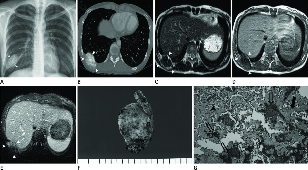
Fig. 1 Radiograph, CT, MRI, and histopathologic specimen images of 31-year-old woman with osteoblastic osteosarcoma of the rib. A. Chest radiograph shows non homogenous calcification (white arrowheads) involving the right 10th rib. B. CT scan of chest shows the tumor mass with central dense calcification (white arrowheads). C. Axial T2-weighted image shows the tumor (white arrowheads) with extensive central signal loss due to dense calcification. D. Axial T1-weighted image shows intermediate signal intensity lesion (white arrowheads) with multiple central signal void areas. E. Axial T1-weighted contrast-enhanced image shows well enhancing portions of the soft tissue mass of the tumor (white arrowheads). F. Gross specimen image shows lobulated mass with mineralized matrix. G. Mineralized (open arrows) or unmineralized (arrow) osteoid is haphazardly distributed and neoplastic hyperchromatic cells (arrowhead) are noted between the osteoid (H&E, × 200).
Reference
-
1. Burt M, Fulton M, Wessner-Dunlap S, Karpeh M, Huvos AG, Bains MS, et al. Primary bony and cartilaginous sarcomas of chest wall: results of therapy. Ann Thorac Surg. 1992; 54:226–232.2. Kim H, Park C, Lee YB, Jin SY, Ro JY, Ayala AG. Case report 643: Osteosarcoma of ribs with giant rosettoid structures. Skeletal Radiol. 1990; 19:609–612.3. Abdulrahman RE, White CS, Templeton PA, Romney B, Moore EH, Aisner SC. Primary osteosarcoma of the ribs: CT findings. Skeletal Radiol. 1995; 24:127–129.4. Chattopadhyay A, Nagendhar Y, Kumar V. Osteosarcoma of the rib. Indian J Pediatr. 2004; 71:543–544.5. Lawson JP, Barwick KW. Case report 162: Periosteal osteosarcoma of rib. Skeletal Radiol. 1981; 7:63–65.6. O'Sullivan P, O'Dwyer H, Flint J, Munk PL, Muller NL. Malignant chest wall neoplasms of bone and cartilage: a pictorial review of CT and MR findings. Br J Radiol. 2007; 80:678–684.7. Lee TJ, Collins J. MR imaging evaluation of disorders of the chest wall. Magn Reson Imaging Clin N Am. 2008; 16:355–379. x
