Deletion of adipose triglyceride lipase abolishes blood flow increase after β3-adrenergic stimulation in visceral adipose tissue of mice
- Affiliations
-
- 1Department of Pharmacology, Korea University College of Medicine, Seoul 02841, Korea
- 2BK21 Graduate Program, Department of Biomedical Sciences, Korea University College of Medicine, Seoul 02841, Korea
- 3Department of Rehabilitation Medicine, Seoul National University Hospital, Seoul 03080, Korea
- KMID: 2516874
- DOI: http://doi.org/10.4196/kjpp.2021.25.4.355
Abstract
- Dynamic changes in adipose tissue blood flow (ATBF) with nutritional status play a role in the regulation of metabolic and endocrine functions. Activation of the sympathetic nervous system via β-adrenergic receptors (β-AR) contributes to the control of postprandial enhancement of ATBF. Herein, we sought to identify the role of each β-AR subtype in the regulation of ATBF in mice. We monitored the changes in visceral epididymal ATBF (VAT BF), induced by local infusion of dobutamine, salbutamol, and CL316,243 (a selective β1-, β2-, and β3-AR agonist, respectively) into VAT of lean CD-1 mice and global adipose triglyceride lipase (ATGL) knockout (KO) mice, using laser Doppler flowmetry. Administration of CL316,243, known to promote lipolysis in adipocytes, significantly increased VAT BF of CD-1 mice to a greater extent compared to that of the vehicle, whereas administration of dobutamine or salbutamol did not produce significant differences in VAT BF. The increase in VAT BF induced by β3-AR stimulation disappeared in ATGL KO mice as opposed to their wild-type (WT) littermates, implying a role of ATGL-mediated lipolysis in the regulation of VAT BF. Different vascular reactivities occurred despite no significant differences in vessel density and adiposity between the groups. Additionally, the expression levels of the angiogenesis-related genes were significantly higher in VAT of ATGL KO mice than in that of WT, implicating an association of ATBF responsiveness with angiogenic activity in VAT. Our findings suggest a potential role of β3-AR signaling in the regulation of VAT BF via ATGL-mediated lipolysis in mice.
Keyword
Figure
-
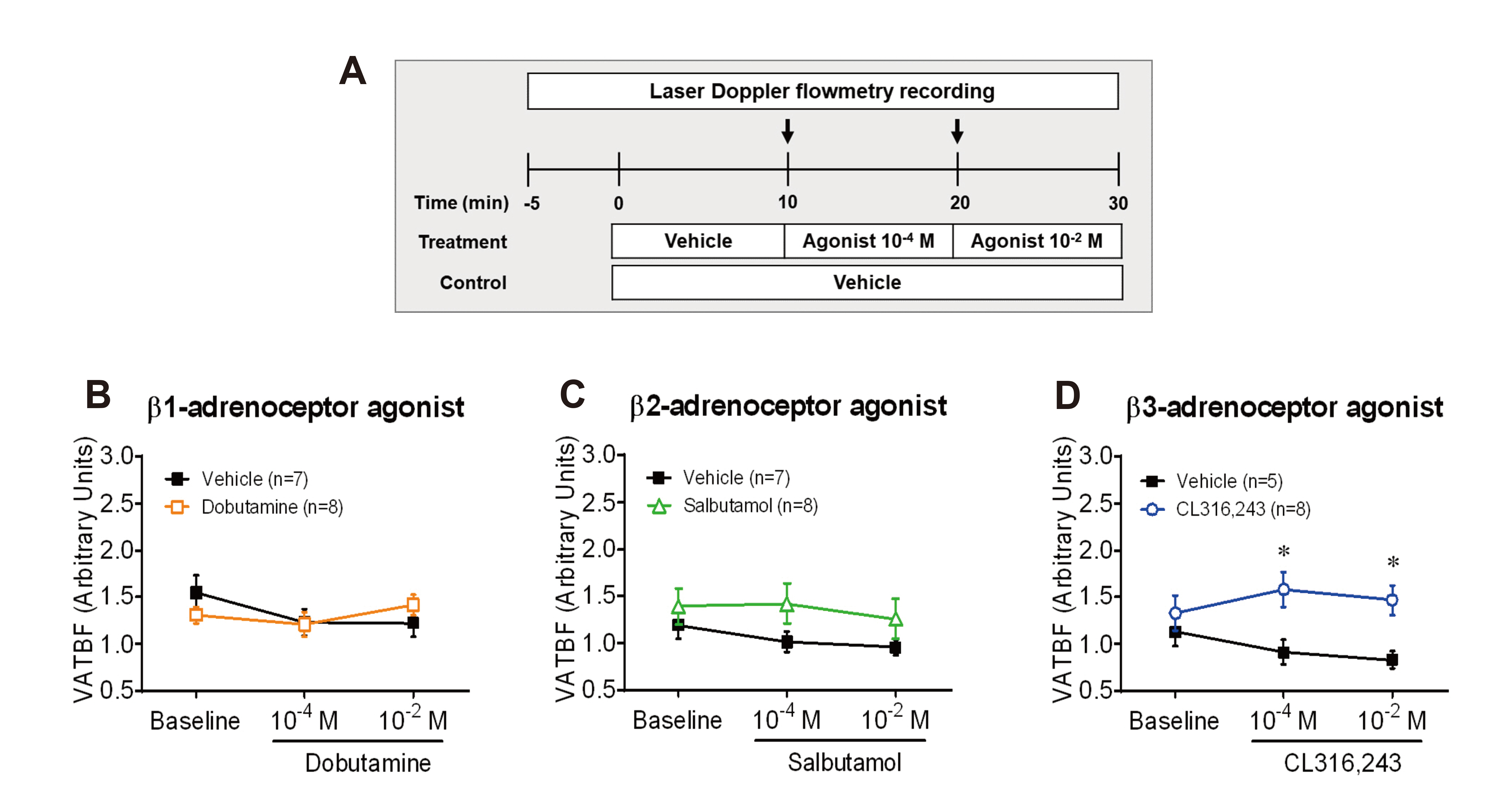
Fig. 1 Effects of β-adrenergic receptor subtypes on the regulation of visceral adipose tissue blood flow (VAT BF) in CD-1 mice. (A) Experimental timeline diagram. After stabilizing VAT BF for 5 min, vehicle and agonists (10–4 and 10–2 M) were infused into VAT a rate of 0.5 µl min–1 for 10 min, respectively. Arrows represent the time point of the switch in the agonist delivery. (B) Changes in VAT BF induced by stimulation of dobutamine, a selective β1-adrenergic receptor agonist. (C) Changes in VAT BF induced by stimulation of salbutamol, a selective β2-adrenergic receptor agonist. (D) Changes in VAT BF induced by stimulation of CL316,243, a selective β3-adrenergic receptor agonist. *p < 0.05.

Fig. 2 Distribution and colocalization of β-adrenergic receptor subtypes in visceral epididymal fat (VAT) of mice. (A) Comparison of the expression levels of Adrb1, Adrb2, and Adrb3 in VAT of CD-1 male mice fed a normal chow diet at 8 weeks of age. The ΔCt method (2–ΔCt) was used to calculate the relative expression level of each gene. (B–D) Comparison of the expression levels of Adrb1 (B), Adrb2 (C), and Adrb3 (D) between fractionated adipocytes and stromal vascular fraction (SVF) in VAT of C57BL/6N male mice fed a normal chow diet at 9 weeks of age. The relative mRNA expression was calculated using the ΔΔCt method (2–ΔΔCt). (E, F) Representative images of double immunofluoresnce staining of whole-mount VAT using ADRB1, ADRB2, or ADRB3 antibodies with PECAM1 antibodies (E) or BODIPY (F) dye in C57BL/6N male mouse fed a normal chow diet at 8 weeks of age. White arrowheads indicate localization of ADRB2 in PECAM1+ vessels. Images are shown at a magnification of ×500. Scale bars, 20 µm. *p < 0.05.
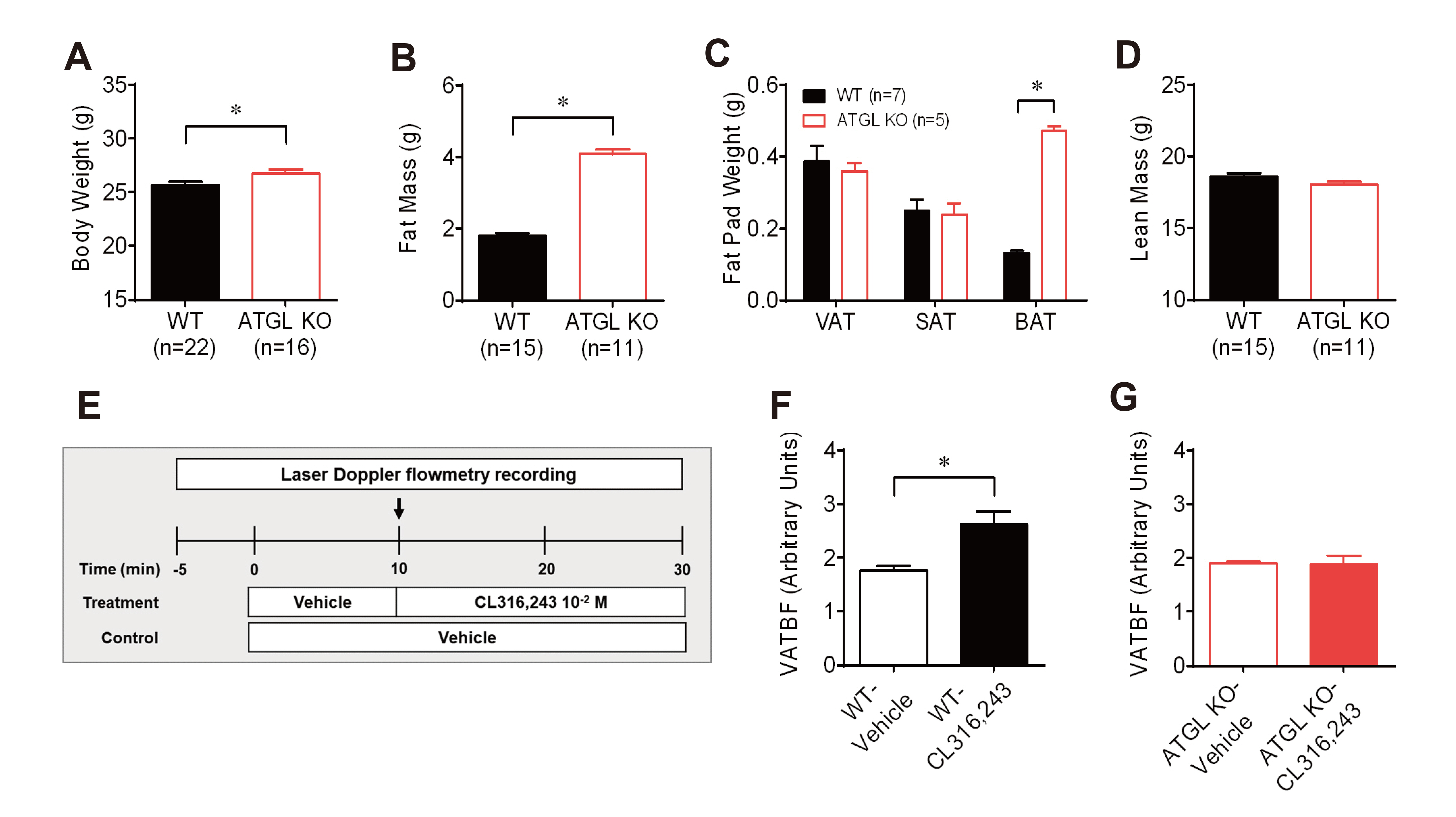
Fig. 3 Changes in visceral adipose tissue blood flow (VAT BF) to β3-adrenergic receptor stimulation in wild-type (WT) and adipose triglyceride lipase (ATGL) knockout (KO) mice. (A–D) Phenotype comparison of body weight (A), total fat mass (B), fat pad weights (C), and total lean mass (D) between WT and ATGL KO mice at 10 weeks of age. (E) Timeline diagram for the experiment on changes in VAT BF by stimulation of β3-adrenergic receptor agonist CL316,243. Arrows represent the time point of the switch in the agonist delivery. (F) Comparison of changes in VAT between the vehicle (n = 8) and CL316,243 (n = 8) infusion in WT mice at time point 30 min. (G) Comparison of changes in VAT between the vehicle (n = 4) and CL316,243 (n = 7) infusion in ATGL KO mice at time point 30 min. *p < 0.05.
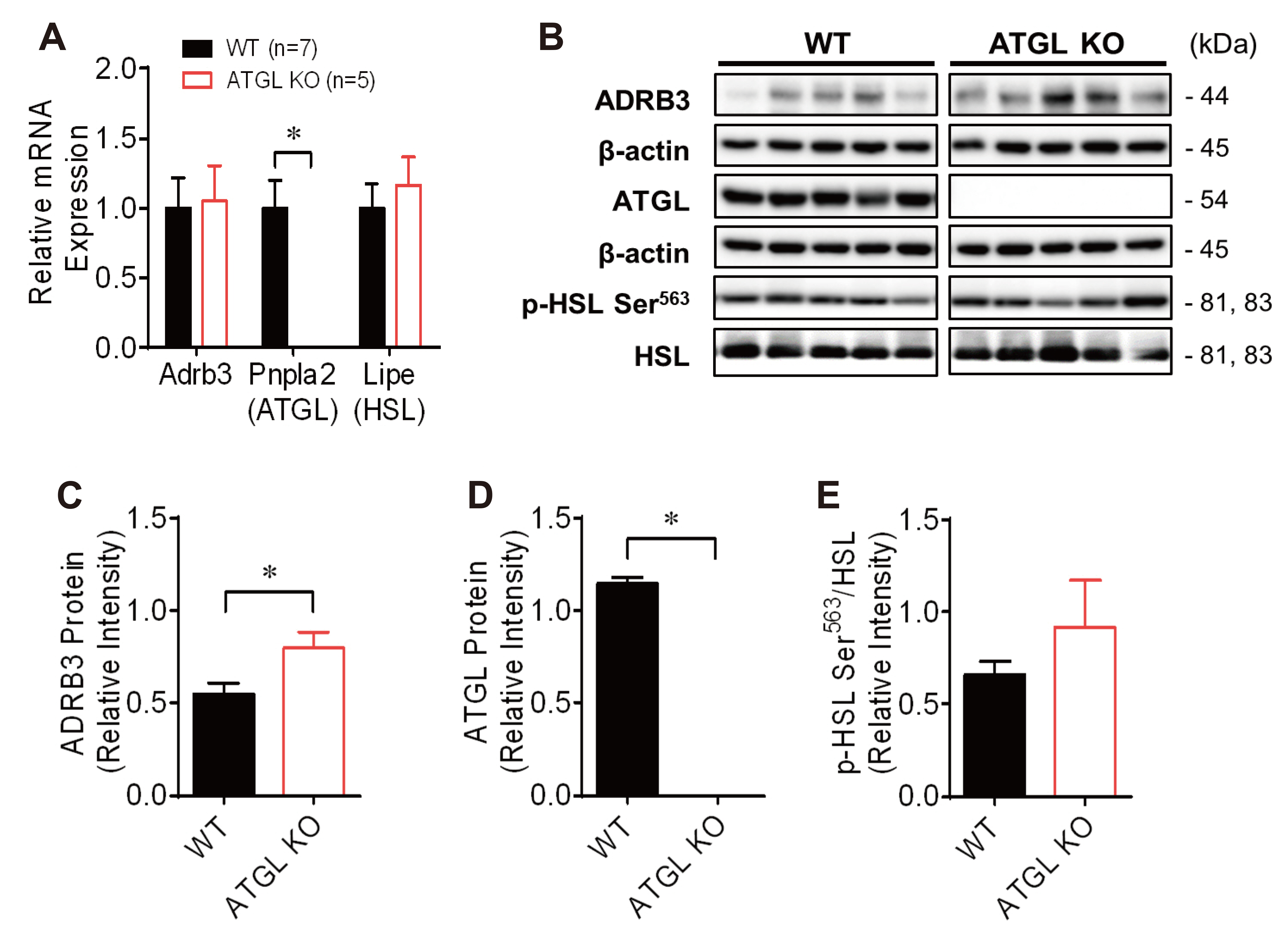
Fig. 4 Expression of β3-adrenergic receptor-mediated lipolysis associated mRNA and protein in visceral adipose tissue (VAT) of wild-type (WT) and adipose triglyceride lipase (ATGL) knockout (KO) mice. (A) Comparison of the gene expression of Adrb3, Pnpla2, and Lipe in VAT of WT and ATGL KO mice. The relative mRNA expression was calculated using the ΔΔCt method (2–ΔΔCt). (B) Western blot analysis of lipolysis associated proteins, including ADRB3, ATGL, HSL, and p-HSL Ser563. (C–E) The relative intensity of ADRB3 (C), ATGL (D), and phosphorylation of HSL on serine 563 (E). Adrb3, beta-3 adrenergic receptor; Pnpla2, Patatin Like Phospholipase Domain Containing 2 known as ATGL; Lipe, Lipase E, Hormone Sensitive Type known as hormone-sensitive lipase (HSL). *p < 0.05: WT (n = 7) vs. ATGL KO (n = 5) mice.

Fig. 5 Comparison of adipose tissue vessel morphology and angiogenesis-related mRNA expression in visceral adipose tissue (VAT) of wild-type (WT) and adipose triglyceride lipase (ATGL) knockout (KO) mice. (A) Representative images of vasculature and adipocytes of whole-mount VAT in WT and ATGL KO mice. Images are shown at a magnification of ×100. Scale bars, 100 µm. (B, C) Quantitative total vessel area (B) and vessel density normalized to average adipocyte size (C) between WT and ATGL KO mice. (D, E) Quantification of mean area of adipocyte (D) and distribution of adipocyte size (E) between WT and ATGL KO mice. (F) Comparison of the expression levels of genes related to angiogenesis in VAT of WT and ATGL KO mice. The relative mRNA expression was calculated using the ΔΔCt method (2–ΔΔCt). Hif1a, hypoxia-inducible factor 1, alpha subunit; Vegfa, vascular endothelial growth factor A; Kdr, kinase insert domain protein receptor; Fgf1, fibroblast growth factor 1; Fgf2, fibroblast growth factor 2; Pecam1, platelet endothelial cell adhesion molecule 1. *p < 0.05.

Fig. 6 Schematic diagram of the role of β3-adrenergic receptor-mediated lipolysis in the regulation of visceral adipose tissue blood flow. β3-adrenergic receptor (AR) signaling contributes to β-AR-mediated enhancement of visceral adipose tissue blood flow (VAT BF) in mice. Adipose triglyceride lipase (ATGL) deficiency negates the enhancement of VAT BF induced by β3-AR stimulation, suggesting the role of ATGL-mediated lipolysis in the regulation of VAT BF. This process may contribute to VAT remodeling in the development of obesity. TAG, triacylglycerol; HSL, hormone-sensitive lipase; MGL, monoglyceride lipase.
Reference
-
1. Cao Y. 2010; Adipose tissue angiogenesis as a therapeutic target for obesity and metabolic diseases. Nat Rev Drug Discov. 9:107–115. DOI: 10.1038/nrd3055. PMID: 20118961.
Article2. Goossens GH, Bizzarri A, Venteclef N, Essers Y, Cleutjens JP, Konings E, Jocken JW, Cajlakovic M, Ribitsch V, Clément K, Blaak EE. 2011; Increased adipose tissue oxygen tension in obese compared with lean men is accompanied by insulin resistance, impaired adipose tissue capillarization, and inflammation. Circulation. 124:67–76. DOI: 10.1161/CIRCULATIONAHA.111.027813. PMID: 21670228.
Article3. Ye J. 2009; Emerging role of adipose tissue hypoxia in obesity and insulin resistance. Int J Obes (Lond). 33:54–66. DOI: 10.1038/ijo.2008.229. PMID: 19050672. PMCID: PMC2650750.
Article4. Unamuno X, Gómez-Ambrosi J, Rodríguez A, Becerril S, Frühbeck G, Catalán V. 2018; Adipokine dysregulation and adipose tissue inflammation in human obesity. Eur J Clin Invest. 48:e12997. DOI: 10.1111/eci.12997. PMID: 29995306.
Article5. Ye J, Gao Z, Yin J, He Q. 2007; Hypoxia is a potential risk factor for chronic inflammation and adiponectin reduction in adipose tissue of ob/ob and dietary obese mice. Am J Physiol Endocrinol Metab. 293:E1118–E1128. DOI: 10.1152/ajpendo.00435.2007. PMID: 17666485.
Article6. Dimitriadis G, Lambadiari V, Mitrou P, Maratou E, Boutati E, Panagiotakos DB, Economopoulos T, Raptis SA. 2007; Impaired postprandial blood flow in adipose tissue may be an early marker of insulin resistance in type 2 diabetes. Diabetes Care. 30:3128–3130. DOI: 10.2337/dc07-0699. PMID: 17890315.
Article7. Karpe F, Fielding BA, Ilic V, Macdonald IA, Summers LK, Frayn KN. 2002; Impaired postprandial adipose tissue blood flow response is related to aspects of insulin sensitivity. Diabetes. 51:2467–2473. DOI: 10.2337/diabetes.51.8.2467. PMID: 12145159.
Article8. McQuaid SE, Hodson L, Neville MJ, Dennis AL, Cheeseman J, Humphreys SM, Ruge T, Gilbert M, Fielding BA, Frayn KN, Karpe F. 2011; Downregulation of adipose tissue fatty acid trafficking in obesity: a driver for ectopic fat deposition? Diabetes. 60:47–55. DOI: 10.2337/db10-0867. PMID: 20943748. PMCID: PMC3012196.9. Bartness TJ, Liu Y, Shrestha YB, Ryu V. 2014; Neural innervation of white adipose tissue and the control of lipolysis. Front Neuroendocrinol. 35:473–493. DOI: 10.1016/j.yfrne.2014.04.001. PMID: 24736043. PMCID: PMC4175185.
Article10. Bowers RR, Festuccia WT, Song CK, Shi H, Migliorini RH, Bartness TJ. 2004; Sympathetic innervation of white adipose tissue and its regulation of fat cell number. Am J Physiol Regul Integr Comp Physiol. 286:R1167–R1175. DOI: 10.1152/ajpregu.00558.2003. PMID: 15142857.
Article11. Cao Y, Wang H, Wang Q, Han X, Zeng W. 2018; Three-dimensional volume fluorescence-imaging of vascular plasticity in adipose tissues. Mol Metab. 14:71–81. DOI: 10.1016/j.molmet.2018.06.004. PMID: 29914852. PMCID: PMC6034070.
Article12. Ardilouze JL, Fielding BA, Currie JM, Frayn KN, Karpe F. 2004; Nitric oxide and beta-adrenergic stimulation are major regulators of preprandial and postprandial subcutaneous adipose tissue blood flow in humans. Circulation. 109:47–52. DOI: 10.1161/01.CIR.0000105681.70455.73. PMID: 14662716.13. Sotornik R, Brassard P, Martin E, Yale P, Carpentier AC, Ardilouze JL. 2012; Update on adipose tissue blood flow regulation. Am J Physiol Endocrinol Metab. 302:E1157–E1170. DOI: 10.1152/ajpendo.00351.2011. PMID: 22318953.
Article14. Frayn KN, Karpe F. 2014; Regulation of human subcutaneous adipose tissue blood flow. Int J Obes (Lond). 38:1019–1026. DOI: 10.1038/ijo.2013.200. PMID: 24166067.
Article15. Collins S. 2012; β-adrenoceptor signaling networks in adipocytes for recruiting stored fat and energy expenditure. Front Endocrinol (Lausanne). 2:102. DOI: 10.3389/fendo.2011.00102. PMID: 22654837. PMCID: PMC3355892.
Article16. Evans BA, Merlin J, Bengtsson T, Hutchinson DS. 2019; Adrenoceptors in white, brown, and brite adipocytes. Br J Pharmacol. 176:2416–2432. DOI: 10.1111/bph.14631. PMID: 30801689. PMCID: PMC6592855.
Article17. Tanaka Y, Horinouchi T, Koike K. 2005; New insights into beta-adrenoceptors in smooth muscle: distribution of receptor subtypes and molecular mechanisms triggering muscle relaxation. Clin Exp Pharmacol Physiol. 32:503–514. DOI: 10.1111/j.1440-1681.2005.04222.x. PMID: 16026507.18. Balligand JL. 2016; Cardiac salvage by tweaking with beta-3-adrenergic receptors. Cardiovasc Res. 111:128–133. DOI: 10.1093/cvr/cvw056. PMID: 27001422.
Article19. Ibrahim MM. 2010; Subcutaneous and visceral adipose tissue: structural and functional differences. Obes Rev. 11:11–18. DOI: 10.1111/j.1467-789X.2009.00623.x. PMID: 19656312.
Article20. Ledoux S, Queguiner I, Msika S, Calderari S, Rufat P, Gasc JM, Corvol P, Larger E. 2008; Angiogenesis associated with visceral and subcutaneous adipose tissue in severe human obesity. Diabetes. 57:3247–3257. DOI: 10.2337/db07-1812. PMID: 18835936. PMCID: PMC2584130.
Article21. Farb MG, Gokce N. 2015; Visceral adiposopathy: a vascular perspective. Horm Mol Biol Clin Investig. 21:125–136. DOI: 10.1515/hmbci-2014-0047. PMID: 25781557. PMCID: PMC4442778.
Article22. Shah RV, Murthy VL, Abbasi SA, Blankstein R, Kwong RY, Goldfine AB, Jerosch-Herold M, Lima JA, Ding J, Allison MA. 2014; Visceral adiposity and the risk of metabolic syndrome across body mass index: the MESA Study. JACC Cardiovasc Imaging. 7:1221–1235. DOI: 10.1016/j.jcmg.2014.07.017. PMID: 25440591. PMCID: PMC4268163.23. Després JP, Lemieux I. 2006; Abdominal obesity and metabolic syndrome. Nature. 444:881–887. DOI: 10.1038/nature05488. PMID: 17167477. PMCID: PMC5935926.
Article24. Kwon H, Kim D, Kim JS. 2017; Body fat distribution and the risk of incident metabolic syndrome: a longitudinal cohort study. Sci Rep. 7:10955. DOI: 10.1038/s41598-017-09723-y. PMID: 28887474. PMCID: PMC5591218.
Article25. Xue Y, Lim S, Bråkenhielm E, Cao Y. 2010; Adipose angiogenesis: quantitative methods to study microvessel growth, regression and remodeling in vivo. Nat Protoc. 5:912–920. DOI: 10.1038/nprot.2010.46. PMID: 20431536.
Article26. Bolsoni-Lopes A, Alonso-Vale MI. 2015; Lipolysis and lipases in white adipose tissue- an update. Arch Endocrinol Metab. 59:335–342. DOI: 10.1590/2359-3997000000067. PMID: 26331321.27. Zechner R, Zimmermann R, Eichmann TO, Kohlwein SD, Haemmerle G, Lass A, Madeo F. 2012; FAT SIGNALS--lipases and lipolysis in lipid metabolism and signaling. Cell Metab. 15:279–291. DOI: 10.1016/j.cmet.2011.12.018. PMID: 22405066. PMCID: PMC3314979.28. Kabon B, Nagele A, Reddy D, Eagon C, Fleshman JW, Sessler DI, Kurz A. 2004; Obesity decreases perioperative tissue oxygenation. Anesthesiology. 100:274–280. DOI: 10.1097/00000542-200402000-00015. PMID: 14739800. PMCID: PMC1395476.
Article29. Pasarica M, Sereda OR, Redman LM, Albarado DC, Hymel DT, Roan LE, Rood JC, Burk DH, Smith SR. 2009; Reduced adipose tissue oxygenation in human obesity: evidence for rarefaction, macrophage chemotaxis, and inflammation without an angiogenic response. Diabetes. 58:718–725. DOI: 10.2337/db08-1098. PMID: 19074987. PMCID: PMC2646071.
Article30. He Q, Gao Z, Yin J, Zhang J, Yun Z, Ye J. 2011; Regulation of HIF-1{alpha} activity in adipose tissue by obesity-associated factors: adipogenesis, insulin, and hypoxia. Am J Physiol Endocrinol Metab. 300:E877–E885. DOI: 10.1152/ajpendo.00626.2010. PMID: 21343542. PMCID: PMC3093977.31. Perrone MG, Notarnicola M, Caruso MG, Tutino V, Scilimati A. 2008; Upregulation of beta3-adrenergic receptor mRNA in human colon cancer: a preliminary study. Oncology. 75:224–229. DOI: 10.1159/000163851. PMID: 18852493.32. Langin D, Dicker A, Tavernier G, Hoffstedt J, Mairal A, Rydén M, Arner E, Sicard A, Jenkins CM, Viguerie N, van Harmelen V, Gross RW, Holm C, Arner P. 2005; Adipocyte lipases and defect of lipolysis in human obesity. Diabetes. 54:3190–3197. DOI: 10.2337/diabetes.54.11.3190. PMID: 16249444.
Article33. Flores-Opazo M, Trieu J, Naim T, Valladares-Ide D, Zbinden-Foncea H, Stapleton D. 2020; Defective fasting-induced PKA activation impairs adipose tissue glycogen degradation in obese Zucker rats. Int J Obes (Lond). 44:500–509. DOI: 10.1038/s41366-019-0327-y. PMID: 30705392.
Article34. Haemmerle G, Moustafa T, Woelkart G, Büttner S, Schmidt A, van de Weijer T, Hesselink M, Jaeger D, Kienesberger PC, Zierler K, Schreiber R, Eichmann T, Kolb D, Kotzbeck P, Schweiger M, Kumari M, Eder S, Schoiswohl G, Wongsiriroj N, Pollak NM, et al. 2011; ATGL-mediated fat catabolism regulates cardiac mitochondrial function via PPAR-α and PGC-1. Nat Med. 17:1076–1085. DOI: 10.1038/nm.2439. PMID: 21857651. PMCID: PMC3244833.
Article35. Haemmerle G, Lass A, Zimmermann R, Gorkiewicz G, Meyer C, Rozman J, Heldmaier G, Maier R, Theussl C, Eder S, Kratky D, Wagner EF, Klingenspor M, Hoefler G, Zechner R. 2006; Defective lipolysis and altered energy metabolism in mice lacking adipose triglyceride lipase. Science. 312:734–737. DOI: 10.1126/science.1123965. PMID: 16675698.
Article36. Onogi Y, Wada T, Kamiya C, Inata K, Matsuzawa T, Inaba Y, Kimura K, Inoue H, Yamamoto S, Ishii Y, Koya D, Tsuneki H, Sasahara M, Sasaoka T. 2017; PDGFRβ regulates adipose tissue expansion and glucose metabolism via vascular remodeling in diet-induced obesity. Diabetes. 66:1008–1021. DOI: 10.2337/db16-0881. PMID: 28122789.
Article37. Song MG, Lee HJ, Jin BY, Gutierrez-Aguilar R, Shin KH, Choi SH, Um SH, Kim DH. 2016; Depot-specific differences in angiogenic capacity of adipose tissue in differential susceptibility to diet-induced obesity. Mol Metab. 5:1113–1120. DOI: 10.1016/j.molmet.2016.09.001. PMID: 27818937. PMCID: PMC5081408.
Article
- Full Text Links
-
- Actions
-
Cited
- CITED
-
- Close
- Share
-
- Similar articles
-
- Sestrin2 Regulates Beneficial β3-Adrenergic Receptor-Mediated Effects Observed in Inguinal White Adipose Tissue and Soleus Muscle
- The Role of Androgen in the Adipose Tissue of Males
- Immune cells in brown adipose tissue are involved in allogeneic immune responses
- Brown Fat and Browning for the Treatment of Obesity and Related Metabolic Disorders
- Effect of sweet pumpkin powder on lipid metabolism in leptin-deficient mice
