Large myxomatous odontogenic tumor in the jaw: a case series
- Affiliations
-
- 1Department of Oral and Maxillofacial Surgery, Dental Research Institute, School of Dentistry, Seoul National University, Seoul, Korea
- KMID: 2515309
- DOI: http://doi.org/10.5125/jkaoms.2021.47.2.112
Abstract
Objectives
Myxomatous odontogenic tumors (MOTs) are the third most common odontogenic tumors in the oral and maxillofacial region. Due to its slow-growing, but locally invasive nature, the tumor is usually detected by accident or only when it becomes a large mass, which causes facial deformity.
Materials and Methods
Current study reports three unusual cases of MOT including huge myxoma involve the mandible in middle-aged man, MOT with ossifying fibroma pattern in mandible, and MOT in maxilla of young female patient. The diagnosis and treatment strategy of MOTs was also summarized and updated.
Results
In reported three cases of patients with large MOTs, surgical treatment was indicated with fibular free flap reconstruction in the mandible and plate reconstruction in the maxilla. The tumors were successfully treated with radical resection and did not show signs of recurrence during the followup period.
Conclusion
Surgical treatment indication depends on size, the position of the lesion, patient systemic condition and surgeon individual experience. In the case of a large tumor, radical resection and reconstruction is the standard surgical strategy. The conservative surgical treatment including enucleation with wide curettage is still under controversy. The recurrence rate for MOTs is significantly high, up to 30%, therefore long-term follow-up is essential.
Figure
-

Fig. 1 Radiograms and clinical view of Case No. 1 with the giant myxomatous odontogenic tumor (arrows) in mandible. A. Preoperative panoramic view. B, C. Preoperative computed tomography image including axial and coronal views, respectively. D. Preoperative facial photo. E. Postoperative panoramic view, patient underwent mass resection and reconstruction surgery with fibular free flap. F. Extraoral view in 5-year follow-up visit, there was no sign of recurrence and patient achieve good mouth-opening. G. Intraoral view, 5-year follow-up.
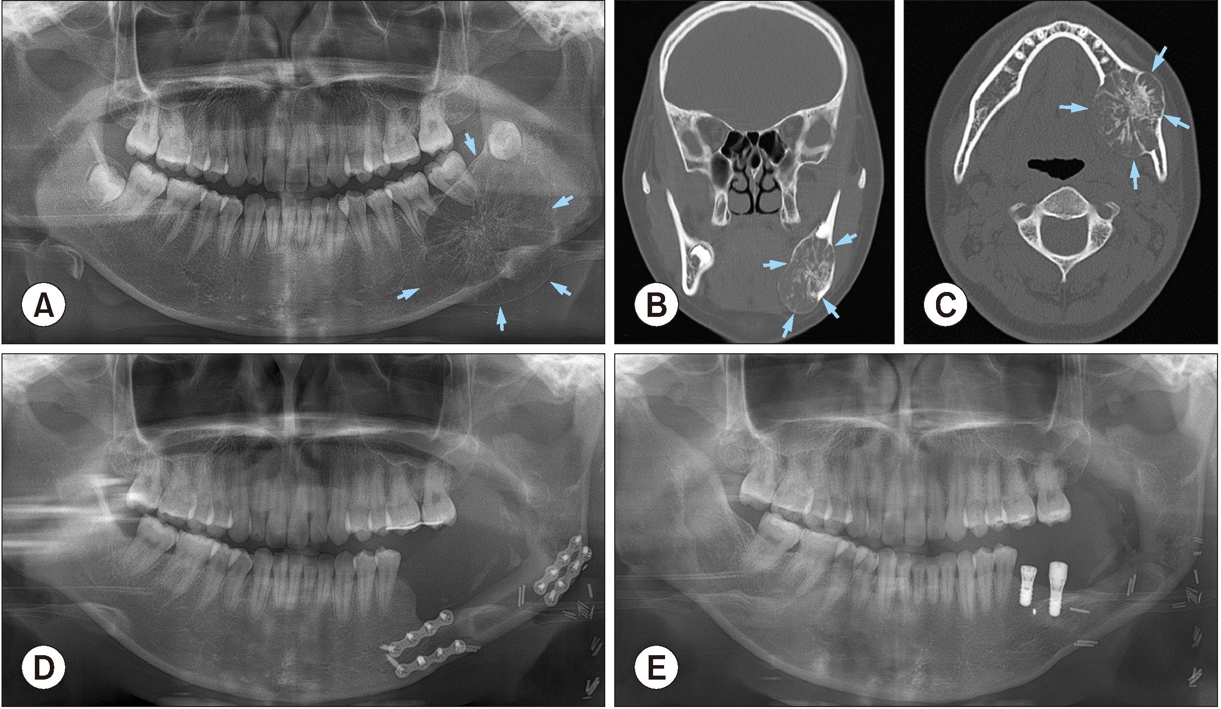
Fig. 2 Radiograms and clinical view of Case No. 2 with the large myxomatous odontogenic tumor (arrows) in mandible. A. Preoperative panoramic view. B, C. Preoperative computed tomography image including coronal and axial views. D. Postoperative panoramic view, the patient underwent resection and reconstructive surgery. E. Five years after odontogenic myxoma treatment surgery, bone augmentation and implant installation were performed.
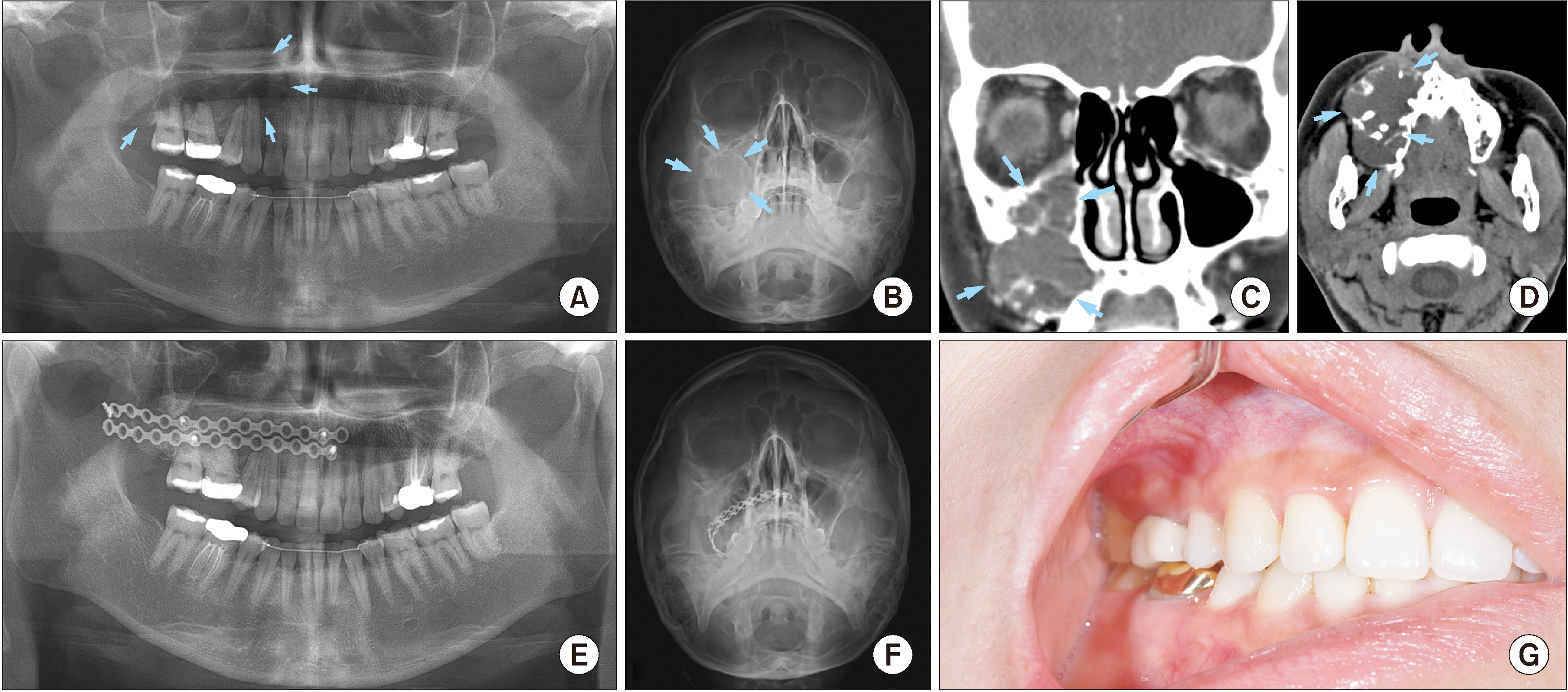
Fig. 3 Radiograms and clinical view of Case No. 3 with the large myxomatous odontogenic tumor (arrows) in mandible. A. Preoperative panoramic view. B. Preoperative Waters’ view. C, D. Preoperative computed tomography image including coronal and axial views. E. Postoperative panoramic view, the patient underwent resection and reconstructive surgery with two plates. F. Postoperative Waters’ view. G. Intraoral view at 1-year follow-up visit.
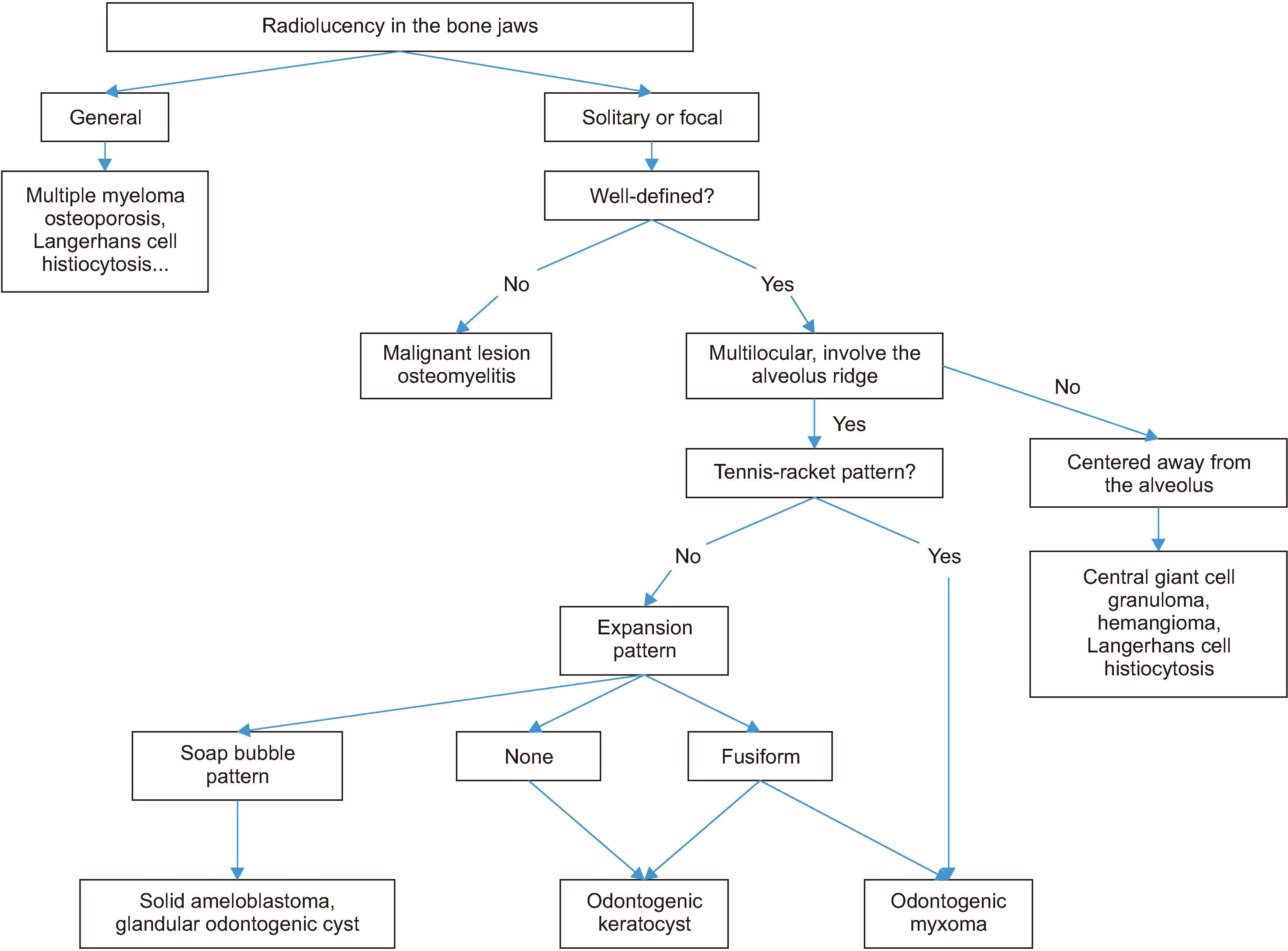
Fig. 4 A flowchart shows the diagnosis strategy of myxomatous odontogenic tumors.
Cited by 1 articles
-
Current options in jaw and facial reconstructions
Soung Min Kim, Jong Ho Lee
J Korean Assoc Oral Maxillofac Surg. 2024;50(6):309-325. doi: 10.5125/jkaoms.2024.50.6.309.
Reference
-
References
1. Kaffe I, Naor H, Buchner A. 1997; Clinical and radiological features of odontogenic myxoma of the jaws. Dentomaxillofac Radiol. 26:299–303. https://doi.org/10.1038/sj.dmfr.4600261 . DOI: 10.1038/sj.dmfr.4600261. PMID: 9482003.
Article2. Buchner A, Merrell PW, Carpenter WM. 2006; Relative frequency of central odontogenic tumors: a study of 1,088 cases from Northern California and comparison to studies from other parts of the world. J Oral Maxillofac Surg. 64:1343–52. https://doi.org/10.1016/j.joms.2006.05.019 . DOI: 10.1016/j.joms.2006.05.019. PMID: 16916667.
Article3. Kansy K, Juergens P, Krol Z, Paulussen M, Baumhoer D, Bruder E, et al. 2012; Odontogenic myxoma: diagnostic and therapeutic challenges in paediatric and adult patients--a case series and review of the literature. J Craniomaxillofac Surg. 40:271–6. https://doi.org/10.1016/j.jcms.2011.04.009 . DOI: 10.1016/j.jcms.2011.04.009. PMID: 21624835.
Article4. Ochsenius G, Ortega A, Godoy L, Peñafiel C, Escobar E. 2002; Odontogenic tumors in Chile: a study of 362 cases. J Oral Pathol Med. 31:415–20. https://doi.org/10.1034/j.1600-0714.2002.00073.x . DOI: 10.1034/j.1600-0714.2002.00073.x. PMID: 12165060.
Article5. Thoma KH, Goldman HM. 1947; Central myxoma of the jaw. Oral Surg Oral Med Oral Pathol. 33:B532–40. https://doi.org/10.1016/0096-6347(47)90315-3 . DOI: 10.1016/0096-6347(47)90315-3. PMID: 20251479.
Article6. Wright JM, Soluk Tekkesin M. 2017; Odontogenic tumors: where are we in 2017? J Istanb Univ Fac Dent. 51(3 Suppl 1):S10–30. https://doi.org/10.17096/jiufd.52886 . DOI: 10.17096/jiufd.52886. PMID: 29354306. PMCID: PMC5750825.
Article7. Mosqueda-Taylor A, Ledesma-Montes C, Caballero-Sandoval S, Portilla-Robertson J, Ruíz-Godoy Rivera LM, Meneses-García A. 1997; Odontogenic tumors in Mexico: a collaborative retrospective study of 349 cases. Oral Surg Oral Med Oral Pathol Oral Radiol Endod. 84:672–5. https://doi.org/10.1016/s1079-2104(97)90371-1 . DOI: 10.1016/s1079-2104(97)90371-1. PMID: 9431538.
Article8. Mori M, Murakami M, Hirose I, Shimozato T. 1975; Histochemical studies of myxoma of the jaws. J Oral Surg. 33:529–36. PMID: 167142.9. Noffke CE, Raubenheimer EJ, Chabikuli NJ, Bouckaert MM. 2007; Odontogenic myxoma: review of the literature and report of 30 cases from South Africa. Oral Surg Oral Med Oral Pathol Oral Radiol Endod. 104:101–9. https://doi.org/10.1016/j.tripleo.2007.01.026 . DOI: 10.1016/j.tripleo.2007.01.026. PMID: 17507265.
Article10. Barnes L, Eveson JW, Reichart P, Sidransky D. 2005. World Health Organization classification of tumours: pathology and genetics of head and neck tumours. IARC Press;Lyon:11. Martínez-Mata G, Mosqueda-Taylor A, Carlos-Bregni R, de Almeida OP, Contreras-Vidaurre E, Vargas PA, et al. 2008; Odontogenic myxoma: clinico-pathological, immunohistochemical and ultrastructural findings of a multicentric series. Oral Oncol. 44:601–7. https://doi.org/10.1016/j.oraloncology.2007.08.009 . DOI: 10.1016/j.oraloncology.2007.08.009. PMID: 17996487.
Article12. Li TJ, Sun LS, Luo HY. 2006; Odontogenic myxoma: a clinicopathologic study of 25 cases. Arch Pathol Lab Med. 130:1799–806. DOI: 10.1043/1543-2165(2006)130[1799:OMACSO]2.0.CO;2. PMID: 17149953.
Article13. Simon EN, Merkx MA, Vuhahula E, Ngassapa D, Stoelinga PJ. 2004; Odontogenic myxoma: a clinicopathological study of 33 cases. Int J Oral Maxillofac Surg. 33:333–7. https://doi.org/10.1016/j.ijom.2003.12.004 . DOI: 10.1016/j.ijom.2003.12.004. PMID: 15145033.
Article14. Keszler A, Dominguez FV, Giannunzio G. 1995; Myxoma in childhood: an analysis of 10 cases. J Oral Maxillofac Surg. 53:518–21. https://doi.org/10.1016/0278-2391(95)90062-4 . DOI: 10.1016/0278-2391(95)90062-4. PMID: 7722719.
Article15. Adekeye EO, Avery BS, Edwards MB, Williams HK. 1984; Advanced central myxoma of the jaws in Nigeria. Clinical features, treatment and pathogenesis. Int J Oral Surg. 13:177–86. https://doi.org/10.1016/s0300-9785(84)80001-0 . DOI: 10.1016/s0300-9785(84)80001-0. PMID: 6430823.
Article16. MacDonald-Jankowski DS, Yeung R, Lee KM, Li TK. 2002; Odontogenic myxomas in the Hong Kong Chinese: clinico-radiological presentation and systematic review. Dentomaxillofac Radiol. 31:71–83. https://doi.org/10.1038/sj.dmfr.4600678 . DOI: 10.1038/sj.dmfr.4600678. PMID: 12076060.
Article17. Vijayabanu B, Sreeja C, Bharath N, Aesha I, Kannan VS, Devi M. 2015; Odontogenic myxoma of maxilla: a rare presentation in an elderly female. J Pharm Bioallied Sci. 7(Suppl 2):S759–62. https://doi.org/10.4103/0975-7406.163550 . DOI: 10.4103/0975-7406.163550. PMID: 26538962. PMCID: PMC4606704.
Article18. Barros RE, Dominguez FV, Cabrini RL. 1969; Myxoma of the jaws. Oral Surg Oral Med Oral Pathol. 27:225–36. https://doi.org/10.1016/0030-4220(69)90177-7 . DOI: 10.1016/s1079-2104(96)80309-x. PMID: 8899782.
Article19. Shivashankara C, Nidoni M, Patil S, Shashikala KT. 2017; Odontogenic myxoma: a review with report of an uncommon case with recurrence in the mandible of a teenage male. Saudi Dent J. 29:93–101. https://doi.org/10.1016/j.sdentj.2017.02.003 . DOI: 10.1016/j.sdentj.2017.02.003. PMID: 28725126. PMCID: PMC5503096.
Article20. Kauke M, Safi AF, Kreppel M, Grandoch A, Nickenig HJ, Zöller JE, et al. 2018; Size distribution and clinicoradiological signs of aggressiveness in odontogenic myxoma-three-dimensional analysis and systematic review. Dentomaxillofac Radiol. 47:20170262. https://doi.org/10.1259/dmfr.20170262 . DOI: 10.1259/dmfr.20170262. PMID: 29082773. PMCID: PMC5965907.
Article21. Mittal Y, Chugh A, Varghese KG, Dwivedi S, Goyal V. 2016; Management of recurrent odontogenic myxoma of mandible: a clinical case report. J Clin Diagn Res. 10:ZD30–1. https://doi.org/10.7860/JCDR/2016/20917.8702 . DOI: 10.7860/JCDR/2016/20917.8702. PMID: 27891488. PMCID: PMC5121826.
Article22. Dunfee BL, Sakai O, Pistey R, Gohel A. 2006; Radiologic and pathologic characteristics of benign and malignant lesions of the mandible. Radiographics. 26:1751–68. https://doi.org/10.1148/rg.266055189 . DOI: 10.1148/rg.266055189. PMID: 17102048.
Article23. Chrcanovic BR, Gomez RS. 2019; Odontogenic myxoma: an updated analysis of 1,692 cases reported in the literature. Oral Dis. 25:676–83. https://doi.org/10.1111/odi.12875 . DOI: 10.1111/odi.12875. PMID: 29683236.
Article
