Increased Wall Enhancement Extent Representing Higher Rupture Risk of Unruptured Intracranial Aneurysms
- Affiliations
-
- 1Department of Radiology, Huashan Hospital Affiliated to Fudan University, Shanghai, China
- 2Department of Neurosurgery, Huashan Hospital Affiliated to Fudan University, Shanghai, China
- KMID: 2513849
- DOI: http://doi.org/10.3340/jkns.2020.0144
Abstract
Objective
: This study aims to investigate the relationship between aneurysm wall enhancement and clinical rupture risks based on the magnetic resonance vessel wall imaging (MR-VWI) quantitative methods.
Methods
: One hundred and eight patients with 127 unruptured aneurysms were prospectively enrolled from Feburary 2016 to October 2017. Aneurysms were divided into high risk (≥10) and intermediate-low risk group (<10) according to the PHASES (Population, Hypertension, Age, Size of aneurysm, Earlier SAH history from another aneurysm, Site of aneurysm) scores. Clinical risk factors, aneurysm morphology, and wall enhancement index (WEI) calculated using 3D MR-VWI were analyzed and compared.
Results
: In comparison of high-risk and intermediated-low risk groups, univariate analysis showed that neck width (4.5±3.3 mm vs. 3.4±1.7 mm, p=0.002), the presence of wall enhancement (100.0% vs. 62.9%, p<0.001), and WEI (1.6±0.6 vs. 0.8±0.8, p<0.001) were significantly associated with high rupture risk. Multivariate regression analysis revealed that WEI was the most important factor in predicting high rupture risk (odds ratio, 2.6; 95% confidence interval, 1.4–4.9; p=0.002). The receiver operating characteristic (ROC) curve analysis can efficiently differentiate higher risk aneurysms (area under the curve, 0.780; p<0.001) which have a reliable WEI cutoff value (1.04; sensitivity, 0.833; specificity, 0.67) predictive of high rupture risk.
Conclusion
: Aneurysms with higher rupture risk based on PHASES score demonstrate increased neck width, wall enhancement, and the enhancement intensity. Higher WEI in unruptured aneurysms has a predictive value for increased rupture risk.
Figure
-

Fig. 1. Three different MR imaging sequences : with ×400% zoomed images. A : TOF MRA. B : Sag CUBE T1 pre-gadolinium. C : Sag CUBE T1 post-gadolinium. MRA : magnetic resonance angiography, TOF : time-of-flight.
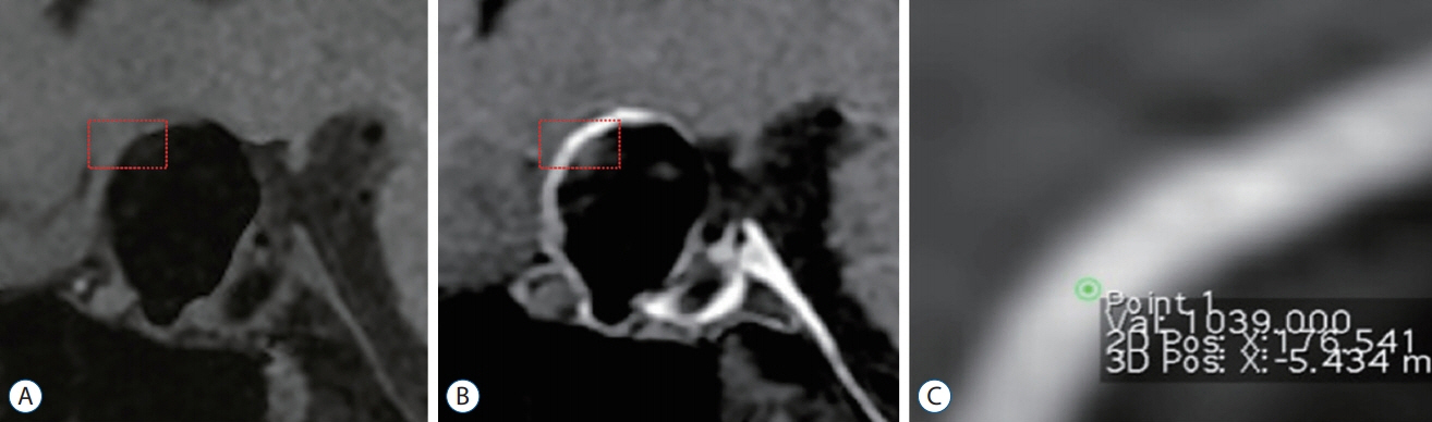
Fig. 2. Representing case. Measurement of signal value (red box). A : Pre-contrast imaging. B : Post-contrast imaging. C : Selected a region of maximum signal using ring tool.
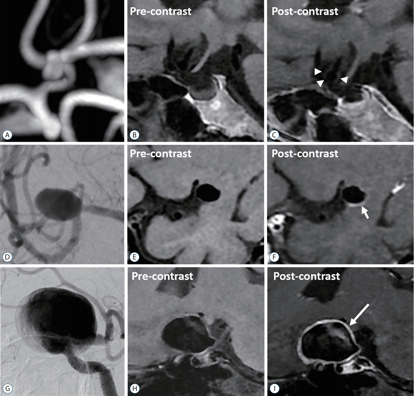
Fig. 3. Representative pre- and post-contrast imaging of aneurysm with different wall enhancement types. A-C : No enhancement (arrowheads). D-F : Partial enhancement (short arrow). G-I : circumferential enhancement (long arrow).
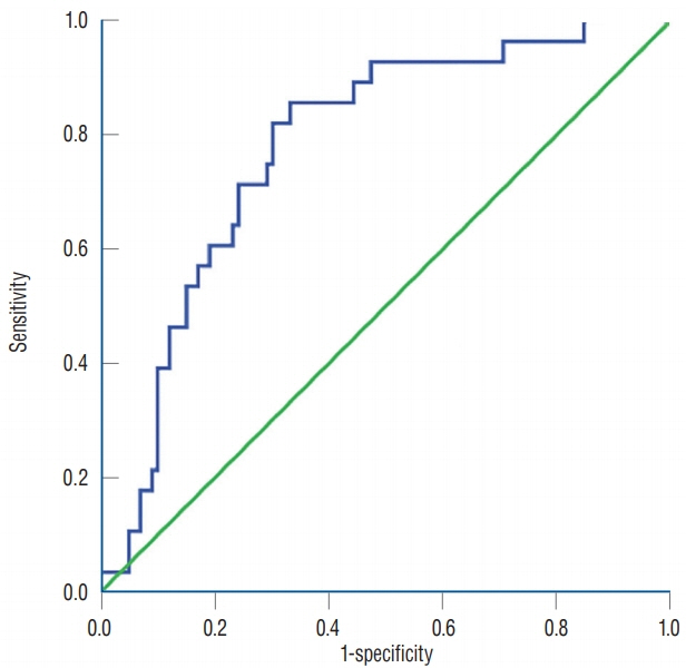
Fig. 4. High rupture risk (PHASES score ≥10) was efficiently differentiated on ROC curve (AUC, 0.780; p<0.001). Higher WEI than cutoff value (1.04; sensitivity, 0.833; specificity, 0.67) predict high rupture risk. PHASES : Population, Hypertension, Age, Size of aneurysm, Earlier SAH history from another aneurysm, Site of aneurysm, ROC : receiver operating characteristics curve, AUC : area under the curve, WEI : wall enhancement index.
Reference
-
References
1. Aoki T, Kataoka H, Ishibashi R, Nozaki K, Egashira K, Hashimoto N. Impact of monocyte chemoattractant protein-1 deficiency on cerebral aneurysm formation. Stroke. 40:942–951. 2009.
Article2. Backes D, Hendrikse J, van der Schaaf I, Algra A, Lindgren AE, Verweij BH, et al. Determinants of gadolinium-enhancement of the aneurysm wall in unruptured intracranial aneurysms. Neurosurgery. 83:719–725. 2018.
Article3. Backes D, Vergouwen MD, Tiel Groenestege AT, Bor AS, Velthuis BK, Greving JP, et al. PHASES score for prediction of intracranial aneurysm growth. Stroke. 46:1221–1226. 2015.
Article4. Chalouhi N, Ali MS, Jabbour PM, Tjoumakaris SI, Gonzalez LF, Rosenwasser RH, et al. Biology of intracranial aneurysms: role of inflammation. J Cereb Blood Flow Metab. 32:1659–1676. 2012.
Article5. Chan DY, Abrigo JM, Cheung TC, Siu DY, Poon WS, Ahuja AT, et al. Screening for intracranial aneurysms? Prevalence of unruptured intracranial aneurysms in Hong Kong Chinese. J Neurosurg. 124:1245–1249. 2016.
Article6. Edjlali M, Gentric JC, Régent-Rodriguez C, Trystram D, Hassen WB, Lion S, et al. Does aneurysmal wall enhancement on vessel wall MRI help to distinguish stable from unstable intracranial aneurysms? Stroke. 45:3704–3706. 2014.
Article7. Edjlali M, Guédon A, Ben Hassen W, Boulouis G, Benzakoun J, Rodriguez-Régent C, et al. Circumferential thick enhancement at vessel wall MRI has high specificity for intracranial aneurysm instability. Radiology. 289:181–187. 2018.
Article8. Endo H, Niizuma K, Fujimura M, Sato K, Inoue T, Osawa S, et al. Ruptured cerebral microaneurysm diagnosed by 3-dimensional fast spinecho T1 imaging with variable flip angles. J Stroke Cerebrovasc Dis. 24:e231–e235. 2015.
Article9. Greving JP, Wermer MJ, Brown RD Jr, Morita A, Juvela S, Yonekura M, et al. Development of the PHASES score for prediction of risk of rupture of intracranial aneurysms: a pooled analysis of six prospective cohort studies. Lancet Neurol. 13:59–66. 2014.
Article10. Jamous MA, Nagahiro S, Kitazato KT, Tamura T, Aziz HA, Shono M, et al. Endothelial injury and inflammatory response induced by hemodynamic changes preceding intracranial aneurysm formation: experimental study in rats. J Neurosurg. 107:405–411. 2007.
Article11. Khan MO, Toro Arana V, Rubbert C, Cornelius JF, Fischer I, Bostelmann R, et al. Association between aneurysm hemodynamics and wall enhancement on 3D vessel wall MRI. J Neurosurg. 2020; [Epub ahead of print].
Article12. Krings T, Mandell DM, Kiehl TR, Geibprasert S, Tymianski M, Alvarez H, et al. Intracranial aneurysms: from vessel wall pathology to therapeutic approach. Nat Rev Neurol. 7:547–559. 2011.
Article13. Larsen N, von der Brelie C, Trick D, Riedel CH, Lindner T, Madjidyar J, et al. Vessel wall enhancement in unruptured intracranial aneurysms: an indicator for higher risk of rupture? High-resolution MR imaging and correlated histologic findings. AJNR Am J Neuroradiol. 39:1617–1621. 2018.
Article14. Linn FH, Rinkel GJ, Algra A, van Gijn J. Incidence of subarachnoid hemorrhage: role of region, year, and rate of computed tomography: a meta-analysis. Stroke. 27:625–629. 1996.15. Liu P, Qi H, Liu A, Lv X, Jiang Y, Zhao X, et al. Relationship between aneurysm wall enhancement and conventional risk factors in patients with unruptured intracranial aneurysms: a black-blood MRI study. Interv Neuroradiol. 22:501–505. 2016.
Article16. Lv N, Karmonik C, Chen S, Wang X, Fang Y, Huang Q, et al. Relationship between aneurysm wall enhancement in vessel wall magnetic resonance imaging and rupture risk of unruptured intracranial aneurysms. Neurosurgery. 84:E385–E391. 2019.
Article17. Marchese E, Vignati A, Albanese A, Nucci CG, Sabatino G, Tirpakova B, et al. Comparative evaluation of genome-wide gene expression profiles in ruptured and unruptured human intracranial aneurysms. J Biol Regul Homeost Agents. 24:185–195. 2010.18. Matouk CC, Mandell DM, Günel M, Bulsara KR, Malhotra A, Hebert R, et al. Vessel wall magnetic resonance imaging identifies the site of rupture in patients with multiple intracranial aneurysms: proof of principle. Neurosurgery. 72:492–496. discussion 496. 2013.
Article19. Miyata H, Imai H, Koseki H, Shimizu K, Abekura Y, Oka M, et al. Vasa vasorum formation is associated with rupture of intracranial aneurysms. J Neurosurg. 1–11. 2019.
Article20. Mocco J, Brown RD Jr, Torner JC, Capuano AW, Fargen KM, Raghavan ML, et al. Aneurysm morphology and prediction of rupture: an international study of unruptured intracranial aneurysms analysis. Neurosurgery. 82:491–496. 2018.
Article21. Nagahata S, Nagahata M, Obara M, Kondo R, Minagawa N, Sato S, et al. Wall enhancement of the intracranial aneurysms revealed by magnetic resonance vessel wall imaging using three-dimensional turbo spin-echo sequence with motion-sensitized driven-equilibrium: a sign of ruptured aneurysm? Clin Neuroradiol. 26:277–283. 2016.
Article22. Omodaka S, Endo H, Niizuma K, Fujimura M, Inoue T, Sato K, et al. Quantitative assessment of circumferential enhancement along the wall of cerebral aneurysms using MR imaging. AJNR Am J Neuroradiol. 37:1262–1266. 2016.
Article23. Peña-Silva RA, Chalouhi N, Wegman-Points L, Ali M, Mitchell I, Pierce GL, et al. Novel role for endogenous hepatocyte growth factor in the pathogenesis of intracranial aneurysms. Hypertension. 65:587–593. 2015.
Article24. Samaniego EA, Roa JA, Hasan D. Vessel wall imaging in intracranial aneurysms. J Neurointerv Surg. 11:1105–1112. 2019.
Article25. Shimonaga K, Matsushige T, Ishii D, Sakamoto S, Hosogai M, Kawasumi T, et al. Clinicopathological insights from vessel wall imaging of unruptured intracranial aneurysms. Stroke. 49:2516–2519. 2018.
Article26. Takemura Y, Hirata Y, Sakata N, Nabeshima K, Takeshita M, Inoue T. Histopathologic characteristics of a saccular aneurysm arising in the nonbranching segment of the distal middle cerebral artery. Pathol Res Pract. 206:391–396. 2010.
Article27. Vergouwen MDI, Backes D, van der Schaaf IC, Hendrikse J, Kleinloog R, Algra A, et al. Gadolinium enhancement of the aneurysm wall in unruptured intracranial aneurysms is associated with an increased risk of aneurysm instability: a follow-up study. AJNR Am J Neuroradiol. 40:1112–1116. 2019.
Article28. Vlak MH, Algra A, Brandenburg R, Rinkel GJ. Prevalence of unruptured intracranial aneurysms, with emphasis on sex, age, comorbidity, country, and time period: a systematic review and meta-analysis. Lancet Neurol. 10:626–636. 2011.
Article29. Wang GX, Wen L, Lei S, Ran Q, Yin JB, Gong ZL, et al. Wall enhancement ratio and partial wall enhancement on MRI associated with the rupture of intracranial aneurysms. J Neurointerv Surg. 10:566–570. 2018.
Article
- Full Text Links
-
- Actions
-
Cited
- CITED
-
- Close
- Share
-
- Similar articles
-
- Management of Unruptured Intracranial Aneurysms
- Guideline for Management of Unruptured Intracranial Aneurysms: Preliminary Report
- Management of Unruptured Intracranial Aneurysms-Natural Course and Surgical Outcomes
- The Clinical Significance and Characteristic Shape of Ruptured 'Very Small'Cerebral Aneurysms
- Angiographic Characteristics of the Intracranial Saccular Aneurysms to Predict the Rupture
