Suppression of Fibrotic Reactions of Chitosan-Alginate Microcapsules Containing Porcine Islets by Dexamethasone Surface Coating
- Affiliations
-
- 1Department of Endocrinology and Metabolism, College of Medicine, The Catholic University of Korea, Seoul, Korea
- 2Department of Polymer Nano Science and Technology, Department of BIN Fusion Technology and BK-21 Polymer BIN Fusion Research Team, Chonbuk National University, Jeonju, Korea
- 3Department of Microbiology and Immunology, Translational Xenotransplantation Research Centre, Cancer Research Institute, Biomedical Research Institute, Seoul National University College of Medicine, Seoul, Korea
- 4Department of Endocrinology and Metabolism, Seoul St. Mary’s Hospital, College of Medicine, The Catholic University of Korea, Seoul, Korea
- KMID: 2513298
- DOI: http://doi.org/10.3803/EnM.2021.879
Abstract
- Background
The microencapsulation is an ideal solution to overcome immune rejection without immunosuppressive treatment. Poor biocompatibility and small molecular antigens secreted from encapsulated islets induce fibrosis infiltration. Therefore, the aims of this study were to improve the biocompatibility of microcapsules by dexamethasone coating and to verify its effect after xenogeneic transplantation in a streptozotocin-induced diabetes mice.
Methods
Dexamethasone 21-phosphate (Dexa) was dissolved in 1% chitosan and was cross-linked with the alginate microcapsule surface. Insulin secretion and viability assays were performed 14 days after microencapsulation. Dexa-containing chitosan-coated alginate (Dexa-chitosan) or alginate microencapsulated porcine islets were transplanted into diabetic mice. The fibrosis infiltration score was calculated from the harvested microcapsules. The harvested microcapsules were stained with trichrome and for insulin and macrophages.
Results
No significant differences in glucose-stimulated insulin secretion and islet viability were noted among naked, alginate, and Dexa-chitosan microencapsulated islets. After transplantation of microencapsulated porcine islets, nonfasting blood glucose were normalized in both the Dexa-chitosan and alginate groups until 231 days. The average glucose after transplantation were lower in the Dexa-chitosan group than the alginate group. Pericapsular fibrosis and inflammatory cell infiltration of microcapsules were significantly reduced in Dexa-chitosan compared with alginate microcapsules. Dithizone and insulin were positive in Dexa-chitosan capsules. Although fibrosis and macrophage infiltration was noted on the surface, some alginate microcapsules were stained with insulin.
Conclusion
Dexa coating on microcapsules significantly suppressed the fibrotic reaction on the capsule surface after transplantation of xenogenic islets containing microcapsules without any harmful effects on the function and survival of the islets.
Keyword
Figure
-
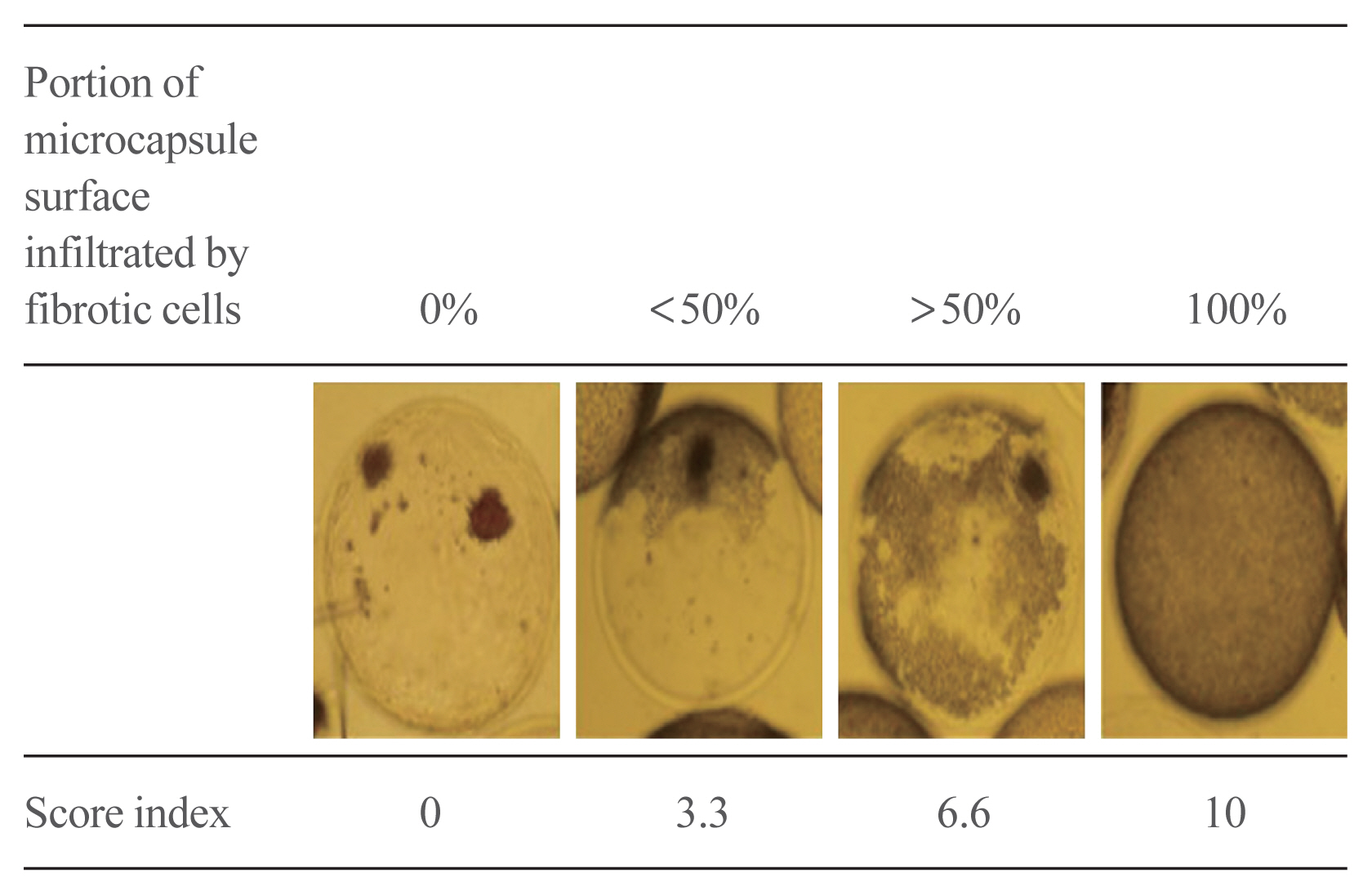
Fig. 1 Classification of microcapsules harvested after graft failure in the peritoneal cavity of galactosyltransferase knock-out mice mice based on the extent of fibrotic cell infiltration to the microcapsule surface.
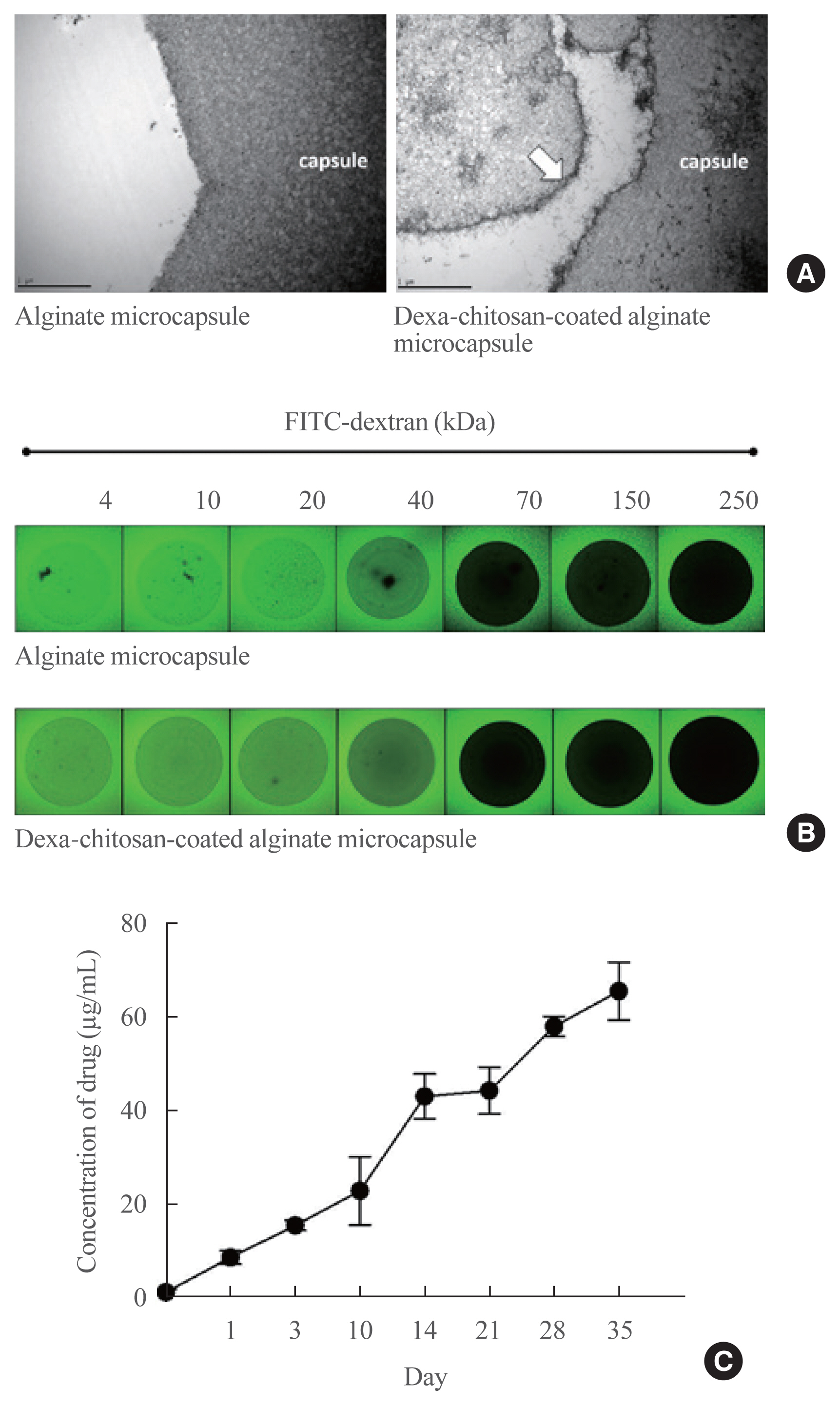
Fig. 2 Characterization of dexamethasone (Dexa)-chitosan-coated alginate microcapsules. (A) Observation of the microcapsule surface by transmission electron microscopy. The left panel is the alginate microcapsule (×10,000 magnification), and the right panel is the Dexa-chitosan-coated alginate microcapsule (white arrow; dexa-chitosan layer). (B) Observation of the diffusion and permeability assessment of microcapsules by fluorescein isothiocyanate (FITC)-labeled dextran. The molecular cutoff of each microcapsule type was between 40 and 70 kDa. (C) Dexa concentration for 36 days after incubation of 1,000 ea Dexa-chitosan-coated alginate microcapsules.
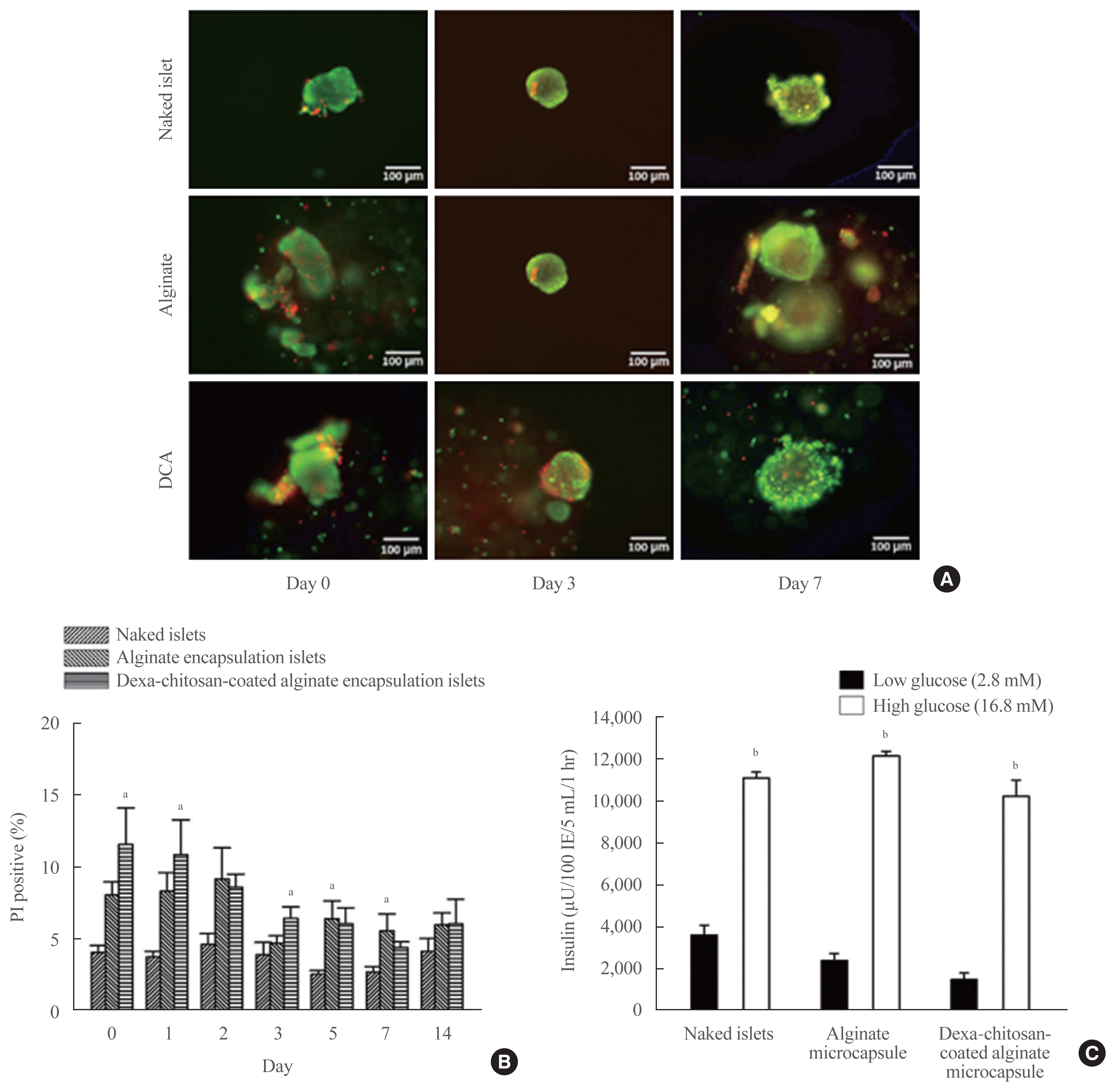
Fig. 3 Viability and secretory function of the microencapsulation islets. (A) Viability of microencapsulated islets based on acridine orange/propidium iodide (AO/PI) staining of naked islets, alginate microcapsules, and dexamethasone (Dexa)-chitosan-coated alginate microcapsules (DCAs) at days 0, 3, and 7. Scale bar=100 μm. (B) PI-positive cells (%) in naked islets, alginate microencapsulated islets and Dexa-chitosan-coated alginate microencapsulated islets over 14 days. (C) Insulin secretory function of microencapsulated islets during glucose-stimulated insulin secretion. Insulin secretion was significantly increased in response to glucose in all groups. aP<0.05 compared with the naked islets in each date group; bP<0.01.
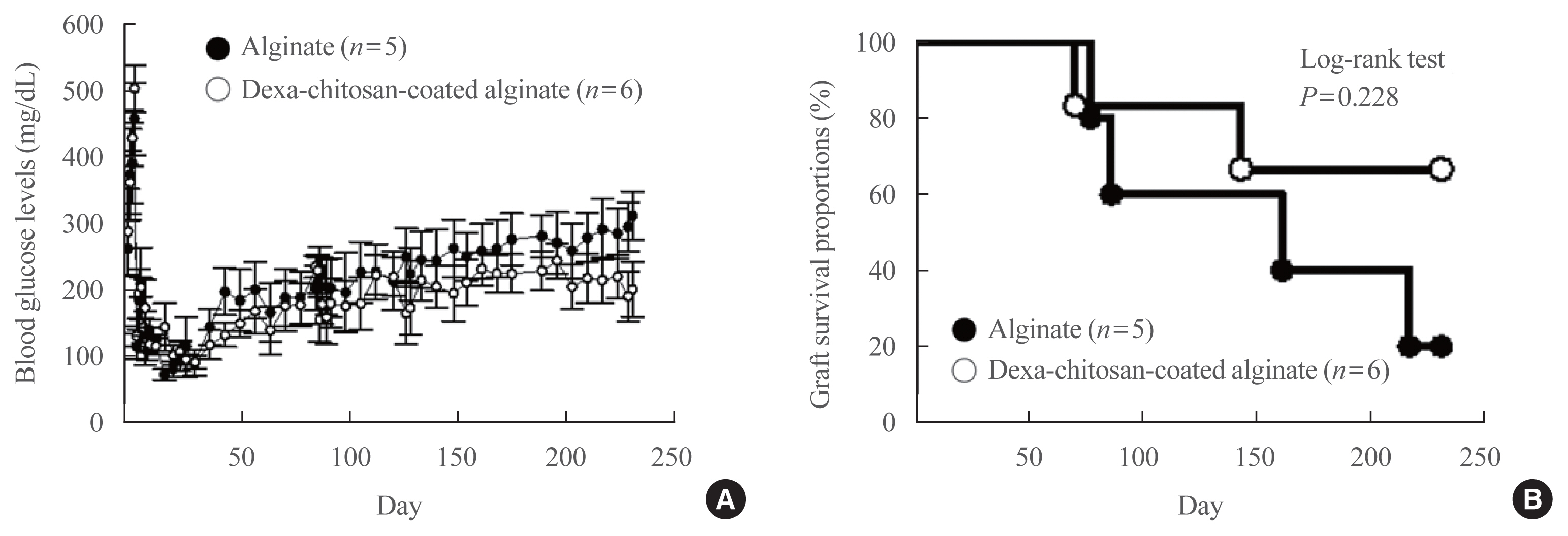
Fig. 4 Blood glucose level and graft survival in galactosyltransferase knock-out mice (GalT KO) mice transplanted with microencapsulated islets. (A) Change in blood glucose levels in GalT KO mice transplanted with alginate microencapsulated islets (n=5) and dexamethasone (Dexa)-chitosan coated alginate microencapsulated islets (n=6). (B) Graft survival proportions in GalT KO mice transplanted with microencapsulated islets. Graft survival proportions of Dexa-chitosan-coated alginate microencapsulated islets (open circle) improved approximately 60% compared with 20% survival for alginate microencapsulated islets (close circle) (log-rank test, P=0.228).
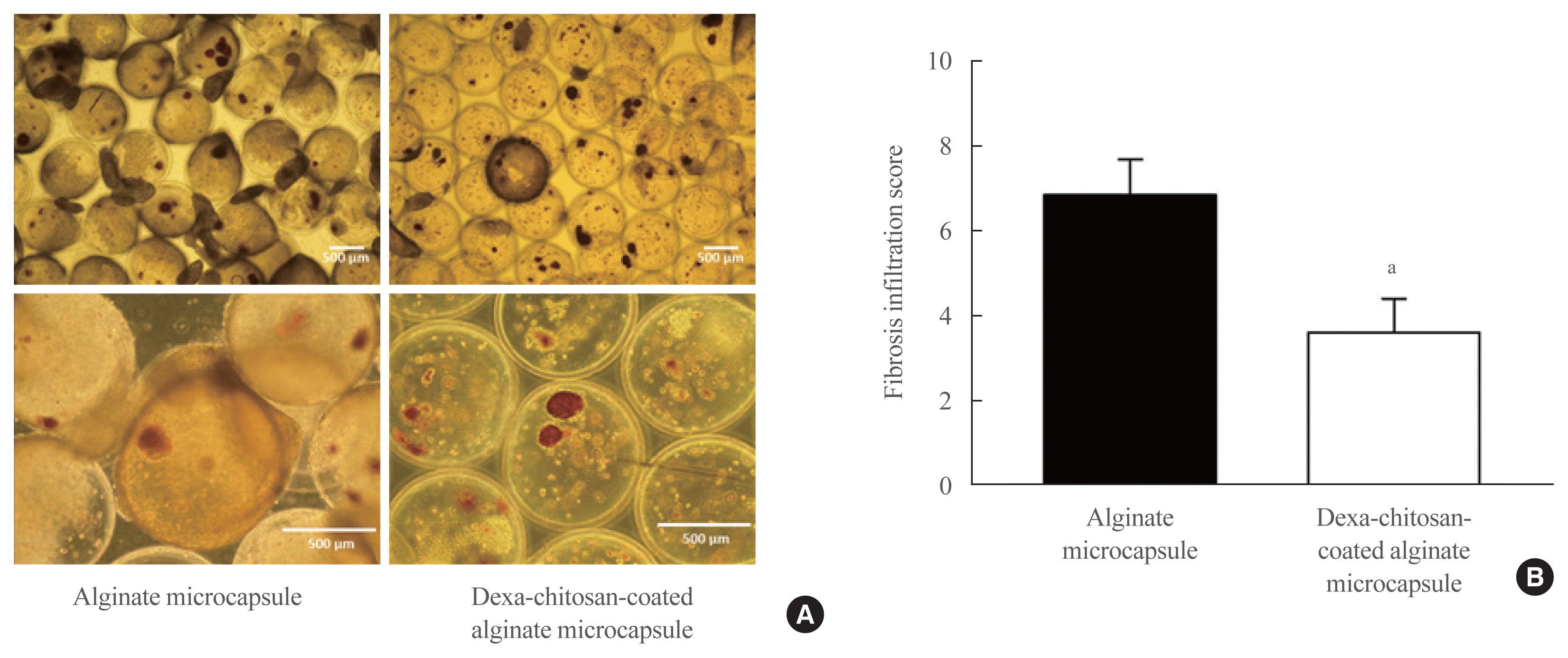
Fig. 5 Dithizone staining of harvested microencapsulated porcine islets and the measurement of fibrotic cell infiltration to the microcapsule surface. (A) The left panel shows harvested porcine islets with alginate microcapsules, and the right panel shows harvested dexamethasone (Dexa)-chitosan-coated alginate microcapsules. (B) Fibrosis infiltration score of microcapsules compared with alginate- and Dexa-chitosan-coated alginate microcapsules at 231 days after 10,000 IEq islet transplantation. aP<0.05 compared with alginate microcapsules.
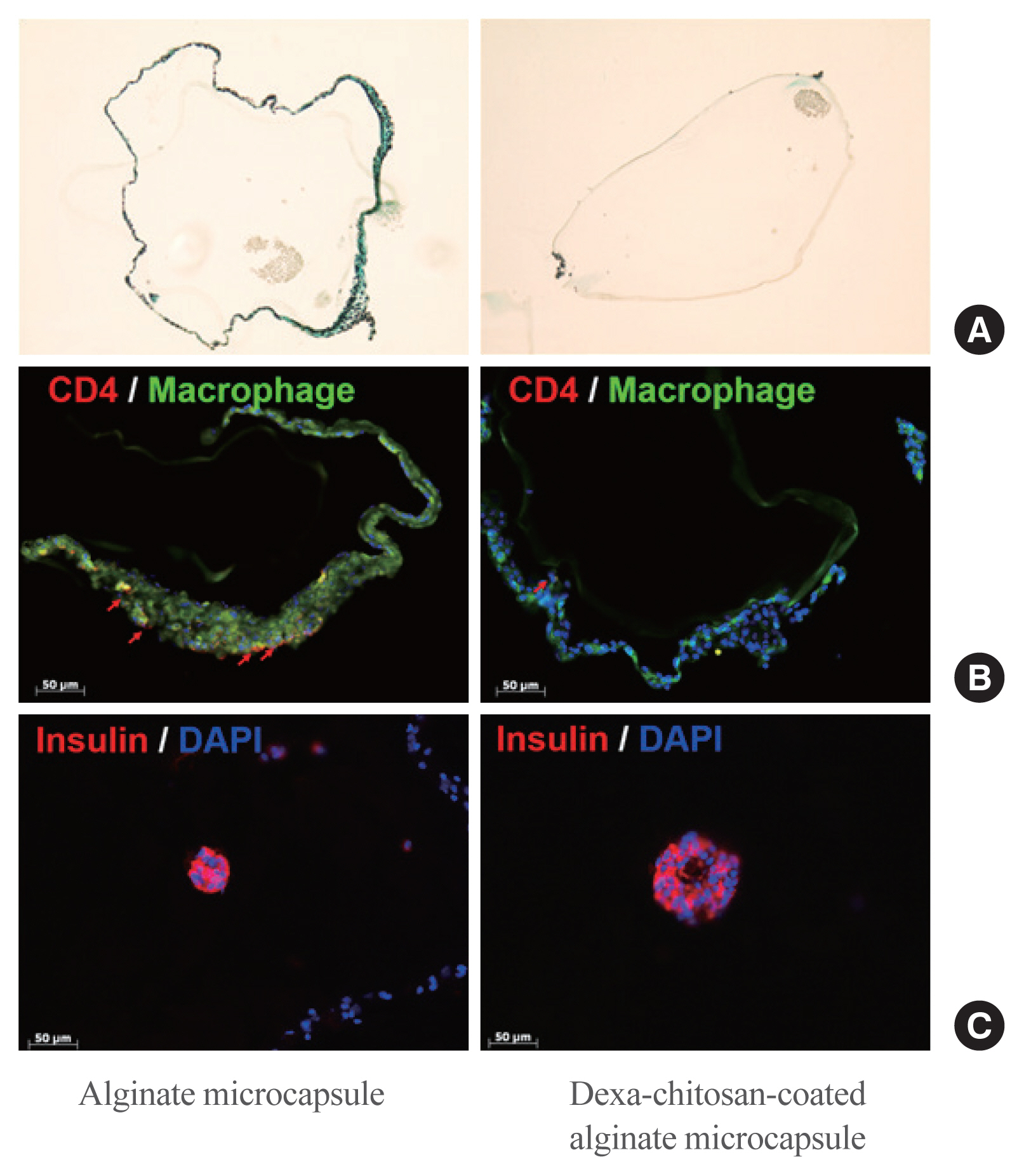
Fig. 6 Trichrome or immunohistochemistry staining of harvested encapsulated porcine islets and measurement of fibrotic cell infiltration to the microcapsule surface. (A) Fibrosis infiltrated stain (green) harvested from alginate microcapsules and dexamethasone (Dexa)-chitosan-coated alginate microcapsules. (B) Macrophages (green) and CD3 (red) were stained in alginate microcapsules. Red arrows indicate CD3-positive cells. Scale bar=50 μm. (C) Porcine islets were stained with insulin antibody (red). Scale bar=50 μm. DAPI, 4′-6-diamidino-2-phenylindole.
Reference
-
1. Atkinson MA, Eisenbarth GS. Type 1 diabetes: new perspectives on disease pathogenesis and treatment. Lancet. 2001; 358:221–9.
Article2. Lipsett M, Aikin R, Castellarin M, Hanley S, Jamal AM, Laganiere S, et al. Islet neogenesis: a potential therapeutic tool in type 1 diabetes. Int J Biochem Cell Biol. 2006; 38:498–503.
Article3. Murdoch TB, McGhee-Wilson D, Shapiro AM, Lakey JR. Methods of human islet culture for transplantation. Cell Transplant. 2004; 13:605–17.
Article4. Sakata N, Sumi S, Yoshimatsu G, Goto M, Egawa S, Unno M. Encapsulated islets transplantation: past, present and future. World J Gastrointest Pathophysiol. 2012; 3:19–26.
Article5. Boker A, Rothenberg L, Hernandez C, Kenyon NS, Ricordi C, Alejandro R. Human islet transplantation: update. World J Surg. 2001; 25:481–6.
Article6. Shapiro AM, Lakey JR, Ryan EA, Korbutt GS, Toth E, Warnock GL, et al. Islet transplantation in seven patients with type 1 diabetes mellitus using a glucocorticoid-free immunosuppressive regimen. N Engl J Med. 2000; 343:230–8.
Article7. Collaborative Islet Transplant Registry. CITR Tenth Annual Reports [Internet]. Rockville: citr;2017. [cited 2020 Nov 25]. Available from: http://citregistry.org/content/reports-publications-presentations .8. Larsen JL, Bennett RG, Burkman T, Ramirez AL, Yamamoto S, Gulizia J, et al. Tacrolimus and sirolimus cause insulin resistance in normal Sprague Dawley rats. Transplantation. 2006; 82:466–70.
Article9. Dai C, Walker JT, Shostak A, Padgett A, Spears E, Wisniewski S, et al. Tacrolimus- and sirolimus-induced human β cell dysfunction is reversible and preventable. JCI Insight. 2020; 5:e130770.
Article10. Dufrane D, Gianello P. Macro- or microencapsulation of pig islets to cure type 1 diabetes. World J Gastroenterol. 2012; 18:6885–93.
Article11. Juste S, Lessard M, Henley N, Menard M, Halle JP. Effect of poly-L-lysine coating on macrophage activation by alginate-based microcapsules: assessment using a new in vitro method. J Biomed Mater Res A. 2005; 72:389–98.12. Lim DJ, Cho JH, Choi YH, Park HS, Hong OK, Kwon HS, et al. Optimization of microencapsulation of the Islets. Tissue Eng Regen Med. 2005; 2:280–6.13. Jiang K, Weaver JD, Li Y, Chen X, Liang J, Stabler CL. Local release of dexamethasone from macroporous scaffolds accelerates islet transplant engraftment by promotion of anti-inflammatory M2 macrophages. Biomaterials. 2017; 114:71–81.
Article14. Wu P, Grainger DW. Drug/device combinations for local drug therapies and infection prophylaxis. Biomaterials. 2006; 27:2450–67.
Article15. Labhasetwar V, Levy RJ. Implants for site-specific drug delivery. J Appl Biomater. 1991; 2:211–2.
Article16. Jones JA, Chang DT, Meyerson H, Colton E, Kwon IK, Matsuda T, et al. Proteomic analysis and quantification of cytokines and chemokines from biomaterial surface-adherent macrophages and foreign body giant cells. J Biomed Mater Res A. 2007; 83:585–96.
Article17. Ramalingam A, Hirai A, Thompson EA. Glucocorticoid inhibition of fibroblast proliferation and regulation of the cyclin kinase inhibitor p21Cip1. Mol Endocrinol. 1997; 11:577–86.
Article18. Shi K, Jiang J, Ma T, Xie J, Duan L, Chen R, et al. Dexamethasone attenuates bleomycin-induced lung fibrosis in mice through TGF-β, Smad3 and JAK-STAT pathway. Int J Clin Exp Med. 2014; 7:2645–50.19. Zawalich WS, Tesz GJ, Yamazaki H, Zawalich KC, Philbrick W. Dexamethasone suppresses phospholipase C activation and insulin secretion from isolated rat islets. Metabolism. 2006; 55:35–42.
Article20. Kaiser G, Gerst F, Michael D, Berchtold S, Friedrich B, Strutz-Seebohm N, et al. Regulation of forkhead box O1 (FOXO1) by protein kinase B and glucocorticoids: different mechanisms of induction of beta cell death in vitro. Diabetologia. 2013; 56:1587–95.
Article21. Kim JW, Sun C, Jeon SY, You YH, Shin JY, Lee SH, et al. Glucocorticoid treatment independently affects expansion and transdifferentiation of porcine neonatal pancreas cell clusters. BMB Rep. 2012; 45:51–6.
Article22. Jin SM, Shin JS, Kim KS, Gong CH, Park SK, Kim JS, et al. Islet isolation from adult designated pathogen-free pigs: use of the newer bovine nervous tissue-free enzymes and a revised donor selection strategy would improve the islet graft function. Xenotransplantation. 2011; 18:369–79.
Article23. O’Neil JJ, Stegemann JP, Nicholson DT, Gagnon KA, Solomon BA, Mullon CJ. The isolation and function of porcine islets from market weight pigs. Cell Transplant. 2001; 10:235–46.
Article24. Pignatello R, Stancampiano AH, Ventura CA, Puglisi G. Dexamethasone sodium phosphate-loaded Chitosan based delivery systems for buccal application. J Drug Target. 2007; 15:603–10.
Article25. Hall KK, Gattas-Asfura KM, Stabler CL. Microencapsulation of islets within alginate/poly(ethylene glycol) gels cross-linked via Staudinger ligation. Acta Biomater. 2011; 7:614–24.
Article26. Sylvestre JP, Guy RH, Delgado-Charro MB. In vitro optimization of dexamethasone phosphate delivery by iontophoresis. Phys Ther. 2008; 88:1177–85.
Article27. Tam SK, Bilodeau S, Dusseault J, Langlois G, Halle JP, Yahia LH. Biocompatibility and physicochemical characteristics of alginate-polycation microcapsules. Acta Biomater. 2011; 7:1683–92.
Article28. Vergani A, D’Addio F, Jurewicz M, Petrelli A, Watanabe T, Liu K, et al. A novel clinically relevant strategy to abrogate autoimmunity and regulate alloimmunity in NOD mice. Diabetes. 2010; 59:2253–64.
Article29. Merani S, Shapiro AM. Current status of pancreatic islet transplantation. Clin Sci (Lond). 2006; 110:611–25.
Article30. Park HS, Ham DS, You YH, Shin JY, Kim JW, Jo JH, et al. Successful xenogenic islet transplantation with Ba2+-alginate encapsulation. Tissue Eng Regen Med. 2010; 7:523–30.31. Singarayar S, Kistler PM, De Winter C, Mond H. A comparative study of the action of dexamethasone sodium phosphate and dexamethasone acetate in steroid-eluting pacemaker leads. Pacing Clin Electrophysiol. 2005; 28:311–5.
Article32. Schneider BL, Schwenter F, Pralong WF, Aebischer P. Prevention of the initial host immuno-inflammatory response determines the long-term survival of encapsulated myoblasts genetically engineered for erythropoietin delivery. Mol Ther. 2003; 7:506–14.
Article33. Zhang WJ, Marx SK, Laue C, Hyder A, Juergensen A, Bickel M, et al. HOE 077 reduces fibrotic overgrowth around the barium alginate microcapsules. Transplant Proc. 2000; 32:206–9.
Article34. Dang TT, Thai AV, Cohen J, Slosberg JE, Siniakowicz K, Doloff JC, et al. Enhanced function of immuno-isolated islets in diabetes therapy by co-encapsulation with an anti-inflammatory drug. Biomaterials. 2013; 34:5792–801.
Article35. Park HS, Kim JW, Lee SH, Yang HK, Ham DS, Sun CL, et al. Antifibrotic effect of rapamycin containing polyethylene glycol-coated alginate microcapsule in islet xenotransplantation. J Tissue Eng Regen Med. 2017; 11:1274–84.
Article36. Azadi SA, Vasheghani-Farahani E, Hashemi-Najafbabadi S, Godini A. Co-encapsulation of pancreatic islets and pentoxifylline in alginate-based microcapsules with enhanced immunosuppressive effects. Prog Biomater. 2016; 5:101–9.
Article
- Full Text Links
-
- Actions
-
Cited
- CITED
-
- Close
- Share
-
- Similar articles
-
- Preadipocyte Culture in Chitosan-Alginate Gel
- Alginate Microencapsulation of Islet Cells Using Electrostatic Droplet Generator
- Experimental Micro Encapsulation of Pancreatic Islets with Air-driven Droplet Generator and Alginate
- Effect of oral ingestion of chitosan and alginate on the removal of orally ingested radiostrontium(Sr) in mice
- Effects of Chitosan on Human Periodontal Ligament Cells in Vitro
