Evaluation of mandibular buccal shelf characteristics in the Colombian population: A cone-beam computed tomography study
- Escobar-Correa N
 1
1 - RamírezBustamante MA1
- SánchezUribe LA1
- Upegui-Zea JC1
- Vergara-Villarreal P2
- RamírezOssa DM1
- Affiliations
-
- 1Department of Orthodontics, Faculty of Dentistry, University of Antioquia, Medellín, Colombia
- 2Department of Orthodontics, Faculty of Dentistry, University of Cartagena, Cartagena, Colombia
- KMID: 2510615
- DOI: http://doi.org/10.4041/kjod.2021.51.1.23
Abstract
Objective
To evaluate the mandibular buccal shelf (MBS) in terms of the angulation and bone depth and thickness according to sex, age, and sagittal and vertical skeletal patterns in a Colombian population using cone-beam computed tomography (CBCT). Accordingly, the optimal site for miniscrew insertion in this area was determined.
Methods
This descriptive, retrospective study included 64 hemi-arches of 34 patients. On CBCT images, the angulation, buccal bone depth (4 and 6 mm from the cementoenamel junction [CEJ] of MBS), and buccal bone thickness (6 and 11 mm from the CEJ of MBS) were measured at the mesial and distal roots of the mandibular first and second molars.
Results
There were no statistically significant differences in the angulation, depth, and thickness of MBS between male and female patients. The values for the bone around the distal root of the mandibular second molar were significantly greater than the other values. The osseous characteristics were significantly better in participants aged 16–24 years. Class III patients exhibited the best osseous characteristics, with the bone depth at 6 mm being significantly different from that in Class I and Class II patients. Although values tended to be greater in patients with low angles, the difference was not statistically significant.
Conclusions
MBS provides an optimal bone surface for miniscrew insertion, with better osseous characteristics at the distal root of the mandibular second molar, 4 mm from CEJ. Adolescent patients, Class III patients, and patients with a low angle exhibit the most favorable osseous characteristics in the MBS area.
Figure
-
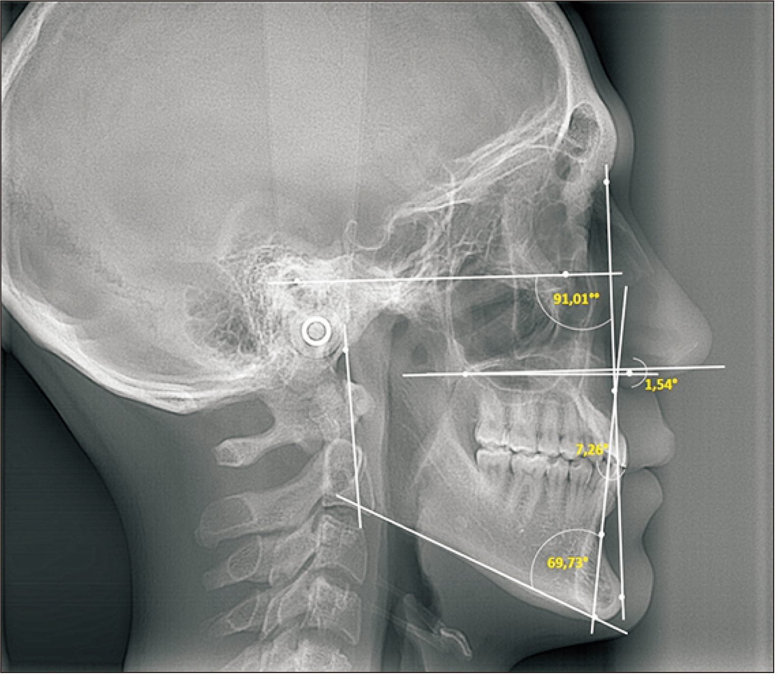
Figure 1 Cephalometric analysis according to the method of Kim.14,15

Figure 2 Reference orientation planes. A, Axial plane (purple line). B, Sagittal plane (blue line). C, Frontal/Coronal plane (orange line).
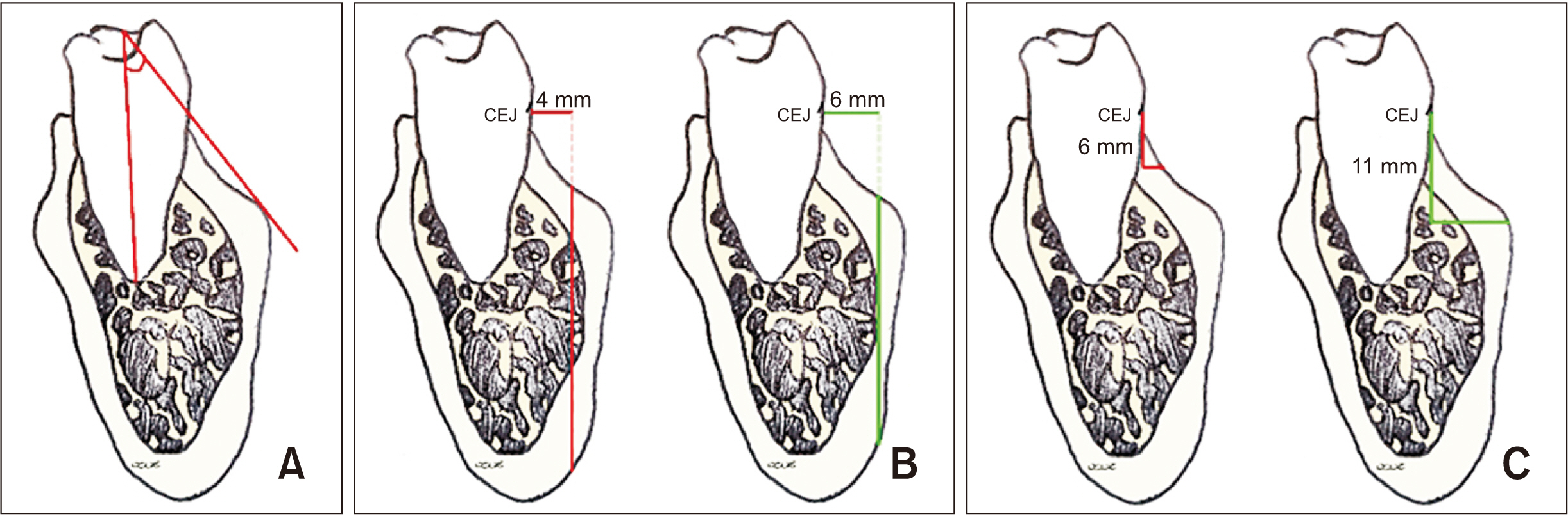
Figure 3 Analysis of the mandibular buccal shelf. A, Angulation: This is measured as the inner angle made by the axial axis of the molar and a tangent to the outermost surface of the buccal shelf. B, Apicocoronal depth: This is measured using two vertical lines drawn toward the outermost part of the cortex, from two horizontal reference lines from cementoenamel junction (CEJ), one at 4 mm and the other at 6 mm parallel to the Y axis. C, Thickness: This is measured using two horizontal lines drawn toward the outermost part of the cortex, from two vertical reference lines from CEJ, one at 6 mm and the other at 11 mm parallel to the Z axis.
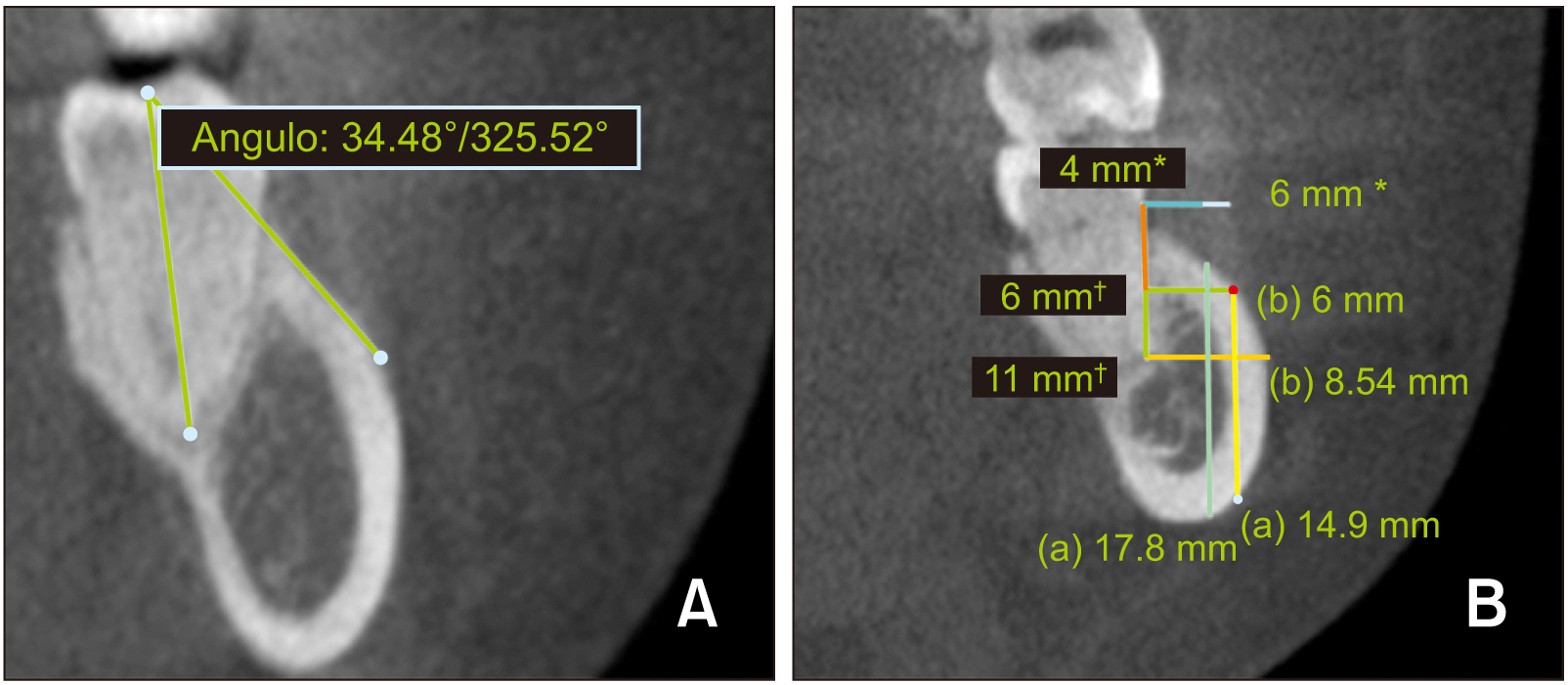
Figure 4 Representative images showing analysis of the mandibular buccal shelf on cone-beam computed tomography images. A, Angulation measurement. B, Apicocoronal depth and thickness measurements. a: Depth measurements. b: Thickness measurements. *References lines for depth measurement at 4 and 6 mm from the cementoenamel junction (CEJ). †Reference lines for thickness measurement at 6 and 11 mm from CEJ.
Reference
-
1. Chang C, Liu SS, Roberts WE. 2015; Primary failure rate for 1680 extra-alveolar mandibular buccal shelf mini-screws placed in movable mucosa or attached gingiva. Angle Orthod. 85:905–10. DOI: 10.2319/092714.695.1. PMID: 25603272.2. Suh HY, Lee SJ, Park HS. 2014; Use of mini-implants to avoid maxillary surgery for Class III mandibular prognathic patient: a long-term post-retention case. Korean J Orthod. 44:342–9. DOI: 10.4041/kjod.2014.44.6.342. PMID: 25473650. PMCID: PMC4250668.3. Kanomi R. 1997; Mini-implant for orthodontic anchorage. J Clin Orthod. 31:763–7. PMID: 9511584.4. Ramírez-Ossa DM, Escobar-Correa N, Ramírez-Bustamante MA, Agudelo-Suárez AA. 2020; An umbrella review of the effectiveness of temporary anchorage devices and the factors that contribute to their success or failure. J Evid Based Dent Pract. 20:101402. DOI: 10.1016/j.jebdp.2020.101402. PMID: 32473811.5. Rao P, Gill A. 2012; Primary stability: the password of implant integration. J Dent Implant. 2:103–9. DOI: 10.4103/0974-6781.102223.6. Migliorati M, Benedicenti S, Signori A, Drago S, Barberis F, Tournier H, et al. 2012; Miniscrew design and bone characteristics: an experimental study of primary stability. Am J Orthod Dentofacial Orthop. 142:228–34. DOI: 10.1016/j.ajodo.2012.03.029. PMID: 22858333.7. Ozdemir F, Tozlu M, Germec-Cakan D. 2013; Cortical bone thickness of the alveolar process measured with cone-beam computed tomography in patients with different facial types. Am J Orthod Dentofacial Orthop. 143:190–6. DOI: 10.1016/j.ajodo.2012.09.013. PMID: 23374925.8. Farnsworth D, Rossouw PE, Ceen RF, Buschang PH. 2011; Cortical bone thickness at common miniscrew implant placement sites. Am J Orthod Dentofacial Orthop. 139:495–503. DOI: 10.1016/j.ajodo.2009.03.057. PMID: 21457860.9. Yang L, Li F, Cao M, Chen H, Wang X, Chen X, et al. 2015; Quantitative evaluation of maxillary interradicular bone with cone-beam computed tomography for bicortical placement of orthodontic mini-implants. Am J Orthod Dentofacial Orthop. 147:725–37. DOI: 10.1016/j.ajodo.2015.02.018. PMID: 26038077.10. Becker K, Unland J, Wilmes B, Tarraf NE, Drescher D. 2019; Is there an ideal insertion angle and position for orthodontic mini-implants in the anterior palate? A CBCT study in humans. Am J Orthod Dentofacial Orthop. 156:345–54. DOI: 10.1016/j.ajodo.2018.09.019. PMID: 31474264.11. Murugesan A, Sivakumar A. 2020; Comparison of bone thickness in infrazygomatic crest area at various miniscrew insertion angles in Dravidian population - A cone beam computed tomography study. Int Orthod. 18:105–14. DOI: 10.1016/j.ortho.2019.12.001. PMID: 31926867.12. Elshebiny T, Palomo JM, Baumgaertel S. 2018; Anatomic assessment of the mandibular buccal shelf for miniscrew insertion in white patients. Am J Orthod Dentofacial Orthop. 153:505–11. DOI: 10.1016/j.ajodo.2017.08.014. PMID: 29602342.13. Nucera R, Lo Giudice A, Bellocchio AM, Spinuzza P, Caprioglio A, Perillo L, et al. 2017; Bone and cortical bone thickness of mandibular buccal shelf for mini-screw insertion in adults. Angle Orthod. 87:745–51. DOI: 10.2319/011117-34.1. PMID: 28598220.14. Kim YH. 1974; Overbite depth indicator with particular reference to anterior open-bite. Am J Orthod. 65:586–611. DOI: 10.1016/0002-9416(74)90255-3. PMID: 4524490.15. Kim YH, Vietas JJ. 1978; Anteroposterior dysplasia indicator: an adjunct to cephalometric differential diagnosis. Am J Orthod. 73:619–33. DOI: 10.1016/0002-9416(78)90223-3. PMID: 276266.16. Baumgaertel S, Hans MG. 2009; Buccal cortical bone thickness for mini-implant placement. Am J Orthod Dentofacial Orthop. 136:230–5. DOI: 10.1016/j.ajodo.2007.10.045. PMID: 19651353.17. Wu TY, Kuang SH, Wu CH. 2009; Factors associated with the stability of mini-implants for orthodontic anchorage: a study of 414 samples in Taiwan. J Oral Maxillofac Surg. 67:1595–9. DOI: 10.1016/j.joms.2009.04.015. PMID: 19615569.18. Huang C, Chang CH, Roberts WE. 2016; 3D Cortical Bone Anatomy of the Mandibular Buccal Shelf: a CBCT study to define sites for extra-alveolar bone screws to treat Class III malocclusion. Int J Orthod Implantol. 41:74–82.19. Kolge NE, Patni VJ, Potnis SS. 2019; Tomographic mapping of buccal shelf area for optimum placement of bone screws: a three-dimensional cone-beam computed tomography evaluation. APOS Trends Orthod. 9:241–5. DOI: 10.25259/APOS_20_2019.20. Alves N, de Oliveira Nascimento CM. 2012; Study of the mucosa thickness of the retromolar triangle to facilitate the installation planning of mini-implants in the region. Int J Odontostomatol. 6:175–9. Portugues. DOI: 10.4067/S0718-381X2012000200010.21. Lin J, White LW, Thavarungkul R. 2017. Creative orthodontics: blending the Damon System & TADs to manage difficult malocclusions. 3rd ed. Yong Chieh Enterprise Co.;Taipei: p. 339.22. Plaza SP, Reimpell A, Silva J, Montoya D. 2019; Relationship between skeletal Class II and Class III malocclusions with vertical skeletal pattern. Dental Press J Orthod. 24:63–72. DOI: 10.1590/2177-6709.24.4.063-072.oar. PMID: 31508708. PMCID: PMC6733235.
- Full Text Links
-
- Actions
-
Cited
- CITED
-
- Close
- Share
-
- Similar articles
-
- Mandibular condyle position in cone beam computed tomography
- Cone beam computed tomography findings of ectopic mandibular third molar in the mandibular condyle: report of a case
- CBCT study of mandibular first molars with a distolingual root in Koreans
- Proximity of the mandibular molar root apex from the buccal bone surface: a cone-beam computed tomographic study
- Anatomical position of the mandibular canal in relation to the buccal cortical bone: relevance to sagittal split osteotomy
