Proximity of the mandibular molar root apex from the buccal bone surface: a cone-beam computed tomographic study
- Affiliations
-
- 1Department of Conservative Dentistry, Kyungpook National University School of Dentistry, Daegu, Korea. skykim@knu.ac.kr
- KMID: 2346766
- DOI: http://doi.org/10.5395/rde.2016.41.3.182
Abstract
OBJECTIVES
The purpose of this study was to evaluate the proximity of the mandibular molar apex to the buccal bone surface in order to provide anatomic information for apical surgery.
MATERIALS AND METHODS
Cone-beam computed tomography (CBCT) images of 127 mandibular first molars and 153 mandibular second molars were analyzed from 160 patients' records. The distance was measured from the buccal bone surface to the root apex and the apical 3.0 mm on the cross-sectional view of CBCT.
RESULTS
The second molar apex and apical 3 mm were located significantly deeper relative to the buccal bone surface compared with the first molar (p < 0.01). For the mandibular second molars, the distance from the buccal bone surface to the root apex was significantly shorter in patients over 70 years of age (p < 0.05). Furthermore, this distance was significantly shorter when the first molar was missing compared to nonmissing cases (p < 0.05). For the mandibular first molars, the distance to the distal root apex of one distal-rooted tooth was significantly greater than the distance to the disto-buccal root apex (p < 0.01). In mandibular second molar, the distance to the apex of C-shaped roots was significantly greater than the distance to the mesial root apex of non-C-shaped roots (p < 0.01).
CONCLUSIONS
For apical surgery in mandibular molars, the distance from the buccal bone surface to the apex and apical 3 mm is significantly affected by the location, patient age, an adjacent missing anterior tooth, and root configuration.
Keyword
Figure
-

Figure 1 Mandibular axial cone beam computed tomography (CBCT) images for measurement. (a) The distance from the first molar apex to the buccal bone surface; (b) The distance from the disto-lingual root apex of the first molar to the buccal bone surface; (c) The distance from the C-shaped second molar apex to the buccal bone surface; (d) The distance from the buccal root surface at the apical 3 mm to the buccal bone surface with a C-shaped root.
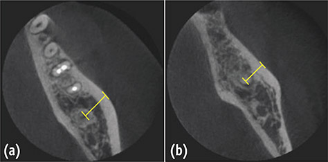
Figure 2 Mandibular axial cone beam computed tomography (CBCT) images for mandibular second molars. (a) The distance between the second molar apex and the buccal bone surface, when the first molar is present; (b) The distance between the second molar apex and the buccal bone surface, when the first molar is absent.
Cited by 1 articles
-
Proximity of maxillary molar apexes to the cortical bone surface and the maxillary sinus
Han Shin Lee, Dokyung Kim, Sung Kyo Kim
Restor Dent Endod. 2022;47(3):e33. doi: 10.5395/rde.2022.47.e33.
Reference
-
1. Ingle JI, Bakland LK, Baumgartner JC. Ingle's endodontics. 6th ed. Hamilton: BC Decker Inc.;2008. p. 705–706.2. Frankle KT, Seibel W, Dumsha TC. Anatomical study of the position of the mesial roots of mandibular molars. J Endod. 1990; 16:480–485.
Article3. Jin GC, Kim KD, Roh BD, Lee CY, Lee SJ. Buccal bone plate thickness of the Asian people. J Endod. 2005; 31:430–434.
Article4. Kratchman S. Intentional replantation. Dent Clin North Am. 1997; 41:603–617.5. Patel S, Horner K. The use of cone beam computed tomography in endodontics. Int Endod J. 2009; 42:755–756.
Article6. Rigolone M, Pasqualini D, Bianchi L, Berutti E, Bianchi SD. Vestibular surgical access to the palatine root of the superior first molar: 'low-dose cone-beam' CT analysis of the pathway and its anatomic variations. J Endod. 2003; 29:773–775.
Article7. Blattner TC, George N, Lee CC, Kumar V, Yelton CD. Efficacy of cone-beam computed tomography as a modality to accurately identify the presence of second mesiobuccal canals in maxillary first and second molars: a pilot study. J Endod. 2010; 36:867–870.
Article8. Siqueira JF Jr. Aetiology of root canal treatment failure: why well-treated teeth can fail. Int Endod J. 2001; 34:1–10.
Article9. Johnson BR, Fayad M. Periradicular surgery. In : Hargreaves KM, Berman LH, editors. Cohen's pathways of the pulp. 11th ed. Philadelphia: Elsevier Inc.;2016. Chapter 9.10. Kim S. . Osteotomy and apical root resection. Philadelphia: Saunders;2000. Chapter 9.11. Khoury F, Hensher R. The bony lid approach for the apical root resection of lower molars. Int J Oral Maxillofac Surg. 1987; 16:166–170.
Article12. Lasaridis N, Zouloumis L, Antoniadis K. Bony lid approach for apicoectomy of mandibular molars. Aust Dent J. 1991; 36:366–368.
Article13. Timock AM, Cook V, McDonald T, Leo MC, Crowe J, Benninger BL, Covell DA Jr. Accuracy and reliability of buccal bone height and thickness measurements from cone-beam computed tomography imaging. Am J Orthod Dentofacial Orthop. 2011; 140:734–744.
Article14. De Deus QD. Frequency, location, and direction of the lateral, secondary, and accessory canals. J Endod. 1975; 1:361–366.
Article15. Seltzer S, Soltanoff W, Bender IB, Ziontz M. Biologic aspects of endodontics, I. Histologic observations of the anatomy and morphology of root apices and surrounding structures. Oral Surg Oral Med Oral Pathol Oral Radiol. 1966; 22:375–385.16. Wada M, Takase T, Nakanuma K, Arisue K, Nagahama F, Yamazaki M. Clinical study of refractory apical periodontitis treated by apicoectomy. Part 1. root canal morphology of resected apex. Int Endod J. 1998; 31:53–56.
Article17. Kim S. Principles of endodontic microsurgery. Dent Clin North Am. 1997; 41:481–497.18. Noyes HJ. Mesial drift. Angle Orthod. 1941; 11:199–200.19. Schropp L, Wenzel A, Kostopoulos L, Karring T. Bone healing and soft tissue contour changes following single-tooth extraction: a clinical and radiographic 12-month prospective study. Int J Periodontics Restorative Dent. 2003; 23:313–323.20. Kingsmill VJ, Boyde A. Variation in the apparent density of human mandibular bone with age and dental status. J Anat. 1998; 192:233–244.
Article21. Kingsmill VJ. Post-extraction remodeling of the adult mandible. Crit Rev Oral Biol Med. 1999; 10:384–404.
Article22. Khosla S, Melton LJ 3rd, Atkinson EJ, O'Fallon WM. Relationship of serum sex steroid levels to longitudinal changes in bone density in young versus elderly men. J Clin Endocrinol Metab. 2001; 86:3555–3561.
Article23. Clarke BL, Khosla S. Physiology of bone loss. Radiol Clin North Am. 2010; 48:483–495.
Article
- Full Text Links
-
- Actions
-
Cited
- CITED
-
- Close
- Share
-
- Similar articles
-
- CBCT study of mandibular first molars with a distolingual root in Koreans
- Proximity of maxillary molar apexes to the cortical bone surface and the maxillary sinus
- Validity of the vertical tube-shift method in determining the relationship between the mandibular third molar roots and the inferior alveolar nerve canal
- Positional relationship between the maxillary sinus floor and the apex of the maxillary first molar using cone beam computed tomograph
- Reliability of panoramic radiography in predicting proximity of third molars to the mandibular canal: A comparison using cone-beam computed tomography
