Anat Cell Biol.
2020 Dec;53(4):512-515. 10.5115/acb.20.153.
A rare case of trifid mandibular canal with bilateral retromolar foramina
- Affiliations
-
- 1Harvard School of Dental Medicine, Harvard University, Boston, MA,, USA
- 2Department of Restorative Dentistry and Biomaterials Sciences, Harvard School of Dental Medicine, Boston MA,, USA
- 3Department of Neurosurgery, Tulane Center for Clinical Neurosciences, Tulane University School of Medicine, New Orleans, LA, USA
- 4Department of Anatomical Sciences, St. George’s University, St. George’s, Grenada
- 5Department of Structural & Cellular Biology, Tulane University School of Medicine, New Orleans, LA, USA
- 6Department of Neurosurgery and Ochsner Neuroscience Institute, Ochsner Health System, New Orleans, LA, USA
- 7Dental and Oral Medical Center, Kurume University School of Medicine, Kurume, Fukuoka, Japan
- 8Division of Gross and Clinical Anatomy, Department of Anatomy, Kurume University School of Medicine, Kurume, Fukuoka, Japan
- KMID: 2509699
- DOI: http://doi.org/10.5115/acb.20.153
Abstract
- There are many reported anatomical variations of the mandibular canal. Consequently, there is great variation in the retromolar area, such as the quantity, size, and location of the retromolar foramen (RMF), the bony entrance of the retromolar canal (RMC). These variations allow for different accessory innervations to the mandibular molars and their adjacent buccal tissue because the RMC contains neurovascular bundles. Consideration of these anatomical variations is crucial for avoiding complications in anesthesia, implant placement, and surgery. However, the rarer canal types are often only imaged by computed tomography (CT) or cone beam computed tomography (CBCT). We present a rare case with bilateral RMF and a unilateral trifid mandibular canal in a cadaver.
Keyword
Figure
-
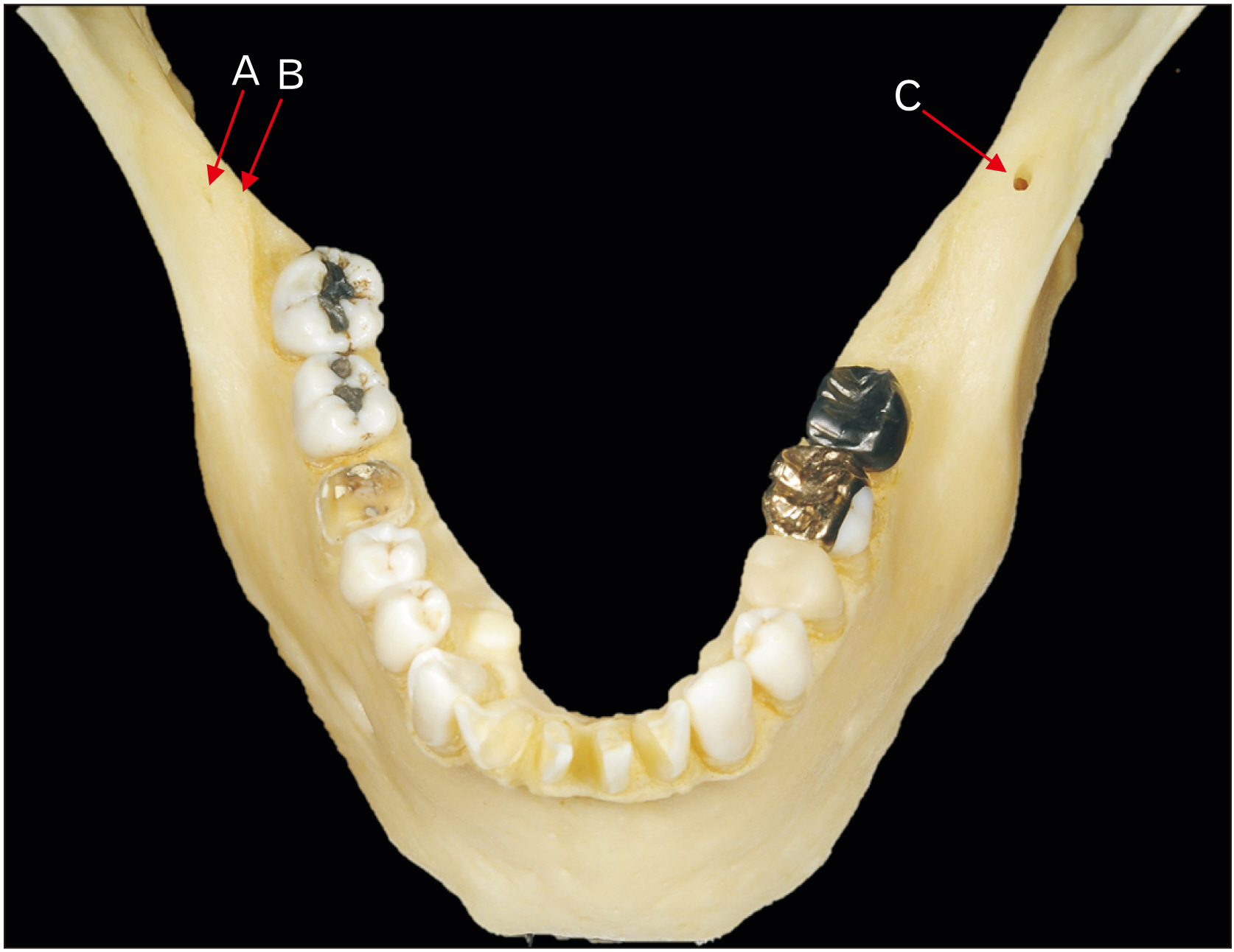
Fig. 1 A dry mandible demonstrating double retromolar foramina on the right (A, B) and a retromolar foramen on the left (C). The three retromolar foramina are labeled.
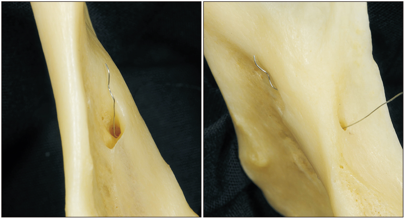
Fig. 2 Continuation of the retromolar foramen and superior canal on the left shown with a metal wire.
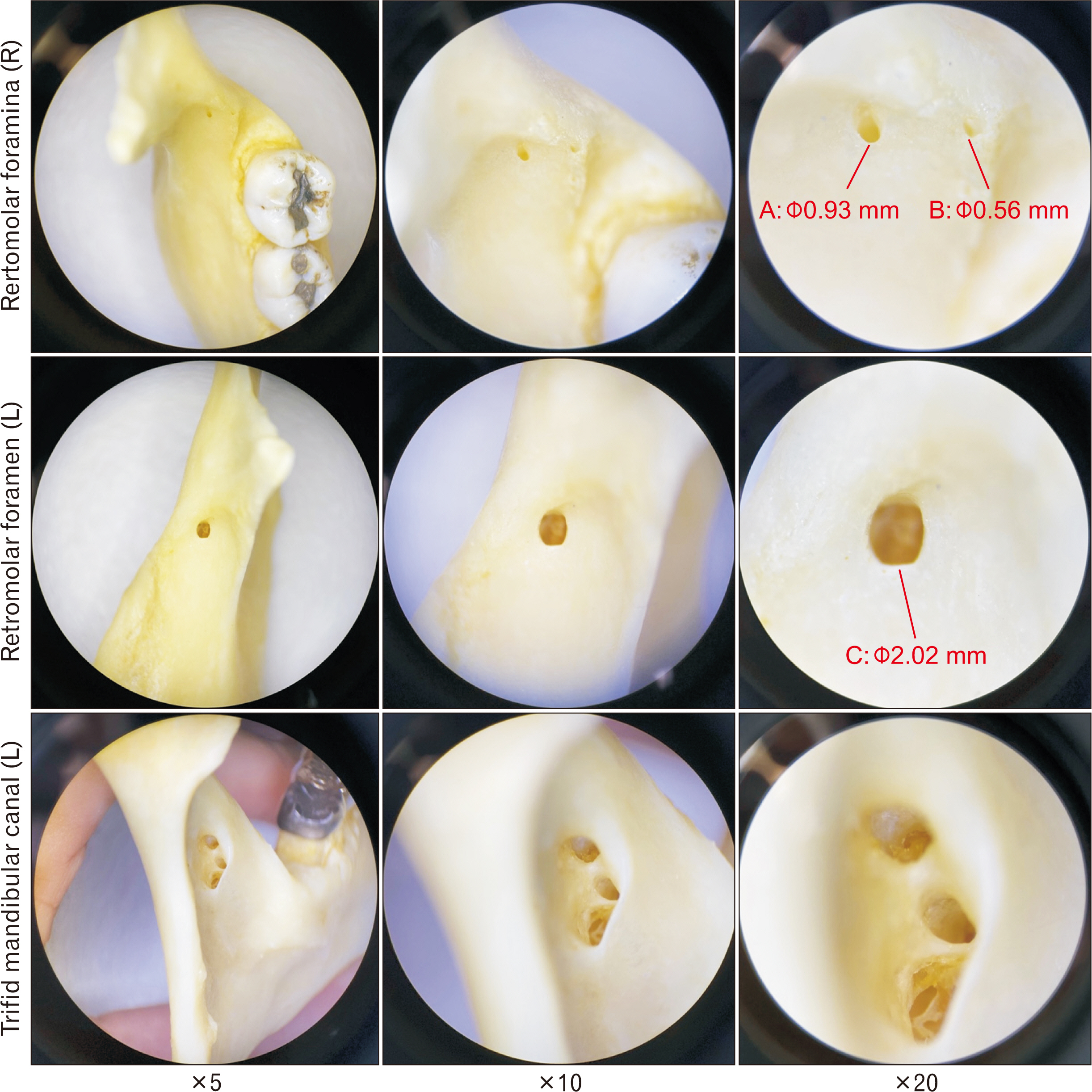
Fig. 3 A closer look at the retromolar foramina under the surgical microscope with ×5, ×10, ×20 magnified view documenting their diameter. Note the trifid mandibular canal is observed from a posterior view.
Reference
-
References
1. Iwanaga J, Watanabe K, Saga T, Tubbs RS, Tanaka K, Kikuta S, Tabira Y, Fisahn C, Kamura Y, Kusukawa J, Yamaki KI. 2017; A novel method for observation of the mandibular foramen: application to a better understanding of dental anatomy. Anat Rec (Hoboken). 300:1875–80. DOI: 10.1002/ar.23639. PMID: 28681490.
Article2. Naitoh M, Hiraiwa Y, Aimiya H, Ariji E. 2009; Observation of bifid mandibular canal using cone-beam computerized tomography. Int J Oral Maxillofac Implants. 24:155–9. PMID: 19344041.3. Rashsuren O, Choi JW, Han WJ, Kim EK. 2014; Assessment of bifid and trifid mandibular canals using cone-beam computed tomography. Imaging Sci Dent. 44:229–36. DOI: 10.5624/isd.2014.44.3.229. PMID: 25279344. PMCID: PMC4182358.
Article4. von Arx T, Hänni A, Sendi P, Buser D, Bornstein MM. 2011; Radiographic study of the mandibular retromolar canal: an anatomic structure with clinical importance. J Endod. 37:1630–5. DOI: 10.1016/j.joen.2011.09.007. PMID: 22099895.
Article5. Ngeow WC, Chai WL. 2020; Feb. 4. The clinical significance of the retromolar canal and foramen in dentistry. Clin Anat. [Epub]. http://dx.doi.org/10.1002/ca.23577. DOI: 10.1002/ca.23577. PMID: 32020669.
Article6. Schejtman R, Devoto FC, Arias NH. 1967; The origin and distribution of the elements of the human mandibular retromolar canal. Arch Oral Biol. 12:1261–8. DOI: 10.1016/0003-9969(67)90127-6. PMID: 5234232.
Article7. Kodera H, Hashimoto I. 1995; [A case of mandibular retromolar canal: elements of nerves and arteries in this canal]. Kaibogaku Zasshi. 70:23–30. Japanese. PMID: 7785408.8. Iwanaga J, Kikuta S, Tanaka T, Kamura Y, Tubbs RS. 2019; Review of risk assessment of major anatomical variations in clinical dentistry: accessory foramina of the mandible. Clin Anat. 32:672–7. DOI: 10.1002/ca.23366. PMID: 30848855.9. Iwanaga J, Kikuta S, Ibaragi S, Watanabe K, Kusukawa J, Tubbs RS. 2020; Clinical anatomy of the accessory mandibular foramen: application to mandibular ramus osteotomy. Surg Radiol Anat. 42:41–7. DOI: 10.1007/s00276-019-02343-3. PMID: 31541271.
Article10. Motamedi MH, Gharedaghi J, Mehralizadeh S, Navi F, Badkoobeh A, Valaei N, Azizi T. 2016; Anthropomorphic assessment of the retromolar foramen and retromolar nerve: anomaly or variation of normal anatomy? Int J Oral Maxillofac Surg. 45:241–4. DOI: 10.1016/j.ijom.2015.10.017. PMID: 26586301.
Article11. Kim HJ, Kang H, Seo YS, Kim DK, Yu SK. 2017; Anatomic evaluation of the retromolar canal by histologic and radiologic analyses. Arch Oral Biol. 81:192–7. DOI: 10.1016/j.archoralbio.2017.05.012. PMID: 28554135.
Article12. Ogawa A, Fukuta Y, Nakasato H, Nakasato S. 2016; Evaluation by dental cone-beam computed tomography of the incidence and sites of branches of the inferior dental canal that supply mandibular third molars. Br J Oral Maxillofac Surg. 54:1116–20. DOI: 10.1016/j.bjoms.2016.08.007. PMID: 27576162.
Article13. Kikuta S, Iwanaga J, Nakamura K, Hino K, Nakamura M, Kusukawa J. 2018; The retromolar canals and foramina: radiographic observation and application to oral surgery. Surg Radiol Anat. 40:647–52. DOI: 10.1007/s00276-018-2005-5. PMID: 29594335.
Article14. Kang JH, Lee KS, Oh MG, Choi HY, Lee SR, Oh SH, Choi YJ, Kim GT, Choi YS, Hwang EH. 2014; The incidence and configuration of the bifid mandibular canal in Koreans by using cone-beam computed tomography. Imaging Sci Dent. 44:53–60. DOI: 10.5624/isd.2014.44.1.53. PMID: 24701459. PMCID: PMC3972406.
Article
- Full Text Links
-
- Actions
-
Cited
- CITED
-
- Close
- Share
-
- Similar articles
-
- Assessment of bifid and trifid mandibular canals using cone-beam computed tomography
- Root canal treatment of a mandibular second premolar with three separate root canals
- Cone beam CT findings of retromolar canals: Report of cases and literature review
- Endodontic treatment of a C-shaped mandibular second premolar with four root canals and three apical foramina: a case report
- Distribution of the lingual foramina in mandibular cortical bone in Koreans
