J Korean Acad Conserv Dent.
2010 Jul;35(4):302-305. 10.5395/JKACD.2010.35.4.302.
Root canal treatment of a mandibular second premolar with three separate root canals
- Affiliations
-
- 1Department of Conservative Dentistry, School of Dentistry, Wonkwang University, Iksan, Korea. mksdd@wonkwang.ac.kr
- KMID: 2176396
- DOI: http://doi.org/10.5395/JKACD.2010.35.4.302
Abstract
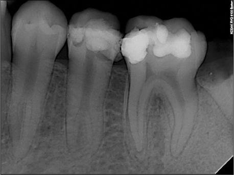
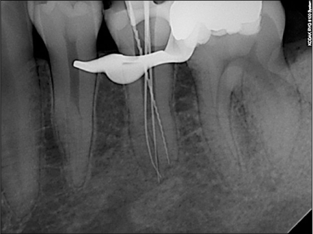
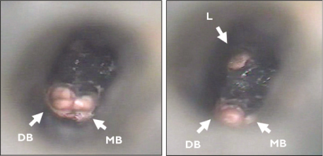
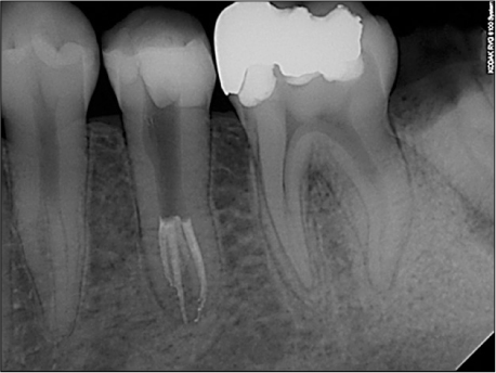
- Mandibular premolars show a wide variety of root canal anatomy. Especially, the occurrence of three canals with three separate foramina in mandibular second premolars is very rare. This case report describes the root canal treatment of an unusual morphological configuration of the root canal system and supplements previous reports of the existence of such configuration in mandibular second premolar.
MeSH Terms
Figure
Reference
-
1. England MC, Hartwell GR, Lance JK. Detection and treatment of multiple canals in mandibular premolars. J Endod. 1991. 17:174–178.
Article2. Cleghorn BM, Christie WH, Dong CC. The root and root canal morphology of the human mandibular second premolar: a literature review. J Endod. 2007. 33:1031–1037.
Article3. Trope M, Elfenbein L, Tronstad L. Mandibular premolars with more than one root canal in different race groups. J Endod. 1986. 12:343–345.
Article4. Vertucci FJ. Root canal morphology of mandibular premolars. J Am Dent Assoc. 1978. 97:47–50.
Article5. Zillich R, Dowson J. Root canal morphology of mandibular first and second premolars. Oral Surg Oral Med Oral Pathol. 1973. 36:738–744.
Article6. Nallapati S. Three canal mandibular first and second premolars: a treatment approach. J Endod. 2005. 31:474–476.7. Slowey RR. Root canal anatomy. Road map to successful endodontics. Dent Clin North Am. 1979. 23:555–573.8. Slowey RR. Radiographic aids in the detection of extra root canal. Oral Surg Oral Med Oral Pathol. 1974. 37:762–772.9. Melton DC, Krell KV, Fuller MW. Anatomical and histological features of C-shaped canals in mandibular second molars. J Endod. 1991. 17:384–388.
Article
- Full Text Links
- Actions
-
Cited
- CITED
-
- Close
- Share
- Similar articles
-
- Endodontic treatment of a C-shaped mandibular second premolar with four root canals and three apical foramina: a case report
- A Study of Root Canals Morphology in Primary Molars using Computerized Tomography
- Assessment of Root and Root Canal Morphology of Human Primary Molars using CBCT
- Predictor factors of 1-rooted mandibular second molars on complicated root and canal anatomies of other mandibular teeth
- A study on the C-shaped root canal system of mandibular second molar


