Predictor factors of 1-rooted mandibular second molars on complicated root and canal anatomies of other mandibular teeth
- Affiliations
-
- 1Department of Endodontics, Faculty of Dentistry, Antalya Bilim University, Antalya, Türkiye
- 2Departent of Endodontics, Faculty of Dentistry, Akdeniz University, Antalya, Türkiye
- KMID: 2554130
- DOI: http://doi.org/10.5395/rde.2024.49.e2
Abstract
Objectives
This study aimed to determine the effects of 1-rooted mandibular second molar (MnSM) teeth on root canal anatomy complexities of the mandibular central incisor (MnCI), mandibular lateral incisor (MnLI), mandibular canine (MnCn), mandibular first premolar (MnFP), mandibular second premolar (MnSP), and mandibular first molar (MnFM) teeth.
Materials and Methods
Cone-beam computed tomography images of 600 patients with full lower dentition were examined. Individuals with 1-rooted MnSMs were determined, and the complexity of root canal anatomy of other teeth was compared with individuals without 1-rooted MnSMs (Group-1; subjects with at least one 1-rooted MnSM, Group-2; subjects with more than a single root in both MnSMs). A second canal in MnCIs, MnLIs, MnCns, MnFPs, and MnSPs indicated a complicated root canal. The presence of a third root in MnFMs was recorded as complicated.
Results
The prevalence of 1-rooted MnSMs was 12.2%, with the C-shaped root type being the most prevalent (9%). There were fewer complicated root canals in MnCIs (p = 0.02), MnLIs (p < 0.001), and MnFPs (p < 0.001) in Group 1. The other teeth showed no difference between the groups (p > 0.05). According to logistic regression analysis, 1-rooted right MnSMs had a negative effect on having complex canal systems of MnLIs and MnFPs. Left MnSMs were explanatory variables on left MnLIs and both MnFPs.
Conclusions
In individuals with single-rooted MnSMs, a less complicated root canal system was observed in all teeth except the MnFMs.
Keyword
Figure
-
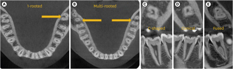
Figure 1 Examples of (A) 1-rooted, and (B) multi-rooted MnSM teeth and 1-rooted types, (C) C-shaped, (D) conical, (E) fused.
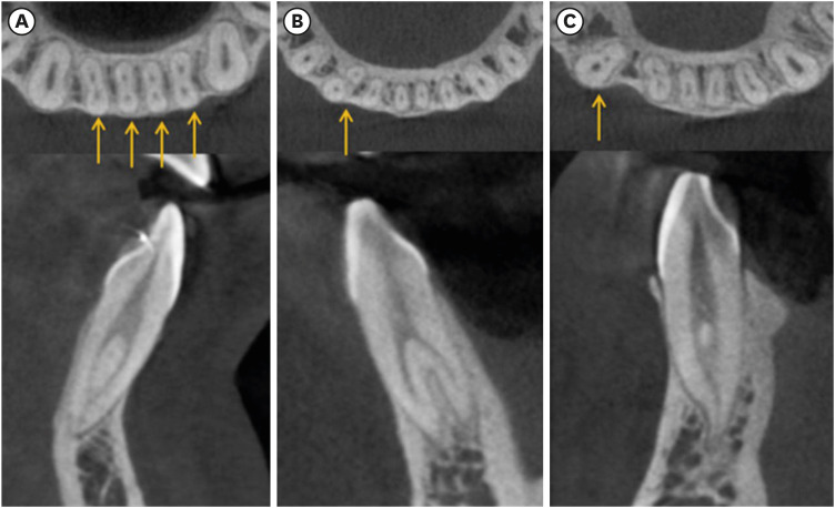
Figure 2 (A) Mandibular central and lateral incisors with complicated root canals, (B) 2-rooted mandibular canine, (C) 1-rooted/complicated canal mandibular canine.
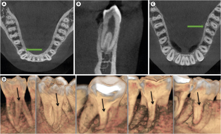
Figure 3 (A, B) Mandibular first premolar with complicated root canals, (C, D) examples of radix entomolaris in the mandibular first molar tooth.
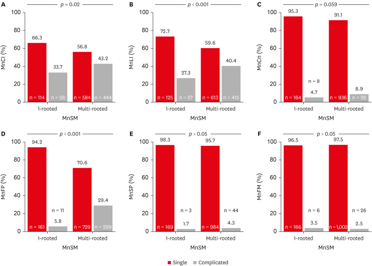
Figure 4 In individuals with 1-rooted and multi-rooted mandibular second molar (MnSM) teeth, the number, percentages, and p values of the root anatomy of other teeth separately. (A) Mandibular central incisor (MnCI), (B) mandibular lateral incisor (MnLI), (C) mandibular canine (MnCn), (D) mandibular first premolar (MnFP), (E) mandibular second premolar (MnSP), (F) mandibular first molar (MnFM).
Reference
-
1. Martins JNR, Gu Y, Marques D, Francisco H, Caramês J. Differences on the root and root canal morphologies between Asian and White ethnic groups analyzed by cone-beam computed tomography. J Endod. 2018; 44:1096–1104. PMID: 29861062.2. Aydın H.. Predictor mandibular dentoalveolar features on the occurrence of 1-rooted/C-shaped mandibular second molar teeth. Eur Endod J. 2023; Nov. 7. [Epub]. DOI: 10.14744/eej.2023.91886.3. Martins JN, Mata A, Marques D, Caramês J. Prevalence of root fusions and main root canal merging in human upper and lower molars: a cone-beam computed tomography in vivo study. J Endod. 2016; 42:900–908. PMID: 27087629.4. Kim SY, Kim BS, Kim Y. Mandibular second molar root canal morphology and variants in a Korean subpopulation. Int Endod J. 2016; 49:136–144. PMID: 25652228.5. Aydın H, Mobaraki S. Comparison of root and canal anatomy of taurodont and normal molar teeth: a retrospective cone-beam computed tomography study. Arch Oral Biol. 2021; 130:105242. PMID: 34411883.6. Fan B, Cheung GS, Fan M, Gutmann JL, Bian Z. C-shaped canal system in mandibular second molars: part I--anatomical features. J Endod. 2004; 30:899–903. PMID: 15564874.7. Kato A, Ziegler A, Higuchi N, Nakata K, Nakamura H, Ohno N. Aetiology, incidence and morphology of the C-shaped root canal system and its impact on clinical endodontics. Int Endod J. 2014; 47:1012–1033. PMID: 24483229.8. Wu YC, Su CC, Tsai YC, Cheng WC, Chung MP, Chiang HS, et al. Complicated root canal configuration of mandibular first premolars is correlated with the presence of the distolingual root in mandibular first molars: a cone-beam computed tomographic study in Taiwanese individuals. J Endod. 2017; 43:1064–1071. PMID: 28416311.9. Wu YC, Cheng WC, Chung MP, Su CC, Weng PW, Cathy Tsai YW, et al. Complicated root canal morphology of mandibular lateral incisors is associated with the presence of distolingual root in mandibular first molars: a cone-beam computed tomographic study in a Taiwanese population. J Endod. 2018; 44:73–79.e1. PMID: 29079050.10. Wu YC, Su WS, Mau LP, Cheng WC, Weng PW, Tsai YC, et al. Association between the presence of distolingual root in mandibular first molars and the presence of C-shaped mandibular second molars: a CBCT study in a Taiwanese population. Quintessence Int. 2020; 51:798–807. PMID: 32954388.11. Yang Y, Jiang C, Chen M, Zeng J, Wu B. Vertucci’s root canal configuration of 11,376 mandibular anteriors and its relationship with distolingual roots in mandibular first molars in a Cantonese population: a cone-beam computed tomography study. BMC Oral Health. 2022; 22:130. PMID: 35429982.12. Aydın H. Correlations between additional roots in maxillary second molars, maxillary first premolars, mandibular first molars and mandibular first premolars: a retrospective cone-beam computed tomography analysis. Odontology. 2022; 110:584–595. PMID: 35098365.13. Wu YC, Cheng WC, Weng PW, Chung MP, Su CC, Chiang HS, et al. The presence of distolingual root in mandibular first molars is correlated with complicated root canal morphology of mandibular central incisors: a cone-beam computed tomographic study in a Taiwanese population. J Endod. 2018; 44:711–716.e1. PMID: 29499856.14. Mashyakhy MH, Chourasia HR, Jabali AH, Bajawi HA, Jamal H, Testarelli L, et al. C-shaped canal configuration in mandibular premolars and molars: prevalence, correlation, and differences: an in vivo study using cone-beam computed tomography. Niger J Clin Pract. 2020; 23:232–239. PMID: 32031099.15. Sinanoglu A, Helvacioglu-Yigit D. Analysis of C-shaped canals by panoramic radiography and cone-beam computed tomography: root-type specificity by longitudinal distribution. J Endod. 2014; 40:917–921. PMID: 24935535.16. Martins JNR, Kishen A, Marques D, Nogueira Leal Silva EJ, Caramês J, Mata A, et al. Preferred reporting items for epidemiologic cross-sectional studies on root and root canal anatomy using cone-beam computed tomographic technology: a systematized assessment. J Endod. 2020; 46:915–935. PMID: 32387077.17. Aydın H. Comparing the crown and root metric properties of double-rooted and single-rooted mandibular canine teeth. Oral Radiol. 2023; 39:301–311. PMID: 35819742.18. Aydin H, Çiloglu Ö. Relationship between root canal merging and presence of C-shaped canal in fused rooted maxillary molar teeth. Braz Dent Sci. 2022; 25:e2964.19. Martins JNR, Marques D, Silva EJNL, Caramês J, Mata A, Versiani MA. Prevalence of C-shaped canal morphology using cone beam computed tomography - a systematic review with meta-analysis. Int Endod J. 2019; 52:1556–1572. PMID: 31215045.20. Ren HY, Zhao YS, Yoo YJ, Zhang XW, Fang H, Wang F, et al. Mandibular molar C-shaped root canals in 5th millennium BC China. Arch Oral Biol. 2020; 117:104773. PMID: 32512259.21. Melton DC, Krell KV, Fuller MW. Anatomical and histological features of C-shaped canals in mandibular second molars. J Endod. 1991; 17:384–388. PMID: 1809802.22. Zheng Q, Zhang L, Zhou X, Wang Q, Wang Y, Tang L, et al. C-shaped root canal system in mandibular second molars in a Chinese population evaluated by cone-beam computed tomography. Int Endod J. 2011; 44:857–862. PMID: 21599707.23. Fan B, Cheung GS, Fan M, Gutmann JL, Fan W. C-shaped canal system in mandibular second molars: part II--radiographic features. J Endod. 2004; 30:904–908. PMID: 15564875.24. Fan B, Min Y, Lu G, Yang J, Cheung GS, Gutmann JL. Negotiation of C-shaped canal systems in mandibular second molars. J Endod. 2009; 35:1003–1008. PMID: 19567323.25. Seo DG, Gu Y, Yi YA, Lee SJ, Jeong JS, Lee Y, et al. A biometric study of C-shaped root canal systems in mandibular second molars using cone-beam computed tomography. Int Endod J. 2012; 45:807–814. PMID: 22432971.26. Martins JNR, Marques D, Mata A, Caramês J. Root and root canal morphology of the permanent dentition in a Caucasian population: a cone-beam computed tomography study. Int Endod J. 2017; 50:1013–1026. PMID: 27883205.
- Full Text Links
-
- Actions
-
Cited
- CITED
-
- Close
- Share
-
- Similar articles
-
- The characteristics of Korean multi-rooted teeth root trunk extracted by periodontal disease
- A retrospective study on incidence of C-shaped canals in mandibular second molars
- Assessment of Root and Root Canal Morphology of Human Primary Molars using CBCT
- The clinical study of the mandibular canal location in mandibular molar areas using dentascan
- A Study of Root Canals Morphology in Primary Molars using Computerized Tomography
