Diagnostic Value of Lesion-specific Measurement of Myocardial Blood Flow Using Hybrid PET/CT
- Affiliations
-
- 1Department of Nuclear Medicine, Chonnam National University Hospital, Gwangju, Korea
- 2Medical Photonics Research Center, Korea Photonics Technology Institute, Gwangju, Korea
- 3Department of Cardiovascular Medicine, Chonnam National University Hospital, Gwangju, Korea
- 4Department of Nuclear Medicine, Chonnam National University Hwasun Hospital, Jeollanam-do, Korea
- KMID: 2509452
- DOI: http://doi.org/10.4250/jcvi.2019.0087
Abstract
- BACKGROUND
We evaluated whether lesion-specific measurement of myocardial blood flow (MBF) and flow reserve (MFR) by hybrid imaging of myocardial perfusion positron emission tomography (PET) and coronary computed tomography (CT) can provide additional diagnostic value.
METHODS
Forty-three patients with stable angina underwent N-13 ammonia PET and coronary CT before invasive coronary angiography (CAG). The lesion-specific MBF was calculated from the average MBF of the myocardial segments downstream of a coronary stenosis using hybrid PET/CT images. The hyperemic MBF, resting MBF, and MFR were measured for the left anterior descending artery (LAD) using conventional and lesion-specific methods. The diagnostic accuracy was compared between the two methods for significant LAD stenoses (≥ 70% reference diameter on CAG).
RESULTS
There were 19 significant LAD stenoses. The sensitivity, specificity, negative predictive value, positive predictive value, and accuracy were 71%, 68%, 74%, 65%, and 70% for conventional hyperemic MBF (optimal cutoff = 2.15 mL/min/g), 79%, 63%, 74%, 65%, and 70% for conventional MFR (optimal cutoff = 1.82), 83%, 74%, 80%, 78%, and 80% for lesion-specific hyperemic MBF (optimal cutoff = 1.75 mL/min/g), and 79%, 79%, 83%, 75%, and 79% for lesion-specific MFR (optimal cutoff = 1.86), respectively. The lesion-specific measurement was more accurate and had a better linear correlation with anatomical stenosis severity for both hyperemic MBF and MFR.
CONCLUSIONS
Lesion-specific measurement using hybrid PET/CT imaging showed significant improvement in the diagnostic accuracy of PET-measured hyperemic MBF and MFR.
Keyword
Figure
-
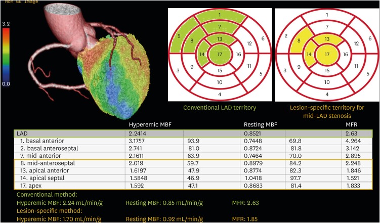
Figure 1 Conventional and lesion-specific measurements of the MBF and MFR in a patient with a significant mid-LAD stenosis (89% reference diameter). For a lesion-specific MBF measurement, the segments downstream of the mid-LAD stenosis (segments 8, 13, 14, and 17) were isolated and their average MBF values were obtained. LAD: left anterior descending artery, MBF: myocardial blood flow, MFR: myocardial flow reserve.
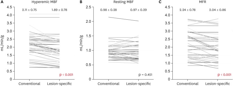
Figure 2 Comparison of MBF and MFR measured by conventional and lesion-specific methods.The hyperemic MBF and MFR significantly decreased after applying lesion-specific measurement, while the resting MBF remained similar. MBF: myocardial blood flow, MFR: myocardial flow reserve.
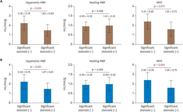
Figure 3 Comparison of the MBF and MFR between LADs with and without significant stenosis for conventional (A) and lesion-specific (B) methods. LAD: left anterior descending artery, MBF: myocardial blood flow, MFR: myocardial flow reserve.
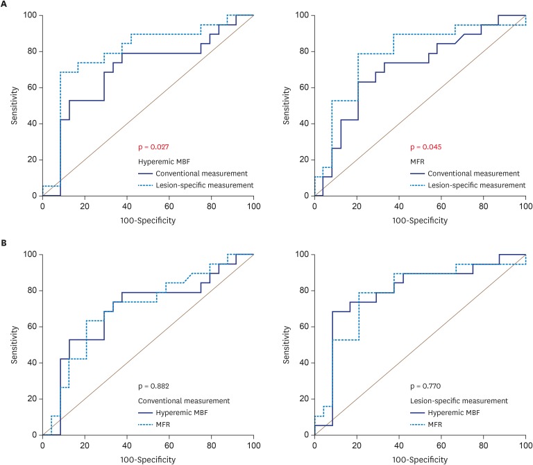
Figure 4 Comparison of the diagnostic accuracy according to the measurement method (A) and MBF parameters (B). MBF: myocardial blood flow, MFR: myocardial flow reserve.
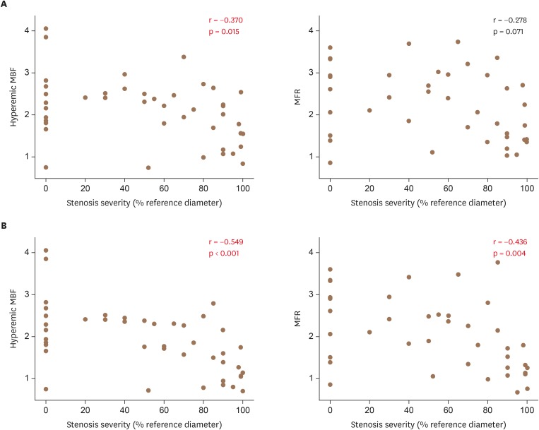
Figure 5 Correlation between the MBF parameters and anatomical stenosis severity for conventional (A) and lesion-specific (B) methods. MBF: myocardial blood flow, MFR: myocardial flow reserve.
Reference
-
1. Fiechter M, Ghadri JR, Gebhard C, et al. Diagnostic value of 13N-ammonia myocardial perfusion PET: added value of myocardial flow reserve. J Nucl Med. 2012; 53:1230–1234. PMID: 22776752.2. Lee JM, Kim CH, Koo BK, et al. Integrated myocardial perfusion imaging diagnostics improve detection of functionally significant coronary artery stenosis by 13N-ammonia positron emission tomography. Circ Cardiovasc Imaging. 2016; 9:e004768. PMID: 27609817.3. Ziadi MC, Dekemp RA, Williams K, et al. Does quantification of myocardial flow reserve using rubidium-82 positron emission tomography facilitate detection of multivessel coronary artery disease? J Nucl Cardiol. 2012; 19:670–680. PMID: 22415819.4. Herzog BA, Husmann L, Valenta I, et al. Long-term prognostic value of 13N-ammonia myocardial perfusion positron emission tomography added value of coronary flow reserve. J Am Coll Cardiol. 2009; 54:150–156. PMID: 19573732.5. Taqueti VR, Hachamovitch R, Murthy VL, et al. Global coronary flow reserve is associated with adverse cardiovascular events independently of luminal angiographic severity and modifies the effect of early revascularization. Circulation. 2015; 131:19–27. PMID: 25400060.6. Gupta A, Taqueti VR, van de Hoef TP, et al. Integrated noninvasive physiological assessment of coronary circulatory function and impact on cardiovascular mortality in patients with stable coronary artery disease. Circulation. 2017; 136:2325–2336. PMID: 28864442.7. Cho SG, Kim HS, Bom HH. Flow-based functional assessment of coronary artery disease by myocardial perfusion positron emission tomography in the era of fractional flow reserve. Ann Nucl Cardiol. 2016; 2:99–105.8. Thomassen A, Petersen H, Johansen A, et al. Quantitative myocardial perfusion by O-15-water PET: individualized vs. standardized vascular territories. Eur Heart J Cardiovasc Imaging. 2015; 16:970–976. PMID: 25944051.9. Dey D, Diaz Zamudio M, Schuhbaeck A, et al. Relationship between quantitative adverse plaque features from coronary computed tomography angiography and downstream impaired myocardial flow reserve by 13N-ammonia positron emission tomography: a pilot study. Circ Cardiovasc Imaging. 2015; 8:e003255. PMID: 26467104.10. Cho SG, Park KS, Kim J, et al. Coronary flow reserve and relative flow reserve measured by N-13 ammonia PET for characterization of coronary artery disease. Ann Nucl Med. 2017; 31:144–152. PMID: 27848160.11. Bom MJ, Schumacher SP, Driessen RS, et al. Impact of individualized segmentation on diagnostic performance of quantitative positron emission tomography for haemodynamically significant coronary artery disease. Eur Heart J Cardiovasc Imaging. 2019; 20:525–532. PMID: 30590493.12. Dilsizian V, Bacharach SL, Beanlands RS, et al. ASNC imaging guidelines/SNMMI procedure standard for positron emission tomography (PET) nuclear cardiology procedures. J Nucl Cardiol. 2016; 23:1187–1226. PMID: 27392702.13. Cerqueira MD, Weissman NJ, Dilsizian V, et al. Standardized myocardial segmentation and nomenclature for tomographic imaging of the heart. A statement for healthcare professionals from the Cardiac Imaging Committee of the Council on Clinical Cardiology of the American Heart Association. Int J Cardiovasc Imaging. 2002; 18:539–542. PMID: 12135124.14. Nørgaard BL, Terkelsen CJ, Mathiassen ON, et al. Coronary CT angiographic and flow reserve-guided management of patients with stable ischemic heart disease. J Am Coll Cardiol. 2018; 72:2123–2134. PMID: 30153968.15. George RT, Arbab-Zadeh A, Cerci RJ, et al. Diagnostic performance of combined noninvasive coronary angiography and myocardial perfusion imaging using 320-MDCT: the CT angiography and perfusion methods of the CORE320 multicenter multinational diagnostic study. AJR Am J Roentgenol. 2011; 197:829–837. PMID: 21940569.16. ICRP. ICRP publication 80: Radiation dose to patients from radiopharmaceuticals (addendum 2 to ICRP publication 53). Ann ICRP. 1998; 28:1–126.17. Trattner S, Halliburton S, Thompson CM, et al. Cardiac-specific conversion factors to estimate radiation effective dose from dose-length product in computed tomography. JACC Cardiovasc Imaging. 2018; 11:64–74. PMID: 28823748.18. Christner JA, Kofler JM, McCollough CH. Estimating effective dose for CT using dose-length product compared with using organ doses: consequences of adopting International Commission on Radiological Protection publication 103 or dual-energy scanning. AJR Am J Roentgenol. 2010; 194:881–889. PMID: 20308486.19. Maddox TM, Stanislawski MA, Grunwald GK, et al. Nonobstructive coronary artery disease and risk of myocardial infarction. JAMA. 2014; 312:1754–1763. PMID: 25369489.20. DeLong ER, DeLong DM, Clarke-Pearson DL. Comparing the areas under two or more correlated receiver operating characteristic curves: a nonparametric approach. Biometrics. 1988; 44:837–845. PMID: 3203132.21. Donato P, Coelho P, Santos C, Bernardes A, Caseiro-Alves F. Correspondence between left ventricular 17 myocardial segments and coronary anatomy obtained by multi-detector computed tomography: an ex vivo contribution. Surg Radiol Anat. 2012; 34:805–810. PMID: 22569833.22. Kim HS, Cho SG, Kim JH, Bom HS. Indirect radionuclide coronary angiography to evaluate gradients of myocardial blood flow and flow reserve through coronary stenosis using N-13 ammonia PET/CT. Chonnam Med J. 2013; 49:69–74. PMID: 24010069.23. Piccinelli M, Cho SG, Garcia EV, et al. Vessel-specific quantification of absolute myocardial blood flow, myocardial flow reserve and relative flow reserve by means of fused dynamic 13NH3 PET and CCTA: ranges in a low-risk population and abnormality criteria. J Nucl Cardiol. 2018; 10. 29. E-pub ahead of print. DOI: 10.1007/s12350-018-01472-3.24. Kim HY, Lim HS, Doh JH, et al. Physiological severity of coronary artery stenosis depends on the amount of myocardial mass subtended by the coronary artery. JACC Cardiovasc Interv. 2016; 9:1548–1560. PMID: 27423225.25. Kim HY, Doh JH, Lim HS, et al. Identification of coronary artery side branch supplying myocardial mass that may benefit from revascularization. JACC Cardiovasc Interv. 2017; 10:571–581. PMID: 28259665.26. Yang DH, Kang SJ, Koo HJ, et al. Incremental value of subtended myocardial mass for identifying FFR-verified ischemia using quantitative CT angiography: comparison with quantitative coronary angiography and CT-FFR. JACC Cardiovasc Imaging. 2019; 12:707–717. PMID: 29361491.27. Gaur S, Øvrehus KA, Dey D, et al. Coronary plaque quantification and fractional flow reserve by coronary computed tomography angiography identify ischaemia-causing lesions. Eur Heart J. 2016; 37:1220–1227. PMID: 26763790.28. van de Hoef TP, Siebes M, Spaan JA, Piek JJ. Fundamentals in clinical coronary physiology: why coronary flow is more important than coronary pressure. Eur Heart J. 2015; 36:3312–9a. PMID: 26033981.29. Kajander S, Ukkonen H, Sipilä H, Teräs M, Knuuti J. Low radiation dose imaging of myocardial perfusion and coronary angiography with a hybrid PET/CT scanner. Clin Physiol Funct Imaging. 2009; 29:81–88. PMID: 19016814.30. Einstein AJ. Effects of radiation exposure from cardiac imaging: how good are the data? J Am Coll Cardiol. 2012; 59:553–565. PMID: 22300689.
- Full Text Links
-
- Actions
-
Cited
- CITED
-
- Close
- Share
-
- Similar articles
-
- Evaluation of Myocardial Blood Flow and Coronary Flow Reserve Using Positron Emission Tomography
- Indirect Radionuclide Coronary Angiography to Evaluate Gradients of Myocardial Blood Flow and Flow Reserve Through Coronary Stenosis Using N-13 Ammonia PET/CT
- Clinical Application of Lesion-specific Measurement of Myocardial Blood Flow in the Left Anterior Descending Artery Using Hybrid Positron Emission Tomography-computed Tomography
- Quantification of Myocardial Perfusion and Function Using SPECT and PET
- Myocardial Perfusion PET
