Spontaneous resolution of serous retinal detachment caused by choroidal mass after a first trimester abortion
- Affiliations
-
- 1Department of Ophthalmology, Keimyung University Dongsan Hospital, Keimyung University School of Medicine, Daegu, Korea
- KMID: 2506525
- DOI: http://doi.org/10.12701/yujm.2020.00087
Abstract
- Pregnancy-related ocular diseases develop mostly in the third trimester of pregnancy. Here, we describe a case of a pregnant woman with a choroidal mass that caused a serous retinal detachment during the first trimester of pregnancy. The patient’s condition resolved spontaneously after an abortion.
Figure
-
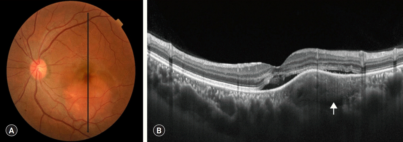
Fig. 1. Fundus photograph and optical coherence tomography of the left eye obtained on the initial visit. (A) An elevated orange-red retina is visible at the posterior pole. (B) Optical coherence tomography reveals a thick choroid, compression of the choriocapillaris, and serous retinal detachment with a choroidal mass (arrow).
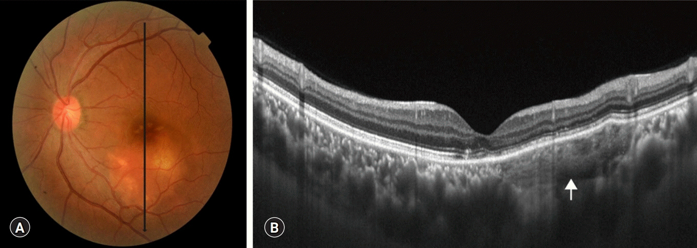
Fig. 2. Fundus photograph and optical coherence tomography of the left eye obtained 1 month after the initial visit. (A) The height of the elevated retina is reduced and patchy yellowish discoloration is seen. (B) The serous retinal detachment is resolved and fine lamellar lines within the choroidal lesion are visible (arrow).
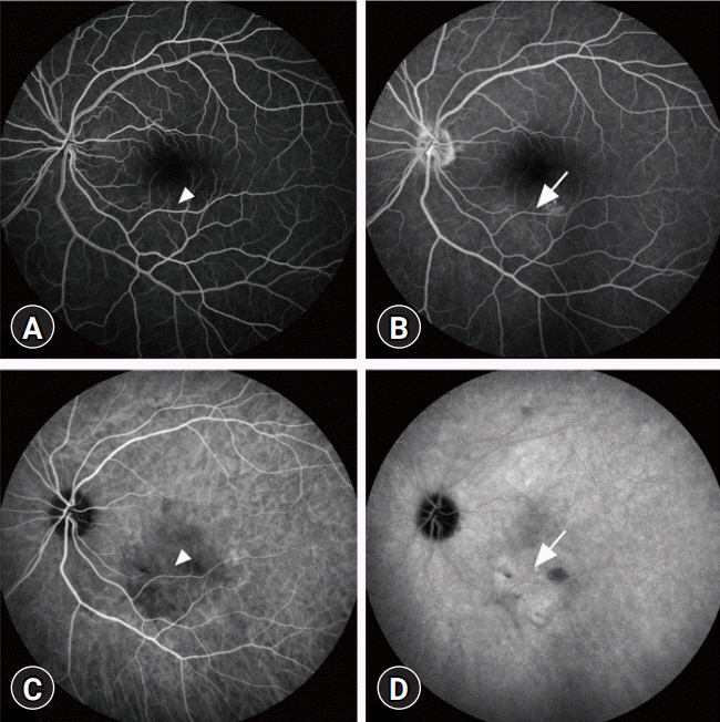
Fig. 3. Fluorescein angiography and indocyanine green angiography of the left eye obtained 1 month after the initial visit. Fluorescein angiography (A, arteriovenous phase; B, late phase) reveals early hyperfluorescence (arrowhead) with late-phase staining (arrow). Indocyanine green angiography (C, early phase; D, late phase) reveals early hypofluorescnece (arrowhead) followed by diffuse confluent fluorescence in the late phase (arrow).
Reference
-
References
1. Thornburg KL, Jacobson SL, Giraud GD, Morton MJ. Hemodynamic changes in pregnancy. Semin Perinatol. 2000; 24:11–4.
Article2. Errera MH, Kohly RP, da Cruz L. Pregnancy-associated retinal diseases and their management. Surv Ophthalmol. 2013; 58:127–42.
Article3. Chatziralli I, Kabanarou SA, Parikakis E, Chatzirallis A, Xirou T, Mitropoulos P. Risk factors for central serous chorioretinopathy: multivariate approach in a case-control study. Curr Eye Res. 2017; 42:1069–73.
Article4. Cohen VM, Rundle PA, Rennie IG. Choroidal hemangiomas with exudative retinal detachments during pregnancy. Arch Ophthalmol. 2002; 120:862–4.5. Sayman Muslubas I, Arf S, Hocaoglu M, Ozdemir H, Karacorlu M. Spontaneous regression of serous retinal detachment associated with circumscribed choroidal hemangioma after childbirth. Retin Cases Brief Rep. 2017; 11:7–11.
Article6. Heimann H, Damato B. Congenital vascular malformations of the retina and choroid. Eye (Lond). 2010; 24:459–67.
Article7. Daruich A, Matet A, Dirani A, Bousquet E, Zhao M, Farman N, et al. Central serous chorioretinopathy: recent findings and new physiopathology hypothesis. Prog Retin Eye Res. 2015; 48:82–118.
Article8. Morikawa M, Cho K, Kojima T, Chiba K, Ishikawa S, Umazume T, et al. Risk factors for central serous chorioretinopathy in pregnant Japanese women. J Obstet Gynaecol Res. 2017; 43:866–72.
Article9. Arevalo JF, Shields CL, Shields JA, Hykin PG, De Potter P. Circumscribed choroidal hemangioma: characteristic features with indocyanine green videoangiography. Ophthalmology. 2000; 107:344–50.
Article10. Rojanaporn D, Kaliki S, Ferenczy SR, Shields CL. Enhanced depth imaging optical coherence tomography of circumscribed choroidal hemangioma in 10 consecutive cases. Middle East Afr J Ophthalmol. 2015; 22:192–7.
Article11. Shields CL, Shields JA, Augsburger JJ. Choroidal osteoma. Surv Ophthalmol. 1988; 33:17–27.
Article12. Gass JD, Guerry RK, Jack RL, Harris G. Choroidal osteoma. Arch Ophthalmol. 1978; 96:428–35.
Article13. Lafaut BA, Mestdagh C, Kohno T, Gaudric A, De Laey JJ. Indocyanine green angiography in choroidal osteoma. Graefes Arch Clin Exp Ophthalmol. 1997; 235:330–7.
Article14. Hussain R, Anantharaman G, Rajesh B, Gopalakrishnan M. Real-time in vivo micromorphology and histopathology of choroidal osteoma using enhanced depth imaging. Indian J Ophthalmol. 2015; 63:453–5.
Article15. Robson SC, Hunter S, Boys RJ, Dunlop W. Serial study of factors influencing changes in cardiac output during human pregnancy. Am J Physiol. 1989; 256(4 Pt 2):H1060–5.
Article
- Full Text Links
-
- Actions
-
Cited
- CITED
-
- Close
- Share
-
- Similar articles
-
- Laser Photocoaculation Treatment in a Case of Circumscribged Choroidal hmangioma Associated with Serous Retinal Detachment
- Takayasu's Arteritis Associated with Serous Retinal Detachment
- Serous Retinal Detachment in Patients with Choroidal Neovascularization Following Photodynamic Therapy
- Transpupillary Thermotherapy in Circumscribed Choroidal Hemangioma
- Effect of Serous Retinal Detachment on the Measurement of Axial Length in Central Serous Chorioretinopathy
