J Korean Ophthalmol Soc.
2007 Oct;48(10):1354-1361. 10.3341/jkos.2007.48.10.1354.
Serous Retinal Detachment in Patients with Choroidal Neovascularization Following Photodynamic Therapy
- Affiliations
-
- 1Department of Ophthalmology, Seoul National University College of Medicine Seoul Artificial Eye Center, Seoul National University Hospital Clinical Research Institute, Seoul, Korea.
- 2Department of Ophthalmology, Seoul National University Boramae Hospital, Seoul, Korea. hjwin@lycos.co.kr
- KMID: 2210989
- DOI: http://doi.org/10.3341/jkos.2007.48.10.1354
Abstract
-
PURPOSE: To quantify the development and resolution of serous retinal detachment in patients with choroidal neovascularization (CNV) following photodynamic therapy (PDT).
METHODS
Six eyes of five patients who developed serous retinal detachment two days after PDT were included in this study. Retinal thickness was measured by optical coherence tomography (OCT) before PDT, and at two days, seven days, and three weeks after PDT. The number of PDT and the greatest linear dimension (GLD) of the CNV lesion were recorded.
RESULTS
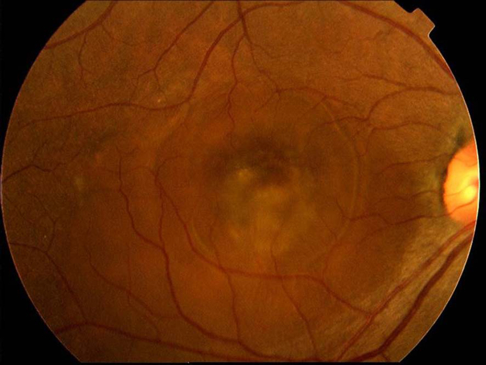
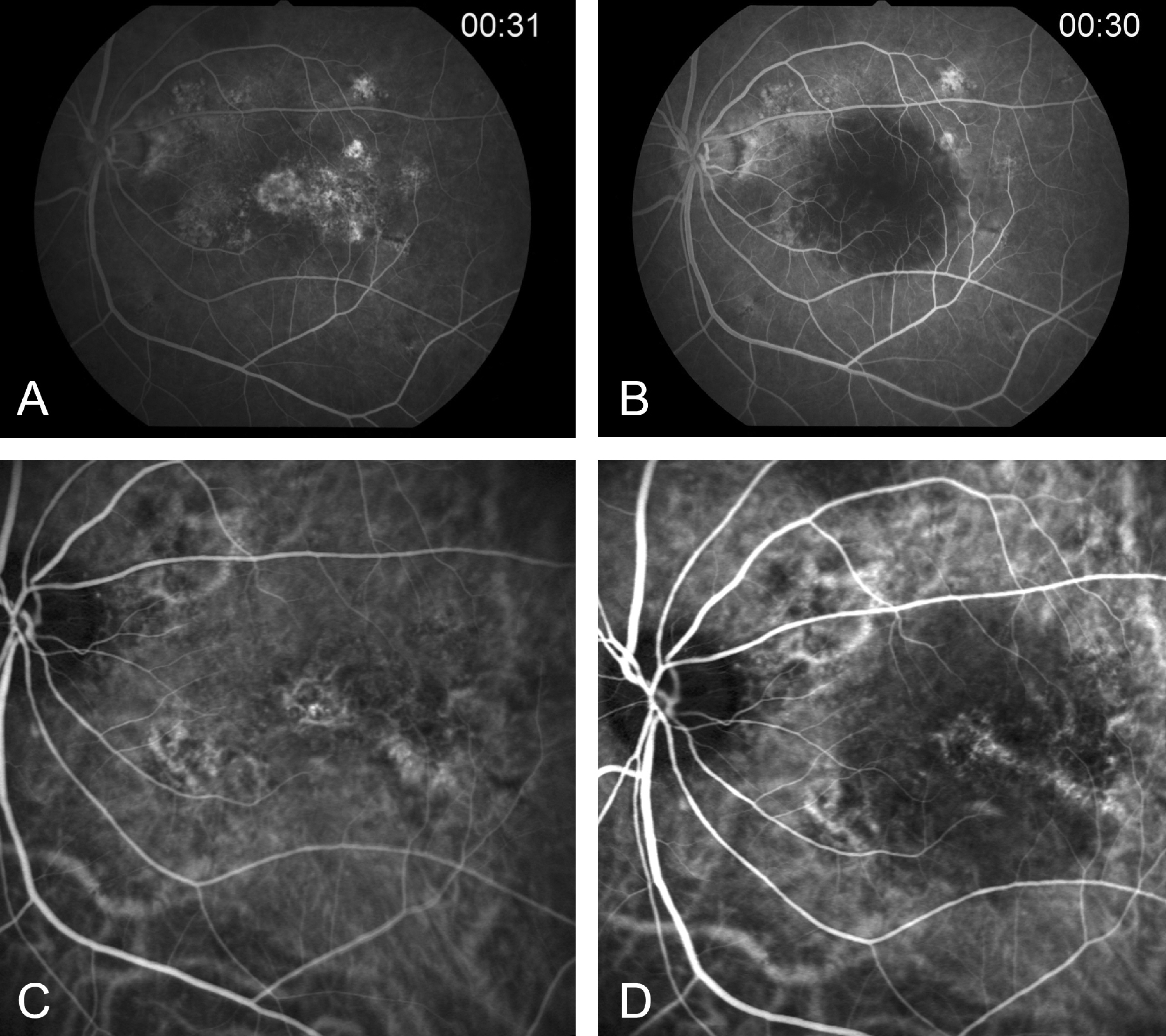
Serous retinal detachment was demonstrated on OCT at two days after PDT. Retinal elevation increased significantly from 313.2 micrometer before PDT to 640.7 micrometer two days after PDT (P<0.01); elevation and decreased to 303.2 micrometer seven days after PDT, and decreased to 223.4 micrometer three weeks after PDT. The mean number of PDT treatments 2.0 (range: 1~3), and the mean GLD before PDT was 3122.8+/-1275.9 micrometer.
CONCLUSIONS
This study suggest that serous retinal detachment in patients with CNV may develop following PDT but may also resolve spontaneously seven days after PDT.
MeSH Terms
Figure
Reference
-
References
1. Bressler NM, Bressler SB, Fine SL. Age-related macular degeneration. Surv Ophthalmol. 1988; 102:374–413.
Article2. Vingerling JR, Dielemans I, Hofman A, et al. The prevalence of age-related maculopathy in the Rotterdam study. Ophthalmology. 1995; 102:205–10.
Article3. Treatment of age-related macular degeneration with photodynamic therapy (TAP) study group. Photodynamic therapy of subfoveal choroidal neovascularization in age-related macular degeneration with verteporfin: One-year results of 2 randomized clinical trials-TAP report 1. Arch Ophthalmol. 1999; 117:1329–45.4. Treatment of age-related macular degeneration with photodynamic therapy (TAP) study group. Photodynamic therapy of subfoveal choroidal neovascularization in age-related macular degeneration with verteporfin: Two-year results of 2 randomized clinical trials-TAP report 2. Arch Ophthalmol. 2001; 119:198–207.5. Verteporfin in photodynamic therapy (VIP) study group. Photodynamic therapy of subfoveal choroidal neovascularization in pathologic myopia with verteporfin: One-year results of a randomized clinical trials-VIP report 1. Ophthalmology. 2001; 108:841–52.6. Verteporfin in photodynamic therapy (VIP) study group. Photodynamic therapy of subfoveal choroidal neovascularization in age-related macular degeneration: Two-year results of a randomized clinical trial including lesions with occult with no classic choroidal neovascularization-VIP report 2. Am J Ophthalmol. 2001; 131:541–60.7. Gelisken F, Inhoffen W, Partsch M, et al. Retinal pigment epithelial tear after photodynamic therapy for choroidal neovascularization. Am J Ophthalmol. 2001; 131:518–20.
Article8. Pece A, Introini U, Bottoni F, Brancato R. Acute retinal pigment epithelial tear after photodynamic therapy. Retina. 2001; 21:661–5.
Article9. Goldstein M, Heilweil G, Barak A, Loewenstein A. Retinal pigment epithelial tear following photodynamic therapy for choroidal neovascularization secondary to AMD. Eye. 2005; 19:1315–24.
Article10. Theodossiadis GP, Panagiotidis D, Georgalas IG, et al. Retinal hemorrhage after photodynamic therapy in patients with subfoveal choroidal neovascularization caused by age-related macular degeneration. Graefes Arch Clin Exp Ophthalmol. 2003; 241:13–8.
Article11. Gelisken F, Inhoffen W, Karim-Zoda K, et al. Subfoveal hemorrhage after verteporfin photodynamic therapy in treatment of choroidal neovascularization. Graefes Arch Clin Exp Ophthalmol. 2005; 243:198–203.
Article12. Fingar V, Kik P, Haydon P, et al. Analysis of acute vascular damage after photodynamic therapy using benzoporphyrin derivative (BPD). Br J Cancer. 1999; 79:1702–8.
Article13. Fogelman AM, Berliner JA, Van Lenten BJ, et al. Lipoprotein receptors and endothelial cells. Semin Thromb Hemost. 1988; 14:206–9.
Article14. Michels S, Schmidt-Erfurth U. Sequence of early vascular events after photodynamic therapy. Invest Ophthalmol Vis Sci. 2003; 44:2147–54.
Article15. Costa RA, Farah ME, Cardillo JA, et al. Immediate indocyanine green angiography and optical coherence tomography evaluation after photodynamic therapy for subfoveal choroidal neovascularization. Retina. 2003; 23:159–65.
Article16. Mennel S, Meyer CH, Eggarter F, et al. Transient serous retinal detachment in classic and occult choroidal neovascularization after photodynamic therapy. Am J Ophthalmol. 2005; 140:758–60.
Article17. Mennel S, Hausmann N, Meyer CH, et al. Transient visual decrease after photodynamic therapy. Ophthalmologe. 2005; 102:58–63.18. Rogers AH, Martidis A, Greenberg PB, Puliafito CA. Optical coherence tomography findings following photodynamic therapy of choroidal neovascularization. Am J Ophthalmol. 2002; 134:566–76.
Article19. Schmidt-Erfurth U, Hasan T, Gragoudas E, et al. Vascular targeting in photodynamic occlusion of subretinal vessels. Ophthalmology. 1994; 101:1953–61.
Article20. Schnurrbusch UEK, Welt K, Horn LC, et al. Histological findings of surgically excised choroidal neovascular membranes after photodynamic therapy. Br J Ophthalmol. 2001; 85:1086–91.
Article
- Full Text Links
- Actions
-
Cited
- CITED
-
- Close
- Share
- Similar articles
-
- Serous Retinal Detachment Following Combined Photodynamic Therapy and Intravitreal Bevacizumab Injection
- Photodynamic Therapy of Choroidal Neovasculariation Associated with Large Serous Pigment Epithelial Detachment
- Laser Photocoaculation Treatment in a Case of Circumscribged Choroidal hmangioma Associated with Serous Retinal Detachment
- Takayasu's Arteritis Associated with Serous Retinal Detachment
- The Result of Photodynamic Therapy in Chronic Central Serous Chorioretinopathy


