J Korean Foot Ankle Soc.
2020 Jun;24(2):75-80. 10.14193/jkfas.2020.24.2.75.
Results of Kidner Procedure Combined with Medial Displacement Calcaneal Osteotomy for the Symptomatic Accessory Navicular with Hindfoot Valgus
- Affiliations
-
- 1Department of Orthopedic Surgery, Yeungnam University Medical Center, Daegu, Korea
- KMID: 2502913
- DOI: http://doi.org/10.14193/jkfas.2020.24.2.75
Abstract
- Purpose
The purpose of this study is to evaluate the results of Kidner procedure combined with medial displacement calcaneal osteotomy (MDCO) in patients with the symptomatic accessory navicular with hindfoot valgus.
Materials and Methods
From January 2014 to January 2019, fifteen patients (15 cases) who had undergone a Kidner procedure combined with MDCO for symptomatic accessory navicular with hindfoot valgus were included. Their mean age was 36.3 years old (19∼61 years old) and there were 6 males and 9 females. The clinical results were evaluated using visual analogue scale (VAS), American Orthopaedic Foot and Ankle Society (AOFAS) midfoot score, and postoperative subjective satisfaction. The radiographic results were evaluated using the talonavicular coverage angle and the anteroposterior talo-first metatarsal angle, the lateral talo-first metatarsal angle, the calcaneal pitch angle, and the hindfoot alignment angle. The postoperative complications were also evaluated.
Results
The VAS and AOFAS midfoot scores continuously improved until 12 months after surgery. Subjective satisfaction after surgery was excellent in 10 cases and good in 5 cases. The hindfoot alignment angle significantly changed after surgery. Pain due to lateral impingement disappeared in five patients, and persisted in one patient. Five patients complained of irritation caused by their fixation devices, and all the symptoms improved after removal of the fixation devices.
Conclusion
Kidner procedure combined with MDCO in patients with the symptomatic accessory navicular with hindfoot valgus showed good clinical results with satisfactory correction of hindfoot valgus. In particular, the clinical results showed continuous improvement until 12 months after surgery.
Figure
-
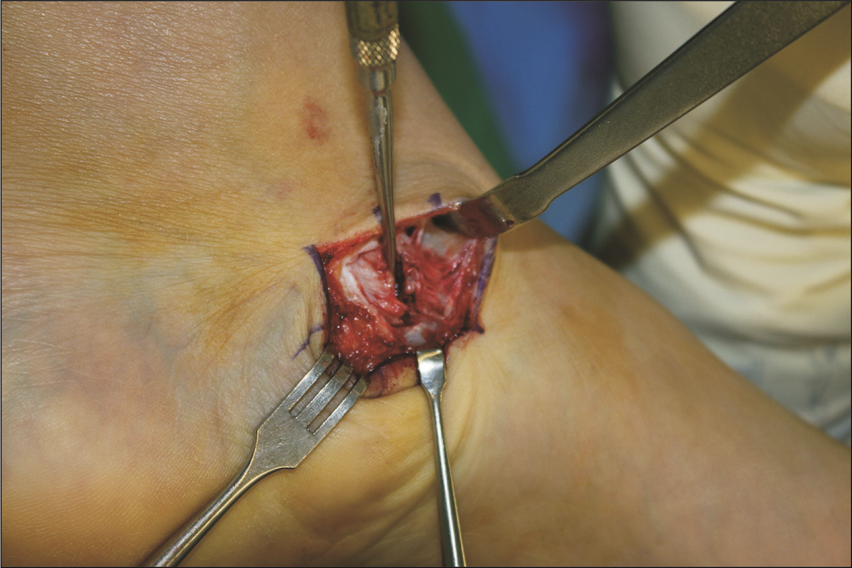
Figure. 1 Intraoperative photograph showed detaching the accessory navicular from the navicular after dissection of periosteum.
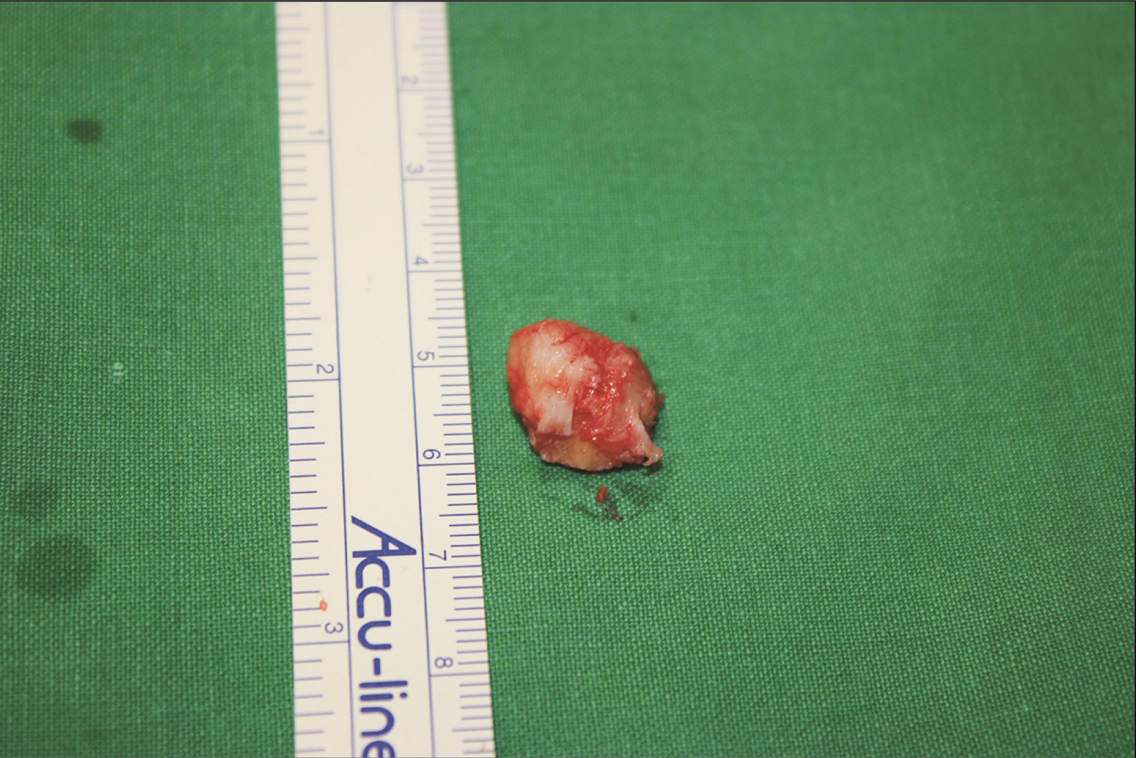
Figure. 2 Intraoperative photograph showed detached accessory navicular.
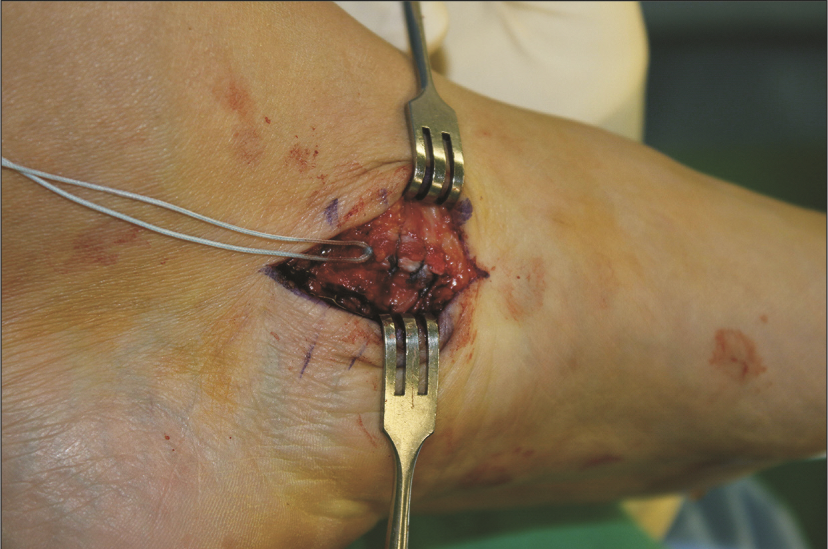
Figure. 3 Intraoperative photograph showed reattaching the tibialis posterior tendon on its original insertion using the suture anchor.
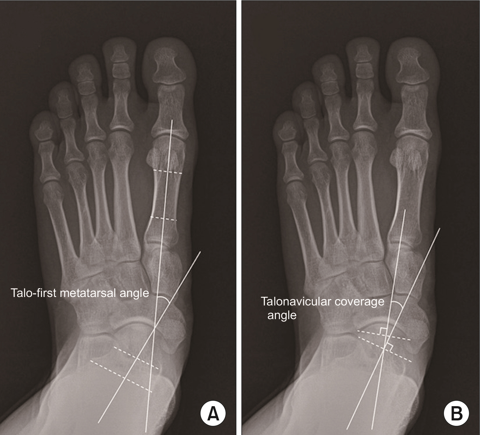
Figure. 4 The anteroposterior weight-bearing radiograph of the foot showed the anterior talo-first metatarsal angle (A) and the talonavicular coverage angle (B).
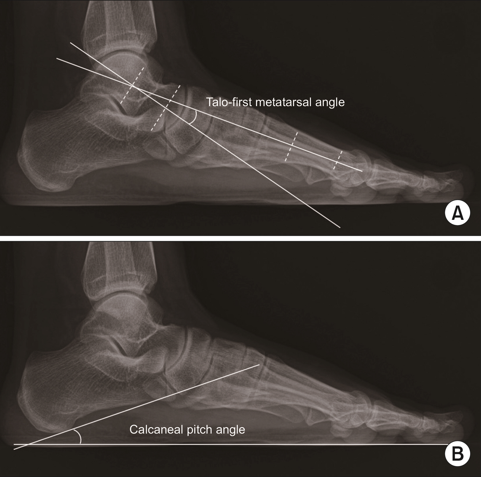
Figure. 5 The lateral weight-bearing radiograph of the foot showed the lateral talo-first metatarsal angle (A) and the calcaneal pitch angle (B).
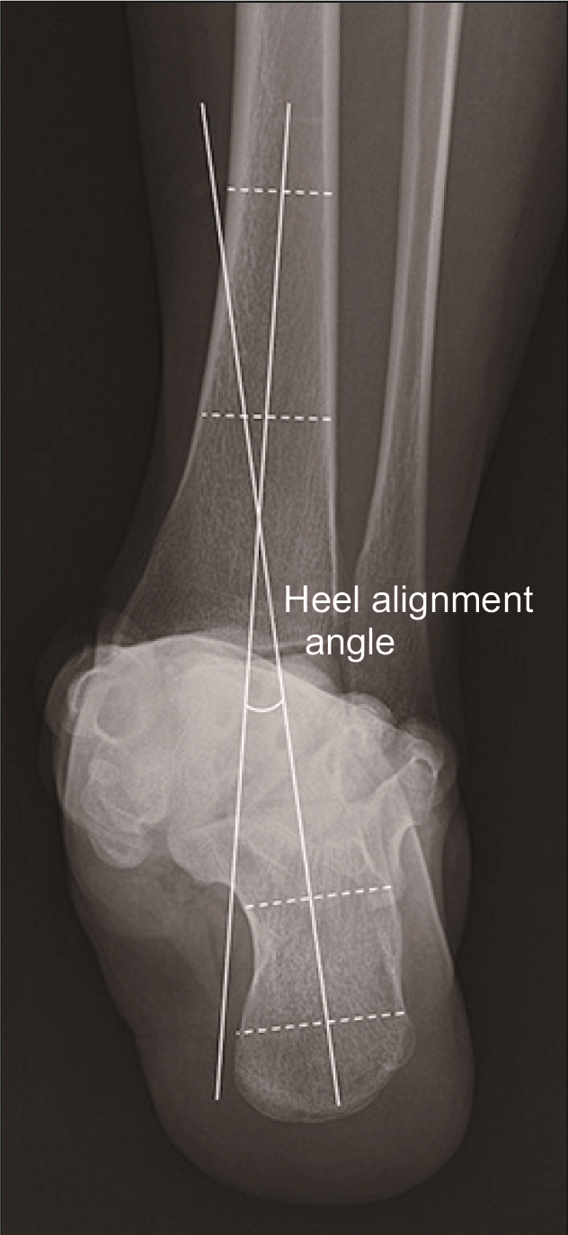
Figure. 6 The hindfoot alignment radiograph showed the hindfoot alignment angle.
Reference
-
1. Ugolini PA, Raikin SM. 2004; The accessory navicular. Foot Ankle Clin. 9:165–80. doi: 10.1016/S1083-7515(03)00176-1. DOI: 10.1016/S1083-7515(03)00176-1. PMID: 15062220.
Article2. Jasiewicz B, Potaczek T, Kacki W, Tesiorowski M, Lipik E. 2008; Results of simple excision technique in the surgical treatment of symptomatic accessory navicular bones. Foot Ankle Surg. 14:57–61. doi: 10.1016/j.fas.2007.12.002. DOI: 10.1016/j.fas.2007.12.002. PMID: 19083616.
Article3. Kidner FC. 1929; The prehallux (accessory scaphoid) in its relation to flat-foot. J Bone Joint Surg Am. 11:831–7.4. Nakayama S, Sugimoto K, Takakura Y, Tanaka Y, Kasanami R. 2005; Percutaneous drilling of symptomatic accessory navicular in young athletes. Am J Sports Med. 33:531–5. doi: 10.1177/0363546504270564. DOI: 10.1177/0363546504270564. PMID: 15722276.
Article5. Chung JW, Chu IT. 2009; Outcome of fusion of a painful accessory navicular to the primary navicular. Foot Ankle Int. 30:106–9. doi: 10.3113/FAI.2009.0106. DOI: 10.3113/FAI.2009.0106. PMID: 19254502.
Article6. Hiller L, Pinney SJ. 2003; Surgical treatment of acquired flatfoot deformity: what is the state of practice among academic foot and ankle surgeons in 2002? Foot Ankle Int. 24:701–5. doi: 10.1177/107110070302400909. DOI: 10.1177/107110070302400909. PMID: 14524521.
Article7. Leonard ZC, Fortin PT. 2010; Adolescent accessory navicular. Foot Ankle Clin. 15:337–47. doi: 10.1016/j.fcl.2010.02.004. DOI: 10.1016/j.fcl.2010.02.004. PMID: 20534360.
Article8. Choi HJ, Lee WC. 2017; Revision surgery for recurrent pain after excision of the accessory navicular and relocation of the tibialis posterior tendon. Clin Orthop Surg. 9:232–8. doi: 10.4055/cios.2017.9.2.232. DOI: 10.4055/cios.2017.9.2.232. PMID: 28567228. PMCID: PMC5435664.
Article9. Vaughan P, Singh D. 2014; Ongoing pain and deformity after an excision of the accessory navicular. Foot Ankle Clin. 19:541–53. doi: 10.1016/j.fcl.2014.06.010. DOI: 10.1016/j.fcl.2014.06.010. PMID: 25129360.
Article10. Flemister AS, Neville CG, Houck J. 2007; The relationship between ankle, hindfoot, and forefoot position and posterior tibial muscle excursion. Foot Ankle Int. 28:448–55. doi: 10.3113/FAI.2007.0448. DOI: 10.3113/FAI.2007.0448. PMID: 17475139.
Article11. Weinfeld SB. 2001; Medial slide calcaneal osteotomy. Technique, patient selection, and results. Foot Ankle Clin. 6:89–94. viidoi: 10.1016/s1083-7515(03)00081-0. DOI: 10.1016/S1083-7515(03)00081-0. PMID: 11385930.
Article12. Arangio GA, Salathé EP. 2001; Medial displacement calcaneal osteotomy reduces the excess forces in the medial longitudinal arch of the flat foot. Clin Biomech (Bristol, Avon). 16:535–9. doi: 10.1016/s0268-0033(01)00011-0. DOI: 10.1016/S0268-0033(01)00011-0. PMID: 11427297.
Article13. Catanzariti AR, Lee MS, Mendicino RW. 2000; Posterior calcaneal displacement osteotomy for adult acquired flatfoot. J Foot Ankle Surg. 39:2–14. doi: 10.1016/s1067-2516(00)80058-7. DOI: 10.1016/S1067-2516(00)80058-7. PMID: 10658945.
Article14. Lawson JP, Ogden JA, Sella E, Barwick KW. 1984; The painful accessory navicular. Skeletal Radiol. 12:250–62. doi: 10.1007/bf00349506. DOI: 10.1007/BF00349506. PMID: 6239377.
Article15. Sella EJ, Lawson JP, Ogden JA. 1986; The accessory navicular synchondrosis. Clin Orthop Relat Res. (209):280–5. DOI: 10.1097/00003086-198608000-00042. PMID: 3731610.
Article16. Garras DN, Hansen PL, Miller AG, Raikin SM. 2012; Outcome of modified Kidner procedure with subtalar arthroereisis for painful accessory navicular associated with planovalgus deformity. Foot Ankle Int. 33:934–9. doi: 10.3113/FAI.2012.0934. DOI: 10.3113/FAI.2012.0934. PMID: 23131438.
Article17. Kanatli U, Yetkin H, Yalcin N. 2003; The relationship between accessory navicular and medial longitudinal arch: evaluation with a plantar pressure distribution measurement system. Foot Ankle Int. 24:486–9. doi: 10.1177/107110070302400606. DOI: 10.1177/107110070302400606. PMID: 12854669.
Article18. Prichasuk S, Sinphurmsukskul O. 1995; Kidner procedure for symptomatic accessory navicular and its relation to pes planus. Foot Ankle Int. 16:500–3. doi: 10.1177/107110079501600807. DOI: 10.1177/107110079501600807. PMID: 8520663.
Article19. Wood WA, Spencer AM. 1970; Incidence of os tibiale externum in clinical pes planus. J Am Podiatry Assoc. 60:276–9. doi: 10.7547/87507315-60-7-276. DOI: 10.7547/87507315-60-7-276. PMID: 5505626.
Article20. Zadek I. 1926; The significance of the accessory tarsal scaphoid. J Bone Joint Surg Am. 8:618–26.21. Veitch JM. 1978; Evaluation of the Kidner procedure in treatment of symptomatic accessory tarsal scaphoid. Clin Orthop Relat Res. (131):210–3. DOI: 10.1097/00003086-197803000-00033. PMID: 657625.
Article22. Ray S, Goldberg VM. 1983; Surgical treatment of the accessory navicular. Clin Orthop Relat Res. (177):61–6. DOI: 10.1097/00003086-198307000-00010. PMID: 6861408.
Article23. Kopp FJ, Marcus RE. 2004; Clinical outcome of surgical treatment of the symptomatic accessory navicular. Foot Ankle Int. 25:27–30. doi: 10.1177/107110070402500106. DOI: 10.1177/107110070402500106. PMID: 14768961.
Article24. Malicky ES, Levine DS, Sangeorzan BJ. 1999; Modification of the Kidner procedure with fusion of the primary and accessory navicular bones. Foot Ankle Int. 20:53–4. doi: 10.1177/107110079902000112. DOI: 10.1177/107110079902000112. PMID: 9921775.
Article25. Cao HH, Tang KL, Lu WZ, Xu JZ. 2014; Medial displacement calcaneal osteotomy with posterior tibial tendon reconstruction for the flexible flatfoot with symptomatic accessory navicular. J Foot Ankle Surg. 53:539–43. doi: 10.1053/j.jfas.2014.04.004. DOI: 10.1053/j.jfas.2014.04.004. PMID: 24856662.
Article26. Park MJ, Lee MC, Seong SC. 2001; A comparative study of the healing of tendon autograft and tendon-bone autograft using patellar tendon in rabbits. Int Orthop. 25:35–9. doi: 10.1007/s002640000199. DOI: 10.1007/s002640000199. PMID: 11374265. PMCID: PMC3620615.
Article27. Tomita F, Yasuda K, Mikami S, Sakai T, Yamazaki S, Tohyama H. 2001; Comparisons of intraosseous graft healing between the doubled flexor tendon graft and the bone-patellar tendon-bone graft in anterior cruciate ligament reconstruction. Arthroscopy. 17:461–76. doi: 10.1053/jars.2001.24059. DOI: 10.1053/jars.2001.24059. PMID: 11337712.
Article
- Full Text Links
-
- Actions
-
Cited
- CITED
-
- Close
- Share
-
- Similar articles
-
- Painful Accessory Navicular
- Revision Surgery for Recurrent Pain after Excision of the Accessory Navicular and Relocation of the Tibialis Posterior Tendon
- Short-Term Results of a Modified Kidner Procedure Using a Suture Bridge Technique for Symptomatic Type II Accessory Navicular
- Surgical Treatment of Symptomatic Accessory Navicular in Adolescent
- Radiographic Characteristics and the Clinical Results of the Operative Treatment of Muller-Weiss Disease
