Clin Orthop Surg.
2017 Jun;9(2):232-238. 10.4055/cios.2017.9.2.232.
Revision Surgery for Recurrent Pain after Excision of the Accessory Navicular and Relocation of the Tibialis Posterior Tendon
- Affiliations
-
- 1Department of Orthopedic Surgery, Inje University Haeundae Paik Hospital, Busan, Korea. cool-cool0829@hanmail.net
- 2Department of Orthopedic Surgery, Institute for Research of Foot and Ankle Diseases, Inje University Seoul Paik Hospital, Seoul, Korea.
- KMID: 2412294
- DOI: http://doi.org/10.4055/cios.2017.9.2.232
Abstract
- BACKGROUND
The results of operative treatments for symptomatic accessory navicular are debatable. In some cases, recurrent pain may develop after the Kidner procedure. The purpose of this study is to review the reasons for recurrent pain after the Kidner procedure and to suggest possible options for revision surgery.
METHODS
We reviewed the clinical and radiological outcomes in 9 patients who underwent revision surgery for recurrent pain after the Kidner procedure. During the revision surgery, the tibialis posterior tendon was reattached to the navicular either by advancing the tendon in 4 patients or by lengthening the tendon in another 4 patients. In the other 1 patient, the flexor digitorum longus tendon was transferred. Surgeries for the accompanying deformities were performed simultaneously in all patients. The results were evaluated using the American Orthopaedic Foot and Ankle Society ankle-hindfoot score and a visual analog scale. The mean follow-up was 2.3 years (range, 1 to 5 years).
RESULTS
The mean American Orthopedic Foot and Ankle Society ankle-hindfoot score improved from 71.25 to 81.50 in the advancement group, and 71.75 to 90.00 in the lengthening group. The mean visual analog scale improved from 7.75 to 4.25 in the advancement group and from 7.50 to 1.75 in the lengthening group.
CONCLUSIONS
Recurrent pain after the Kidner procedure was associated with pes planovalgus or hindfoot valgus deformity. In revision surgery, correction of the associated deformities and reattachment of the tibialis posterior tendon after lengthening may need to be considered.
MeSH Terms
-
Adolescent
Adult
Female
Flatfoot
Foot/surgery
Foot Diseases/*surgery
Humans
Male
Middle Aged
Osteotomy/*adverse effects
Pain Measurement
*Pain, Postoperative/epidemiology/surgery
*Reoperation/adverse effects/methods
Retrospective Studies
Tarsal Bones/*abnormalities/surgery
*Tendon Transfer/adverse effects/methods
Tendons/surgery
Young Adult
Figure
-
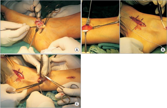
Fig. 1 (A) The intraoperative finding revealed degeneration of the tibialis posterior tendon. (B) To lengthen the tibialis posterior tendon, a 6-cm longitudinal incision was made on the posterior aspect of the medial border of the tibia. (C) The tibialis posterior tendon was lengthened by 2 cm and repaired with interrupted 3-0 nylon sutures and the distal end of the tibialis posterior tendon was securely fixed using 2.7-mm suture anchors.
Cited by 1 articles
-
Results of Kidner Procedure Combined with Medial Displacement Calcaneal Osteotomy for the Symptomatic Accessory Navicular with Hindfoot Valgus
Chul Hyun Park
J Korean Foot Ankle Soc. 2020;24(2):75-80. doi: 10.14193/jkfas.2020.24.2.75.
Reference
-
1. Kiter E, Gunal I, Karatosun V, Korman E. The relationship between the tibialis posterior tendon and the accessory navicular. Ann Anat. 2000; 182(1):65–68. PMID: 10668560.
Article2. Grogan DP, Gasser SI, Ogden JA. The painful accessory navicular: a clinical and histopathological study. Foot Ankle. 1989; 10(3):164–169. PMID: 2613130.
Article3. Bennett GL, Weiner DS, Leighley B. Surgical treatment of symptomatic accessory tarsal navicular. J Pediatr Orthop. 1990; 10(4):445–449. PMID: 2358479.
Article4. Chung JW, Chu IT. Outcome of fusion of a painful accessory navicular to the primary navicular. Foot Ankle Int. 2009; 30(2):106–109. PMID: 19254502.
Article5. Kiter E, Gunal I, Turgut A, Kose N. Evaluation of simple excision in the treatment of symptomatic accessory navicular associated with flat feet. J Orthop Sci. 2000; 5(4):333–335. PMID: 10982680.
Article6. Kopp FJ, Marcus RE. Clinical outcome of surgical treatment of the symptomatic accessory navicular. Foot Ankle Int. 2004; 25(1):27–30. PMID: 14768961.
Article7. Macnicol MF, Voutsinas S. Surgical treatment of the symptomatic accessory navicular. J Bone Joint Surg Br. 1984; 66(2):218–226. PMID: 6707058.
Article8. Sullivan JA, Miller WA. The relationship of the accessory navicular to the development of the flat foot. Clin Orthop Relat Res. 1979; (144):233–237. PMID: 535230.
Article9. Nakayama S, Sugimoto K, Takakura Y, Tanaka Y, Kasanami R. Percutaneous drilling of symptomatic accessory navicular in young athletes. Am J Sports Med. 2005; 33(4):531–535. PMID: 15722276.
Article10. Malicky ES, Levine DS, Sangeorzan BJ. Modification of the Kidner procedure with fusion of the primary and accessory navicular bones. Foot Ankle Int. 1999; 20(1):53–54. PMID: 9921775.
Article11. Scott AT, Sabesan VJ, Saluta JR, Wilson MA, Easley ME. Fusion versus excision of the symptomatic Type II accessory navicular: a prospective study. Foot Ankle Int. 2009; 30(1):10–15. PMID: 19176179.
Article12. Kitaoka HB, Alexander IJ, Adelaar RS, Nunley JA, Myerson MS, Sanders M. Clinical rating systems for the anklehindfoot, midfoot, hallux, and lesser toes. Foot Ankle Int. 1994; 15(7):349–353. PMID: 7951968.
Article13. Ibrahim T, Beiri A, Azzabi M, Best AJ, Taylor GJ, Menon DK. Reliability and validity of the subjective component of the American Orthopaedic Foot and Ankle Society clinical rating scales. J Foot Ankle Surg. 2007; 46(2):65–74. PMID: 17331864.
Article14. Lee WC, Moon JS, Lee HS, Lee K. Alignment of ankle and hindfoot in early stage ankle osteoarthritis. Foot Ankle Int. 2011; 32(7):693–699. PMID: 21972764.
Article15. Ray S, Goldberg VM. Surgical treatment of the accessory navicular. Clin Orthop Relat Res. 1983; (177):61–66. PMID: 6861408.
Article16. Veitch JM. Evaluation of the Kidner procedure in treatment of symptomatic accessory tarsal scaphoid. Clin Orthop Relat Res. 1978; (131):210–213.
Article17. Sella EJ, Lawson JP, Ogden JA. The accessory navicular synchondrosis. Clin Orthop Relat Res. 1986; (209):280–285. PMID: 3731610.
Article18. Vaughan P, Singh D. Ongoing pain and deformity after an excision of the accessory navicular. Foot Ankle Clin. 2014; 19(3):541–553. PMID: 25129360.
Article
- Full Text Links
-
- Actions
-
Cited
- CITED
-
- Close
- Share
-
- Similar articles
-
- Surgical Treatment of Symptomatic Accessory Navicular in Adolescent
- Painful Accessory Navicular
- Accessory Navicular in Sports Players
- Morphological variations and accessory ossicles in the peroneal and tibialis muscles
- A Irreducible Ankle Fracture and Dislocation Due to Injured Tibialis Posterior Tendon Interposition: A Case Report
