Unilateral maxillary central incisor root resorption after orthodontic treatment for Angle Class II, division 1 malocclusion with significant maxillary midline deviation: A possible correlation with root proximity to the incisive canal
- Affiliations
-
- 1Department of Orthodontic Science, Graduate School of Medical and Dental Sciences, Tokyo Medical and Dental University, Tokyo, Japan
- KMID: 2502631
- DOI: http://doi.org/10.4041/kjod.2020.50.3.216
Abstract
- Root resorption can be caused by several factors, including contact with the cortical bone. Here we report a case involving a 21-year-old female with Angle Class II, division 1 malocclusion who exhibited significant root resorption in the maxillary right central incisor after orthodontic treatment. The patient presented with significant left-sided deviation of the maxillary incisors due to lingual dislocation of the left lateral incisor and a Class II molar relationship. Cephalometric analysis demonstrated a Class I skeletal relationship (A pointnasion- B point, 2.5o) and proclined maxillary anterior teeth (upper incisor to sella-nasion plane angle, 113.4o). The primary treatment objectives were the achievement of stable occlusion with midline agreement between the maxillary and mandibular dentitions and appropriate maxillary anterior tooth axes and molar relationship. A panoramic radiograph obtained after active treatment showed significant root resorption in the maxillary right central incisor; therefore, we performed cone-beam computed tomography, which confirmed root resorption along the cortical bone around the incisive canal. The findings from this case, where different degrees of root resorption were observed despite comparable degrees of orthodontic movement in the bilateral maxillary central incisors, suggest that the incisive canal could be an inducing factor for root
Figure
-
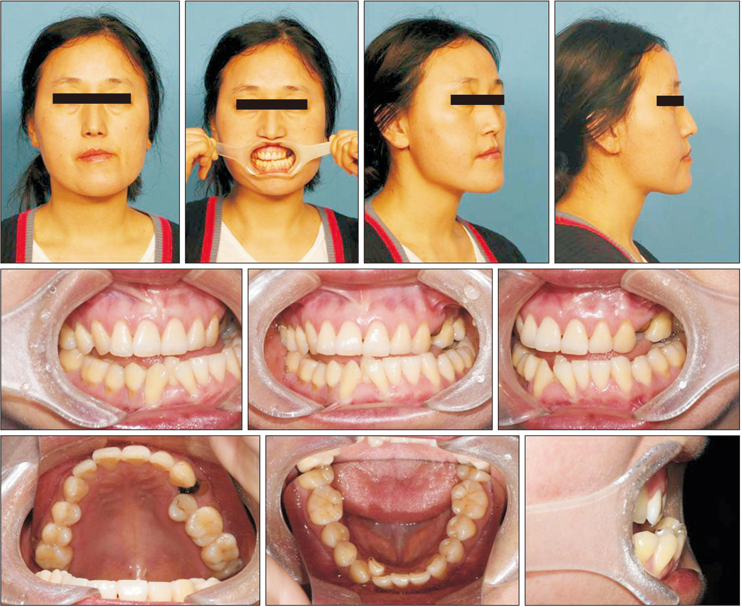
Figure 1 Pretreatment facial and intraoral photographs.
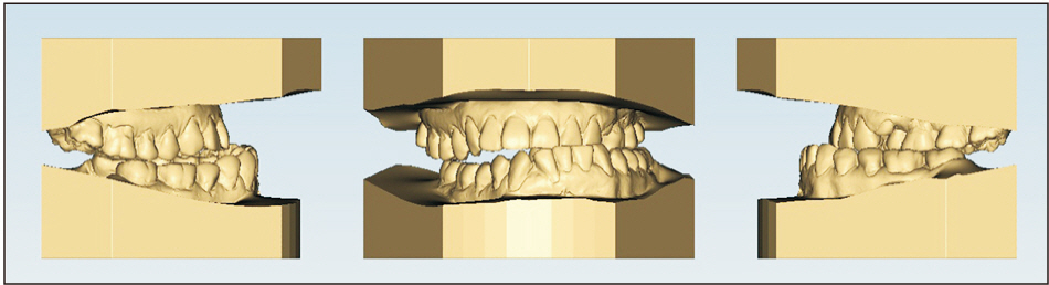
Figure 2 Pretreatment dental casts.
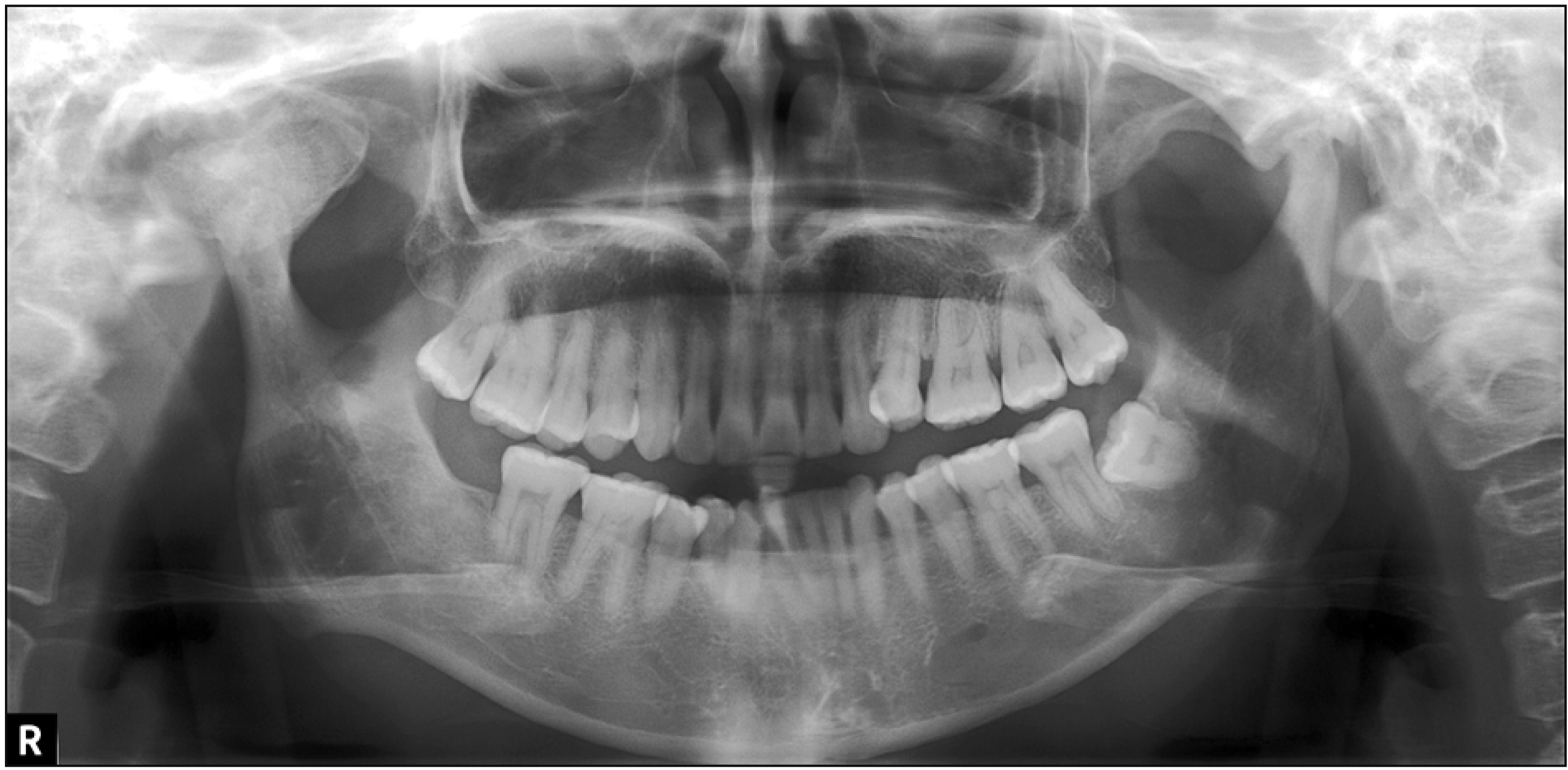
Figure 3 Pretreatment radiographs. A, Lateral cephalogram. B, Posteroanterior cephalogram. C, Panoramic radiograph.
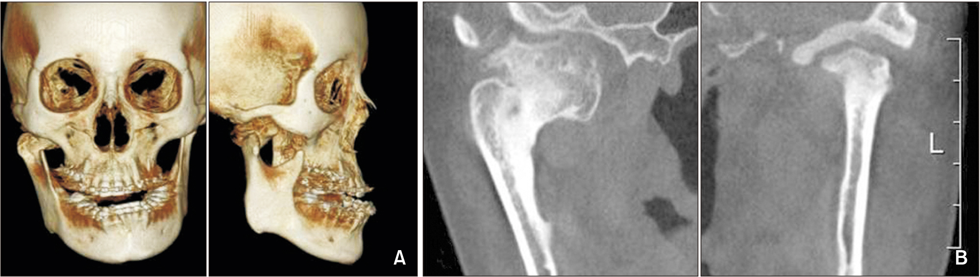
Figure 4 Pretreatment cone-beam computed tomography images. A, Transverse image at the apex level of the maxillary incisor roots. B, Parasagittal image of the maxillary right central incisor. C, Parasagittal image of the maxillary left central incisor.
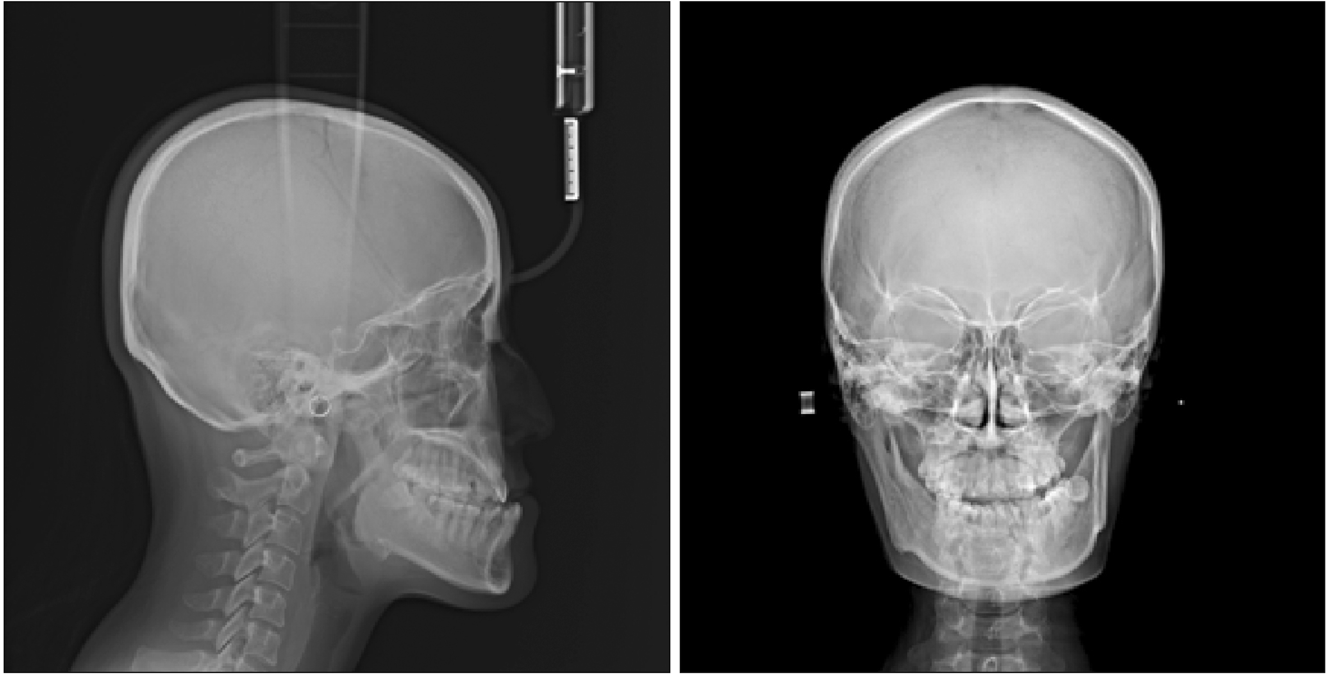
Figure 5 Treatment progress. A, After 1 month of active treatment. B, After 9 months of active treatment. C, After 13 months of active treatment.
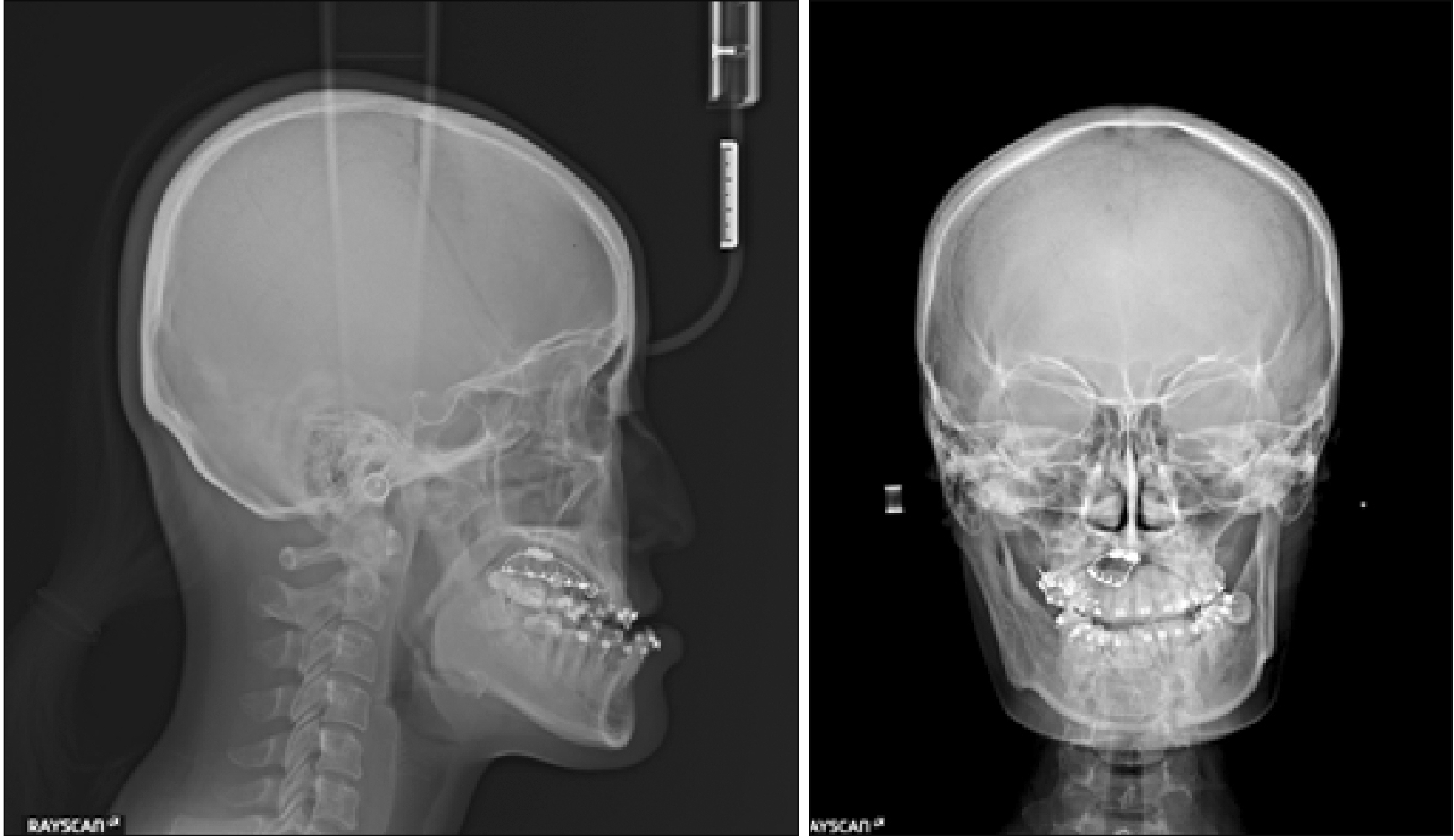
Figure 6 Post-treatment facial and intraoral photographs.
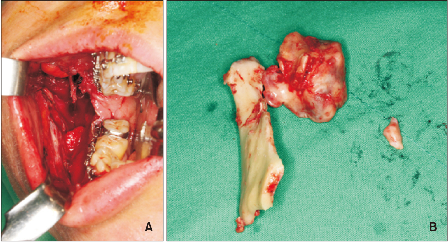
Figure 7 Post-treatment dental casts.
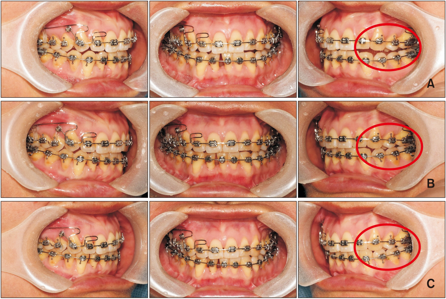
Figure 8 Post-treatment radiographs. A, Lateral cephalogram. B, Posteroanterior cephalogram. C, Panoramic radiograph.

Figure 9 Superimposed tracings of pre- (black line) and post-treatment (red line) lateral cephalograms. A, Superimposition on the sella-nasion plane at the sella. B, Superimposition on the palatal plane at anterior nasal spine. C, Superimposition on the mandibular plane at the menton.

Figure 10 Angle between the maxillary incisor midline (i.e., the line connecting the midpoints of the crowns and root apices of the left and right maxillary central incisors) and the facial midline. A, Before orthodontic treatment. B, After orthodontic treatment.
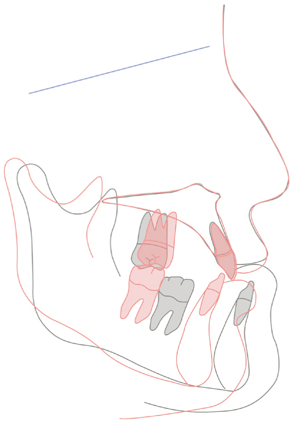
Figure 11 Post-treatment cone-beam computed tomography images. A, Transverse image at the apical level of the maxillary incisor roots. B, Parasagittal image of the maxillary right central incisor. C, Parasagittal image of the maxillary left central incisor.

Figure 12 Superimposed three-dimensional models of pre- and post-treatment maxillary central incisors. A, Right. B, Left. Light blue, Post-treatment maxillary left central incisor; light green, post-treatment maxillary right central incisor; gray, pre-treatment incisors (left and right).
Figure 13 Amount of displacement of the maxillary incisors during active treatment. A color scale is provided to indicate the amount of displacement. A, Frontal view. B, Posterior view. C, Right view. D, Left view.
Figure 14 Post-retention facial and intraoral photographs.
Cited by 1 articles
-
Combined anterior and posterior miniscrews increase apical root resorption of maxillary incisors in protrusion and premolar extraction cases
Zhizun Wang, Li Mei, Zhenxing Tang, Dong Wu, Yue Zhou, Ehab A. Abdulghani, Yuan Li, Wei Zheng, Yu Li
Korean J Orthod. 2025;55(1):26-36. doi: 10.4041/kjod24.136.
Reference
-
1. Weltman B, Vig KW, Fields HW, Shanker S, Kaizar EE. 2010; Root resorption associated with orthodontic tooth movement: a systematic review. Am J Orthod Dentofacial Orthop. 137:462–76. discussion 12ADOI: 10.1016/j.ajodo.2009.06.021. PMID: 20362905.
Article2. Li F, Hu HK, Chen JW, Liu ZP, Li GF, He SS, et al. 2011; Comparison of anchorage capacity between implant and headgear during anterior segment retraction: a systematic review. Angle Orthod. 81:915–22. DOI: 10.2319/101410-603.1. PMID: 21299412.3. Otis LL, Hong JS, Tuncay OC. 2004; Bone structure effect on root resorption. Orthod Craniofac Res. 7:165–77. DOI: 10.1111/j.1601-6343.2004.00282.x. PMID: 15359503.
Article4. Horiuchi A, Hotokezaka H, Kobayashi K. 1998; Correlation between cortical plate proximity and apical root resorption. Am J Orthod Dentofacial Orthop. 114:311–8. DOI: 10.1016/S0889-5406(98)70214-8. PMID: 9743137.
Article5. Chung CJ, Choi YJ, Kim KH. 2015; Approximation and contact of the maxillary central incisor roots with the incisive canal after maximum retraction with temporary anchorage devices: report of 2 patients. Am J Orthod Dentofacial Orthop. 148:493–502. DOI: 10.1016/j.ajodo.2015.04.033. PMID: 26321347.
Article6. Cho EA, Kim SJ, Choi YJ, Kim KH, Chung CJ. 2016; Morphologic evaluation of the incisive canal and its proximity to the maxillary central incisors using computed tomography images. Angle Orthod. 86:571–6. DOI: 10.2319/063015-433.1. PMID: 26624251.
Article7. Roscoe MG, Meira JB, Cattaneo PM. 2015; Association of orthodontic force system and root resorption: a systematic review. Am J Orthod Dentofacial Orthop. 147:610–26. DOI: 10.1016/j.ajodo.2014.12.026. PMID: 25919107.
Article8. Fox N. 2005; Longer orthodontic treatment may result in greater external apical root resorption. Evid Based Dent. 6:21. DOI: 10.1038/sj.ebd.6400304. PMID: 15789049.
Article9. Iizuka T, Ishikawa F. 1958; Normal standards for various cephalometric analysis in Japanese adult. J Stomatol Soc. 25:260–72. DOI: 10.5357/koubyou.25.260.10. Park JH, Tai K, Owtad P. 2015; 3-Dimensional cone-beam computed tomography superimposition: a review. Semin Orthod. 21:263–73. DOI: 10.1053/j.sodo.2015.07.007.
Article11. Segal GR, Schiffman PH, Tuncay OC. 2004; Meta analysis of the treatment-related factors of external apical root resorption. Orthod Craniofac Res. 7:71–8. DOI: 10.1111/j.1601-6343.2004.00286.x. PMID: 15180086.
Article12. Owman-Moll P, Kurol J. 2000; Root resorption after orthodontic treatment in high- and low-risk patients: analysis of allergy as a possible predisposing factor. Eur J Orthod. 22:657–63. DOI: 10.1093/ejo/22.6.657. PMID: 11212601.13. Nieto-Nieto N, Solano JE, Yañez-Vico R. 2017; External apical root resorption concurrent with orthodontic forces: the genetic influence. Acta Odontol Scand. 75:280–7. DOI: 10.1080/00016357.2017.1294260. PMID: 28358285.
Article14. Baumrind S, Korn EL, Boyd RL. 1996; Apical root resorption in orthodontically treated adults. Am J Orthod Dentofacial Orthop. 110:311–20. DOI: 10.1016/S0889-5406(96)80016-3. PMID: 8814033.
Article15. Bishara SE, Vonwald L, Jakobsen JR. 1999; Changes in root length from early to mid-adulthood: resorption or apposition? Am J Orthod Dentofacial Orthop. 115:563–8. DOI: 10.1016/S0889-5406(99)70281-7. PMID: 10229891.
Article16. Malmgren O, Goldson L, Hill C, Orwin A, Petrini L, Lundberg M. 1982; Root resorption after orthodontic treatment of traumatized teeth. Am J Orthod. 82:487–91. DOI: 10.1016/0002-9416(82)90317-7. PMID: 6961819.
Article17. Sameshima GT, Sinclair PM. 2004; Characteristics of patients with severe root resorption. Orthod Craniofac Res. 7:108–14. DOI: 10.1111/j.1601-6343.2004.00284.x. PMID: 15180089.
Article18. Lee RY, Årtun J, Alonzo TA. 1999; Are dental anomalies risk factors for apical root resorption in orthodontic patients? Am J Orthod Dentofacial Orthop. 116:187–95. DOI: 10.1016/S0889-5406(99)70217-9. PMID: 10434093.
Article19. Han G, Huang S, Von den Hoff JW, Zeng X, Kuijpers-Jagtman AM. 2005; Root resorption after orthodontic intrusion and extrusion: an intraindividual study. Angle Orthod. 75:912–8. DOI: 10.1043/0003-3219(2005)75[912:RRAOIA]2.0.CO;2. PMID: 16448231.20. Parker RJ, Harris EF. 1998; Directions of orthodontic tooth movements associated with external apical root resorption of the maxillary central incisor. Am J Orthod Dentofacial Orthop. 114:677–83. DOI: 10.1016/S0889-5406(98)70200-8. PMID: 9844208.
Article21. Pan Y, Chen S. 2019; Contact of the incisive canal and upper central incisors causing root resorption after retraction with orthodontic mini-implants: A CBCT study. Angle Orthod. 89:200–5. DOI: 10.2319/042318-311.1. PMID: 30484326.
Article22. Thakur AR, Burde K, Guttal K, Naikmasur VG. 2013; Anatomy and morphology of the nasopalatine canal using cone-beam computed tomography. Imaging Sci Dent. 43:273–81. DOI: 10.5624/isd.2013.43.4.273. PMID: 24380067. PMCID: PMC3873316.
Article23. Al-Amery SM, Nambiar P, Jamaludin M, John J, Ngeow WC. 2015; Cone beam computed tomography assessment of the maxillary incisive canal and foramen: considerations of anatomical variations when placing immediate implants. PLoS One. 10:e0117251. DOI: 10.1371/journal.pone.0117251. PMID: 25679505. PMCID: PMC4332502.
Article24. Matsumura T, Ishida Y, Kawabe A, Ono T. 2017; Quantitative analysis of the relationship between maxillary incisors and the incisive canal by cone-beam computed tomography in an adult Japanese population. Prog Orthod. 18:24. DOI: 10.1186/s40510-017-0181-1. PMID: 28762152. PMCID: PMC5554776.
Article
- Full Text Links
-
- Actions
-
Cited
- CITED
-
- Close
- Share
-
- Similar articles
-
- A radiographic study on root resorption in the malocclusion patients before orthodontic treatment
- Crown-root angulations of the maxillary anterior teeth according to malocclusions: A cone-beam computed tomography study in Korean population
- Cone-beam computed tomography-based diagnosis and treatment simulation for a patient with a protrusive profile and a gummy smile
- Orthodontic treatment of an impacted maxillary central incisor with dilacerations
- A study on the pattern of the root resorption following maxillary incisor retraction
