Cone-beam computed tomography-based diagnosis and treatment simulation for a patient with a protrusive profile and a gummy smile
- Affiliations
-
- 1Department of Orthodontic Science, Graduate School of Medical and Dental Sciences, Tokyo Medical and Dental University, Tokyo, Japan. uesuorts@gmail.com
- KMID: 2418330
- DOI: http://doi.org/10.4041/kjod.2018.48.3.189
Abstract
- For patients with bimaxillary protrusion, significant retraction and intrusion of the anterior teeth are sometimes essential to improve the facial profile. However, severe root resorption of the maxillary incisors occasionally occurs after treatment because of various factors. For instance, it has been reported that approximation or invasion of the incisive canal by the anterior tooth roots during retraction may cause apical root damage. Thus, determination of the position of the maxillary incisors is key for orthodontic diagnosis and treatment planning in such cases. Cone-beam computed tomography (CBCT) may be useful for simulating the post-treatment position of the maxillary incisors and surrounding structures in order to ensure safe teeth movement. Here, we present a case of Class II malocclusion with bimaxillary protrusion, wherein apical root damage due to treatment was minimized by pretreatment evaluation of the anatomical structures and simulation of the maxillary central incisor movement using CBCT. Considerable retraction and intrusion of the maxillary incisors, which resulted in a significant improvement in the facial profile and smile, were achieved without severe root resorption. Our findings suggest that CBCT-based diagnosis and treatment simulation may facilitate safe and dynamic orthodontic tooth movement, particularly in patients requiring maximum anterior tooth retraction.
Keyword
MeSH Terms
Figure
-
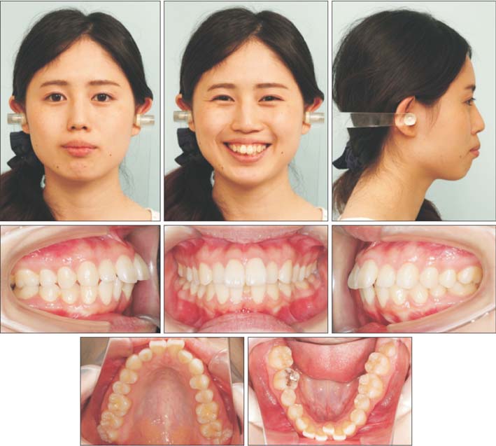
Figure 1 Pretreatment facial and intraoral photographs for a young woman with Class II malocclusion and bimaxillary protrusion.
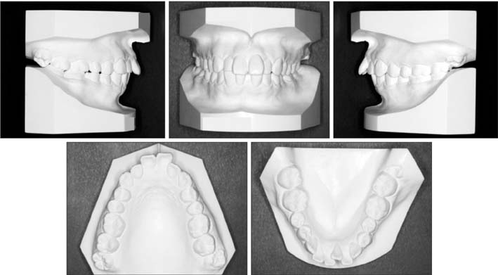
Figure 2 Pretreatment dental casts.
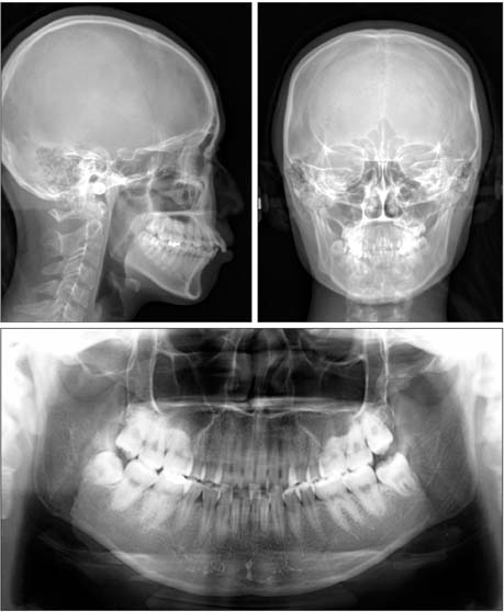
Figure 3 Pretreatment lateral and anteroposterior cephalograms and an orthopantomogram.
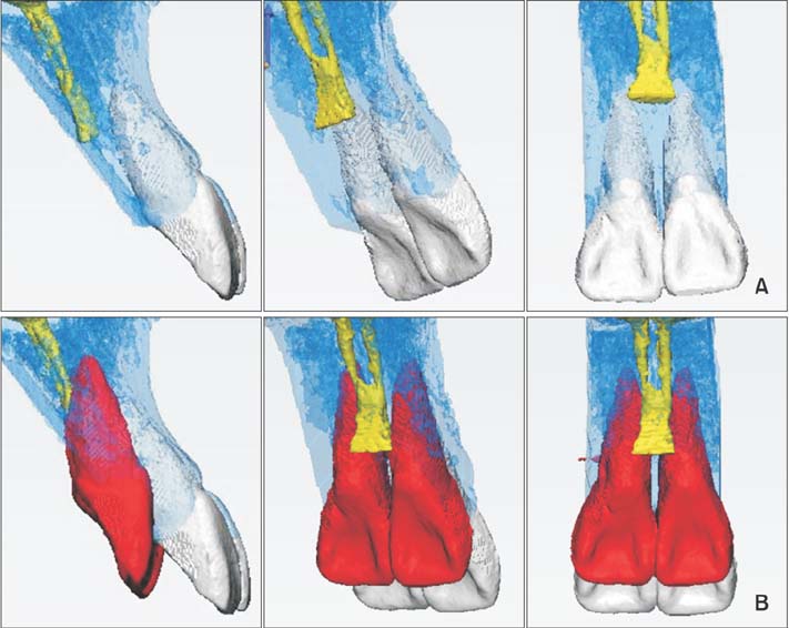
Figure 4 Three-dimensional anatomical structures in the maxillary anterior region. A, Positional relationships among the maxillary incisors, alveolar bone, and incisive canal. B, Simulation of retraction and intrusion of the maxillary central incisors. Gray, pretreatment incisors; blue, alveolar bone; yellow, incisive canal; red, simulation of the incisor movement.
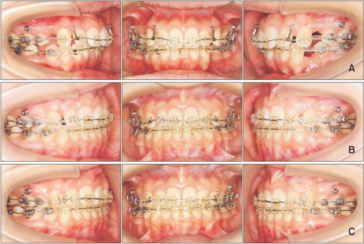
Figure 5 Intraoral photographs obtained during the course of orthodontic treatment. A, Initiation of space closure by leveling, anterior tooth retraction, partial canine retraction, and mesial movement of the mandibular right first molar. B, Completion of canine retraction. Anterior tooth retraction and intrusion. C, Achievement of space closure and initiation of detailing.
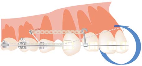
Figure 6 Anterior retraction with a compensating curve of working wires and long hooks.
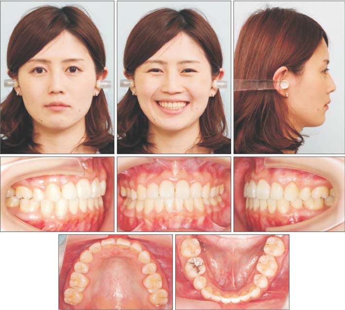
Figure 7 Post-treatment facial and intraoral photographs.
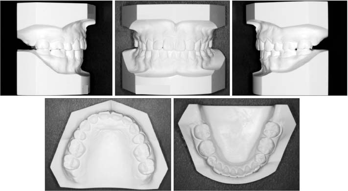
Figure 8 Post-treatment dental casts.
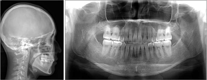
Figure 9 A post-treatment lateral cephalogram and an orthopantomogram.
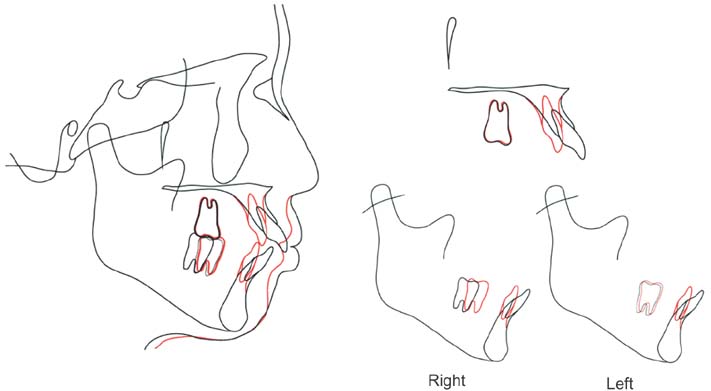
Figure 10 Superimposed tracings of pretreatment (black) and post-treatment (red) cephalograms.
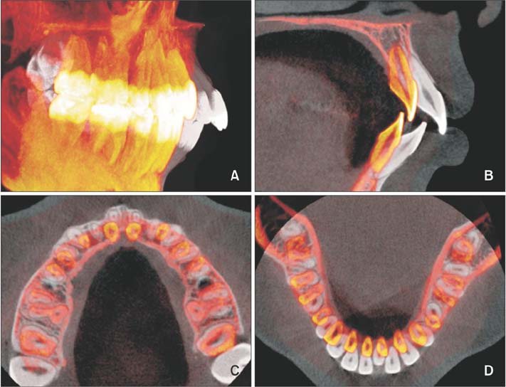
Figure 11 Three-dimensional cone beam computed tomography superimposition registered to the cranial base after orthodontic treatment. A, Changes in the tooth position throughout treatment. B, Midsagittal image of the center of the maxillary right central incisor. C, Horizontal image of the posttreatment midroot level of the maxillary incisors. D, Horizontal image of the post-treatment midroot level of the mandibular incisors. Black, pretreatment; red, post-treatment.
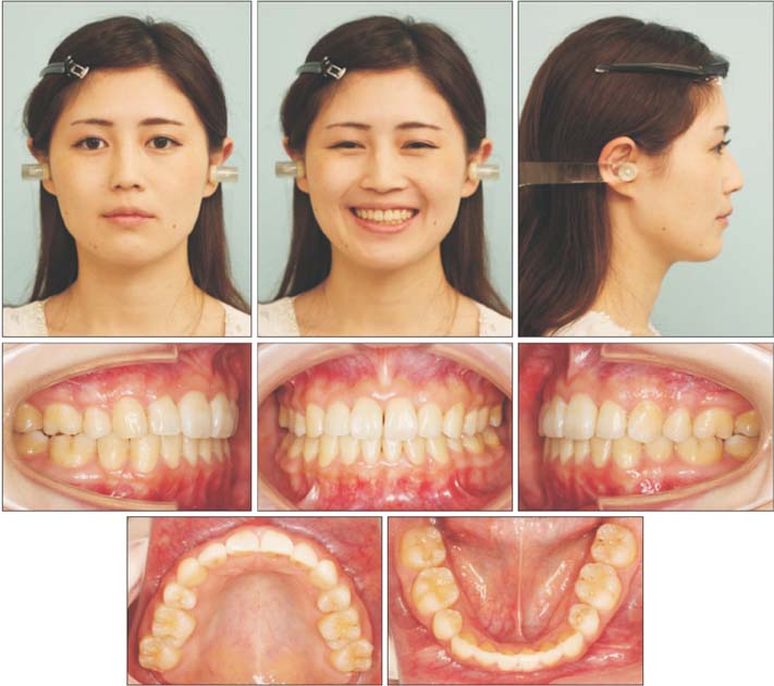
Figure 12 Post-retention facial and intraoral photographs.
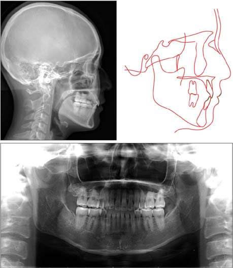
Figure 13 A post-retention lateral cephalogram and an orthopantomogram and superimposed tracings of posttreatment (red) and post-retention (green) cephalograms.
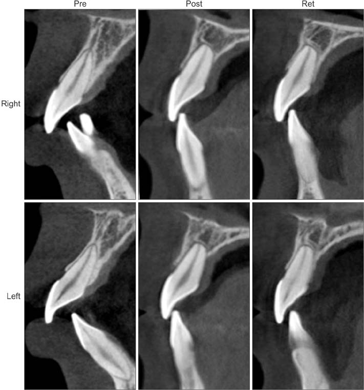
Figure 14 Changes in the central incisors and surrounding alveolar bone after orthodontic treatment and retention. Mild root resorption is noted. Pre, Pretreatment; post, post-treatment; Ret, postretention.
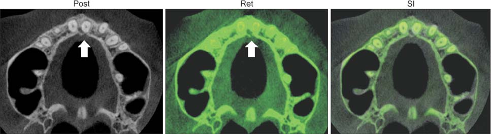
Figure 15 Cone-beam computed tomography images of the incisive canal after orthodontic treatment and retention. Although the left central incisor is contacting the incisive canal on the post-treatment image (left arrow), the incisive canal is surrounded by cortical bone on the post-retention image (right arrow). Gray, post-treatment; green, post-retention. Post, Post-treatment; Ret, post-retention; SI, superimposition.
Cited by 1 articles
-
Effect of extraction treatment on upper airway dimensions in patients with bimaxillary skeletal protrusion relative to their vertical skeletal pattern
Ha-Nul Cho, Hyun Joo Yoon, Jae Hyun Park, Young-Guk Park, Su-Jung Kim
Korean J Orthod. 2021;51(3):166-178. doi: 10.4041/kjod.2021.51.3.166.
Reference
-
1. Leonardi R, Annunziata A, Licciardello V, Barbato E. Soft tissue changes following the extraction of premolars in nongrowing patients with bimaxillary protrusion. A systematic review. Angle Orthod. 2010; 80:211–216.
Article2. Ayaz M, Kharbanda OP. Successful treatment of Class II malocclusion with bidental protrusion using standard edgewise prescription. Contemp Clin Dent. 2016; 7:75–78.
Article3. Parker RJ, Harris EF. Directions of orthodontic tooth movements associated with external apical root resorption of the maxillary central incisor. Am J Orthod Dentofacial Orthop. 1998; 114:677–683.
Article4. Han G, Huang S, Von den, Zeng X, Kuijpers-Jagtman AM. Root resorption after orthodontic intrusion and extrusion: an intraindividual study. Angle Orthod. 2005; 75:912–918.5. Liou EJ, Chang PM. Apical root resorption in orthodontic patients with en-masse maxillary anterior retraction and intrusion with miniscrews. Am J Orthod Dentofacial Orthop. 2010; 137:207–212.
Article6. Weltman B, Vig KW, Fields HW, Shanker S, Kaizar EE. Root resorption associated with orthodontic tooth movement: a systematic review. Am J Orthod Dentofacial Orthop. 2010; 137:462–476.
Article7. Nakano Y, Yamaguchi M, Fujita S, Asano M, Saito K, Kasai K. Expressions of RANKL/RANK and M-CSF/c-fms in root resorption lacunae in rat molar by heavy orthodontic force. Eur J Orthod. 2011; 33:335–343.
Article8. Martins DR, Tibola D, Janson G, Maria FR. Effects of intrusion combined with anterior retraction on apical root resorption. Eur J Orthod. 2012; 34:170–175.
Article9. Zawawi KH, Malki GA. Radiographic comparison of apical root resorption after orthodontic treatment between bidimensional and Roth straight-wire techniques. J Orthod Sci. 2014; 3:106–110.
Article10. Roscoe MG, Meira JB, Cattaneo PM. Association of orthodontic force system and root resorption: a systematic review. Am J Orthod Dentofacial Orthop. 2015; 147:610–626.
Article11. Chung CJ, Choi YJ, Kim KH. Approximation and contact of the maxillary central incisor roots with the incisive canal after maximum retraction with temporary anchorage devices: Report of 2 patients. Am J Orthod Dentofacial Orthop. 2015; 148:493–502.
Article12. Song WC, Jo DI, Lee JY, Kim JN, Hur MS, Hu KS, et al. Microanatomy of the incisive canal using threedimensional reconstruction of microCT images: an ex vivo study. Oral Surg Oral Med Oral Pathol Oral Radiol Endod. 2009; 108:583–590.
Article13. Liang X, Jacobs R, Martens W, Hu Y, Adriaensens P, Quirynen M, et al. Macro- and micro-anatomical, histological and computed tomography scan characterization of the nasopalatine canal. J Clin Periodontol. 2009; 36:598–603.
Article14. Matsumura T, Ishida Y, Kawabe A, Ono T. Quantitative analysis of the relationship between maxillary incisors and the incisive canal by cone-beam computed tomography in an adult Japanese population. Prog Orthod. 2017; 18:24.
Article15. Upadhyay M, Yadav S, Nagaraj K, Patil S. Treatment effects of mini-implants for en-masse retraction of anterior teeth in bialveolar dental protrusion patients: a randomized controlled trial. Am J Orthod Dentofacial Orthop. 2008; 134:18–29.
Article16. Lee KJ, Park YC, Hwang CJ, Kim YJ, Choi TH, Yoo HM, et al. Displacement pattern of the maxillary arch depending on miniscrew position in sliding mechanics. Am J Orthod Dentofacial Orthop. 2011; 140:224–232.
Article
- Full Text Links
-
- Actions
-
Cited
- CITED
-
- Close
- Share
-
- Similar articles
-
- Treatment of gummy smile using botulinum toxin: a review
- Three-dimensional imaging modalities in endodontics
- Analysis of Beam Hardening of Modulation Layers for Dual Energy Cone-beam CT
- Management of root canal perforation by using cone-beam computed tomography
- Improvements of facial profile and smile aesthetic using temporary anchorage devices and botulinum toxin: a case report
