Evaluation of growth changes induced by functional appliances in children with Class II malocclusion: Superimposition of lateral cephalograms on stable structures
- Affiliations
-
- 1Section of Orthodontics, Department of Odontology, Faculty of Health and Medical Sciences, University of Copenhagen, Copenhagen, Denmark
- 2Department of Orthodontics, School of Dentistry, Seoul National University, Seoul, Korea
- KMID: 2502626
- DOI: http://doi.org/10.4041/kjod.2020.50.3.170
Abstract
Objective
To compare short- and long-term dentoalveolar, skeletal, and rotational changes evaluated by Björk’s structural method of superimposition between children with Class II malocclusion treated by functional appliances and untreated matched controls.
Methods
Seventy-nine prepubertal or pubertal children (mean age, 11.57 ± 1.40 years) with Class II malocclusion were included. Thirty-four children were treated using an activator with a high-pull headgear (Z-activator), while 28 were treated using an activator without a headgear (E-activator). Seventeen untreated children were included as controls. Lateral cephalograms were obtained before treatment (T1), after functional appliance treatment (T2), and after retention in the postpubertal phase (T3). Changes from T1 to T2 and T1 to T3 were compared between the treated groups and control group using multiple linear regression analysis.
Results
Relative to the findings in the control group at T2, the sagittal jaw relationship (subspinalenasion- pogonion, p < 0.001), maxillary prognathism (sella-nasion-subspinale, p < 0.05), and condylar growth (p < 0.001) exhibited significant improvements in the Z- and E-activator groups, which also showed a significantly increased maxillary incisor retraction (p < 0.001) and decreased overjet (p < 0.001). Only the E-activator group exhibited significant backward rotation of the maxilla at T2 (p < 0.01). The improvements in the sagittal jaw relationship (p < 0.01) and dental relationship (p < 0.001) remained significant at T3. Condylar growth and jaw rotations were not significant at T3.
Conclusions
Functional appliance treatment in children with Class II malocclusion can significantly improve the sagittal jaw relationship and dental relationships in the long term.
Figure
-
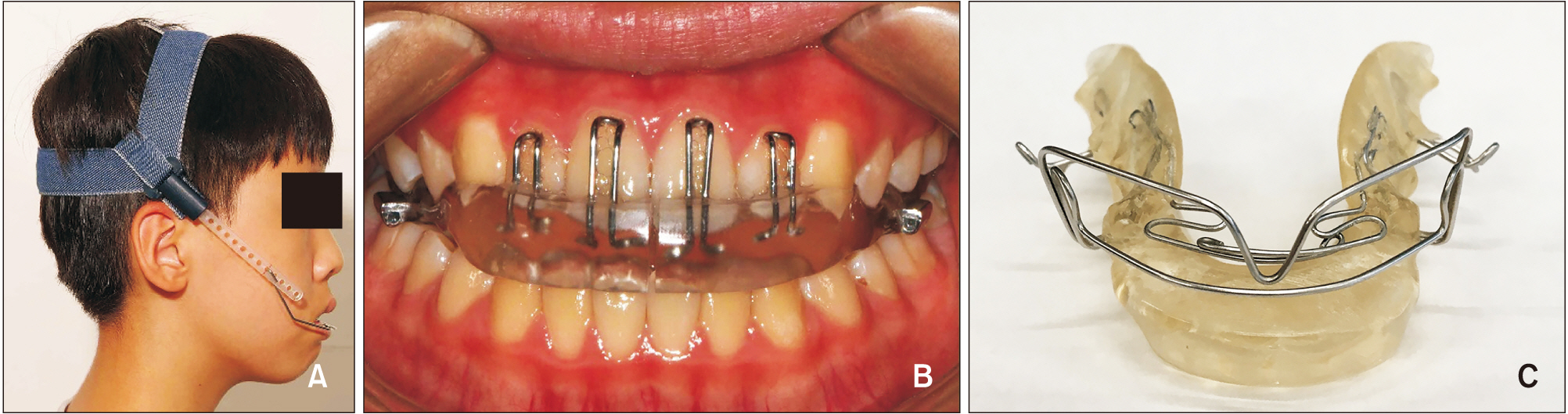
Figure 1 Functional appliances used in the present study. A and B, Z-activator (Teuscher activator with a high-pull headgear)10. C, E-activator (modified Andresen activator, also known as an Ergenzinger activator).11

Figure 2 Cephalometric landmarks used for measurements.14 n, Nasion; s, sella; cd, condylion; ba, basion; ar, articulare; gn, gnathion; pg, pogonion; sm, supramentale; isi, incisal superior incisor; iii, incisal inferior incisor; ss, subspinale; sp, spinale; MBLar, mandibular base line; Rlar, ramus line; ILi, incisal line inferior; ILs, incisal line superior; ML, mandibular line; NL, nasal line.

Figure 3 Illustration of superimposition on the stable structures in the anterior cranial base between two stages.9 NSLRef, Nasion-sella line at before treatment (T1) transferred to the following lateral cephalogram as a reference line when the two sets are superimposed on the stable structures in the anterior cranial base; MaxillaRef, a reference line drawn in the maxilla when the two sets are superimposed on the stable structures in the maxilla; MandibleRef, a reference line drawn in the mandible when the two sets are superimposed on the stable structures in the mandible.
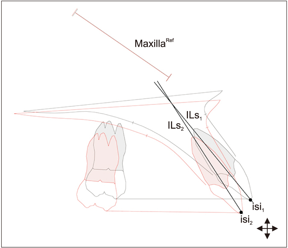
Figure 4 Illustration of superimposition on the stable structures in the maxilla between two stages.9 MaxillaRef, Maxillary reference line; ILs1, incisal line superior stage 1; ILs2, incisal line superior stage 2; isi1, incisal superior stage 1; isi2, incisal superior stage 2.
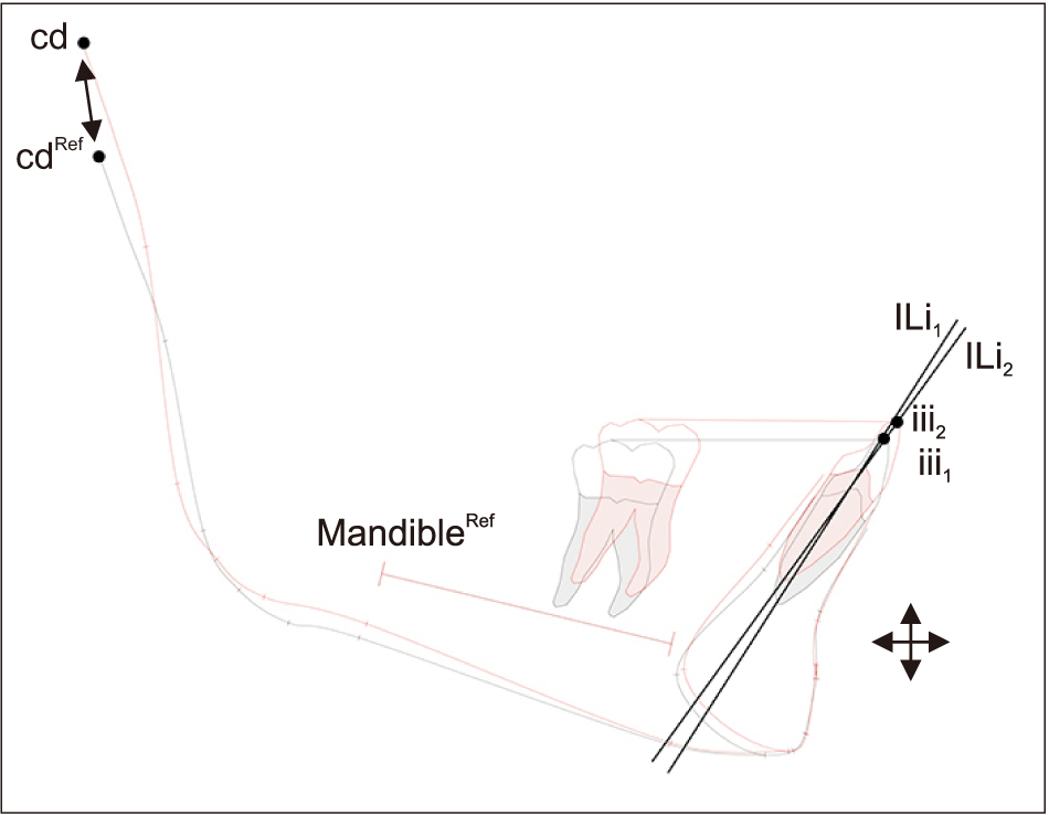
Figure 5 Illustration of superimposition on the stable structures in the mandible between two stages.9 cd, Condylion; cdRef, condylion at baseline (T1); MandibleRef, mandibular reference line; ILi1, incisal line inferior stage 1; ILi2, incisal line inferior stage 2; iii1, incisal inferior stage 1; iii2, incisal inferior stage 2.
Reference
-
1. Cozza P, Baccetti T, Franchi L, De Toffol L, McNamara JA Jr. 2006; Mandibular changes produced by functional appliances in Class II malocclusion: a systematic review. Am J Orthod Dentofacial Orthop. 129:599.e1–12. discussion e1–6. DOI: 10.1016/j.ajodo.2005.11.010. PMID: 16679196.
Article2. Zelderloo A, Cadenas de Llano-Pérula M, Verdonck A, Fieuws S, Willems G. 2017; Cephalometric appraisal of Class II treatment effects after functional and fixed appliances: a retrospective study. Eur J Orthod. 39:334–41. DOI: 10.1093/ejo/cjw064. PMID: 27742730.
Article3. Batista KB, Thiruvenkatachari B, Harrison JE, O'Brien KD. 2018; Orthodontic treatment for prominent upper front teeth (Class II malocclusion) in children and adolescents. Cochrane Database Syst Rev. 3:CD003452. DOI: 10.1002/14651858.CD003452.pub4. PMID: 29534303. PMCID: PMC6494411.
Article4. Tulloch JF, Proffit WR, Phillips C. 2004; Outcomes in a 2-phase randomized clinical trial of early Class II treatment. Am J Orthod Dentofacial Orthop. 125:657–67. DOI: 10.1016/j.ajodo.2004.02.008. PMID: 15179390.
Article5. Cross JJ. 1977; Facial growth: before, during, and following orthodontic treatment. Am J Orthod. 71:68–78. DOI: 10.1016/0002-9416(77)90177-4. PMID: 264365.
Article6. Arat ZM, Türkkahraman H, English JD, Gallerano RL, Boley JC. 2010; Longitudinal growth changes of the cranial base from puberty to adulthood. A comparison of different superimposition methods. Angle Orthod. 80:537–44. DOI: 10.2319/080709-447.1. PMID: 20482360.7. Björk A. 1969; Prediction of mandibular growth rotation. Am J Orthod. 55:585–99. DOI: 10.1016/0002-9416(69)90036-0. PMID: 5253957.
Article8. Björk A, Skieller V. 1972; Facial development and tooth eruption. An implant study at the age of puberty. Am J Orthod. 62:339–83. DOI: 10.1016/S0002-9416(72)90277-1. PMID: 4506491.9. Björk A, Skieller V. 1983; Normal and abnormal growth of the mandible. A synthesis of longitudinal cephalometric implant studies over a period of 25 years. Eur J Orthod. 5:1–46. DOI: 10.1093/ejo/5.1.1. PMID: 6572593.10. Teuscher U. 1978; A growth-related concept for skeletal class II treatment. Am J Orthod. 74:258–75. DOI: 10.1016/0002-9416(78)90202-6. PMID: 281130.
Article11. Andresen V. 1936; The Norwegian system of functional gnatho-orthopedics. Acta Gnathol. 1:5–36.12. Baccetti T, Franchi L, McNamara JA Jr. 2005; The cervical vertebral maturation (CVM) method for the assessment of optimal treatment timing in dentofacial orthopedics. Semin Orthod. 11:119–29. DOI: 10.1053/j.sodo.2005.04.005.
Article13. Siersb©¡k-Nielsen S, Solow B. 1982; Intra- and interexaminer variability in head posture recorded by dental auxiliaries. Am J Orthod. 82:50–7. DOI: 10.1016/0002-9416(82)90546-2. PMID: 6961777.14. Solow B, Tallgren A. 1976; Head posture and craniofacial morphology. Am J Phys Anthropol. 44:417–35. DOI: 10.1002/ajpa.1330440306. PMID: 937521.
Article15. Sandham A. 1986; Cervical vertebral anomalies in cleft lip and palate. Cleft Palate J. 23:206–14. PMID: 3524906.16. Huggare J. 1991; Association between morphology of the first cervical vertebra, head posture, and craniofacial structures. Eur J Orthod. 13:435–40. DOI: 10.1093/ejo/13.6.435. PMID: 1817068.
Article17. Oh E, Ahn SJ, Sonnesen L. 2018; Ethnic differences in craniofacial and upper spine morphology in children with skeletal Class II malocclusion. Angle Orthod. 88:283–91. DOI: 10.2319/083017-584.1. PMID: 29337630.
Article18. Sonnesen L, Kjaer I. 2007; Cervical vertebral body fusions in patients with skeletal deep bite. Eur J Orthod. 29:464–70. DOI: 10.1093/ejo/cjm043. PMID: 17693430.
Article19. Dahlberg G. 1940. Statistical methods for medical and biological students. Interscience;New York: p. 122–32.20. Houston WJ. 1983; The analysis of errors in orthodontic measurements. Am J Orthod. 83:382–90. DOI: 10.1016/0002-9416(83)90322-6. PMID: 6573846.
Article21. Papageorgiou SN, Koretsi V, Jäger A. 2017; Bias from historical control groups used in orthodontic research: a meta-epidemiological study. Eur J Orthod. 39:98–105. DOI: 10.1093/ejo/cjw035. PMID: 27129869.
Article22. Oh E, Ahn SJ, Sonnesen L. 2019; Ethnic differences in craniofacial and upper spine morphology between European and Asian children with skeletal Class III malocclusion. Am J Orthod Dentofacial Orthop. 156:502–11. DOI: 10.1016/j.ajodo.2018.10.024. PMID: 31582122.
Article23. Arntsen T, Sonnesen L. 2011; Cervical vertebral column morphology related to craniofacial morphology and head posture in preorthodontic children with Class II malocclusion and horizontal maxillary overjet. Am J Orthod Dentofacial Orthop. 140:e1–7. DOI: 10.1016/j.ajodo.2010.10.021. PMID: 21724066.
Article24. Sonnesen L, Kjaer I. 2008; Anomalies of the cervical vertebrae in patients with skeletal Class II malocclusion and horizontal maxillary overjet. Am J Orthod Dentofacial Orthop. 133:188.e15–20. DOI: 10.1016/j.ajodo.2007.07.018. PMID: 18249281.
Article25. Melsen B. 1969; Time of closure of the spheno-occipital synchondrosis determined on dry skulls. A radiographic craniometric study. Acta Odontol Scand. 27:73–90. DOI: 10.3109/00016356909033580. PMID: 5256953.
Article26. Solow B. 1980; The dentoalveolar compensatory mechanism: background and clinical implications. Br J Orthod. 7:145–61. DOI: 10.1179/bjo.7.3.145. PMID: 6934010.
Article27. Petrovic AG, Stutzmann JJ. Graber LW, Graber TM, editors. 1986. The concept of mandibular tissue-level growth potential and the responsiveness to a functional appliance. Orthodontics, state of the art, essence of the science. Mosby Co;St. Louis: p. 59–74.28. Türkkahraman H, Sayin MO. 2016; Effects of activator and activator headgear treatment: comparison with untreated Class II subjects. Eur J Orthod. 28:27–34. DOI: 10.1093/ejo/cji062. PMID: 16093256.
Article
- Full Text Links
-
- Actions
-
Cited
- CITED
-
- Close
- Share
-
- Similar articles
-
- Logic for the use of stable structural superimposition method and introduction of its application
- Study of Functional Appliance for Treatments of Children and Adolescents with Class II Malocclusion
- An evaluation of treatment effects of modified teuscher appliance in Class II division 1 malocclusion
- Long-term pharyngeal airway changes after bionator treatment in adolescents with skeletal Class II malocclusions
- A study of the etiology of unilateral Class II, division 1 malocclusion
