Three-dimensional cone beam computed tomography analysis of temporomandibular joint response to the Twin-block functional appliance
- Affiliations
-
- 1Jiangsu Key Laboratory of Oral Disease, Nanjing Medical University, Nanjing, China. zhangweibing@njmu.edu.cn
- 2Department of Orthodontics, Affiliated Hospital of Stomatology, Nanjing Medical University, Nanjing, China.
- KMID: 2471869
- DOI: http://doi.org/10.4041/kjod.2020.50.2.86
Abstract
OBJECTIVE
To propose a three-dimensional (3D) method for evaluating temporomandibular joint (TMJ) changes during Twin-block treatment.
METHODS
Seventeen patients with Class II division 1 malocclusion treated using Twin-block and nine untreated patients with a similar malocclusion were included in this research. We collected their cone beam computed tomography (CBCT) data from before and 8 months after treatment. Segmentations were constructed using ITK-SNAP. Condylar volume and superficial area were measured using 3D Slicer. The 3D landmarks were identified on CBCT images by using Dolphin software to assess the condylar positional relationship. 3D models of the mandible and glenoid fossa of the patients were constructed and registered via voxel-based superimposition using 3D Slicer. Thereafter, skeletal changes could be visualized using 3DMeshMetric in any direction of the superimposition on a color-coded map. All the superimpositions were measured using the same scale on the distance color-coded map, in which red color represents overgrowth and blue color represents resorption.
RESULTS
Significant differences were observed in condylar volume, superficial area, and condylar position in both groups after 8 months. Compared with the control group (CG), the Twin-block group exhibited more obvious condyle-fossa modifications and joint positional changes. Moreover, on the color-coded map, more obvious condyle-fossa modifications could be observed in the posterior and superior directions in the Twin-block group than in the CG.
CONCLUSIONS
We successfully established a 3D method for measuring and evaluating TMJ changes caused by Twin-block treatment. The treatment produced a larger condylar size and caused condylar positional changes.
MeSH Terms
Figure
-
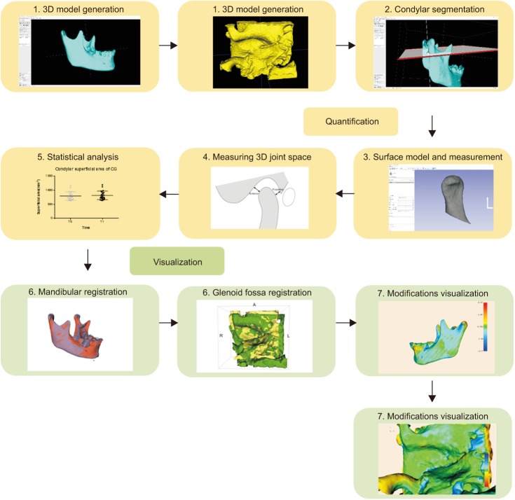
Figure 1 Technical route of the experiment. 1, Construction of the three-dimensional (3D) segmentations of the mandible and glenoid fossa. 2, Performing condylar segmentation. 3, Generating the surface model and performing volumetric measurements of the condyle. 4, Measuring the joint space by using 3D landmarks. 5, Performing the statistical analysis. 6, Registering the condyle-fossa based on the voxels. 7, Visualization of condyle-fossa modifications using the distance color-coded map.
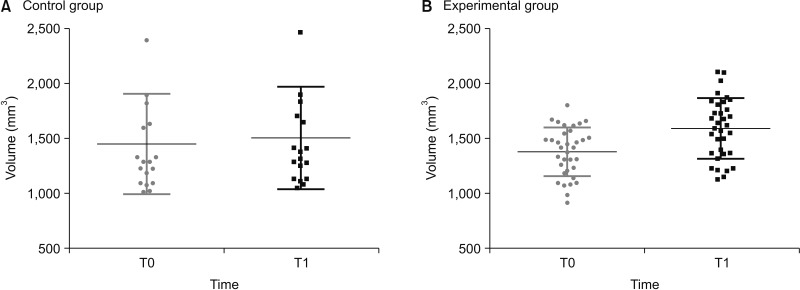
Figure 2 Condylar volume increases significantly 8 months after treatment (T1) compared to before treatment (T0) in both control (A) and experimental groups (B).
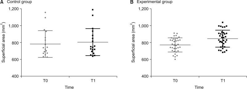
Figure 3 Condylar superficial area increases significantly 8 months after treatment (T1) compared to before treatment (T0) in both control (A) and experimental groups (B).
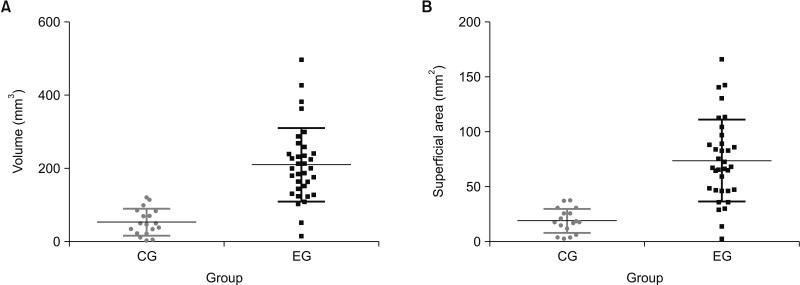
Figure 4 The absolute increases in condylar volume (A) and superficial area (B) are significantly higher in the experimental group (EG) than in the control group (CG) after 8 months of treatment.
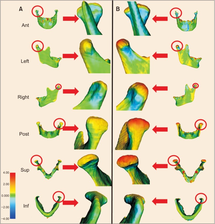
Figure 5 One example of six-directional views of superimposition in the control (A) and experimental (B) groups (CG and EG, respectively). More obvious skeletal remodeling is observed in the superior and posterior directions of the condyle in the EG than in the CG. All the skeletal superimpositions are visualized on a color-coded map and measured using the same scale, in which red color represents overgrowth and blue color represents resorption.Ant, Anterior; Post, posterior; Sup, superior; Inf, inferior.
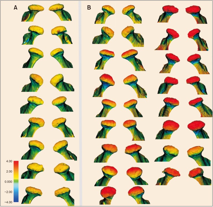
Figure 6 The superior view of all condylar superimpositions in the control (A) and experimental (B) groups (CG and EG, respectively). More obvious skeletal remodeling is observed in the superior direction of the condyle in the EG than in the CG. All the skeletal superimpositions are visualized on a color-coded map and measured using the same scale, in which red color represents overgrowth and blue color represents resorption.
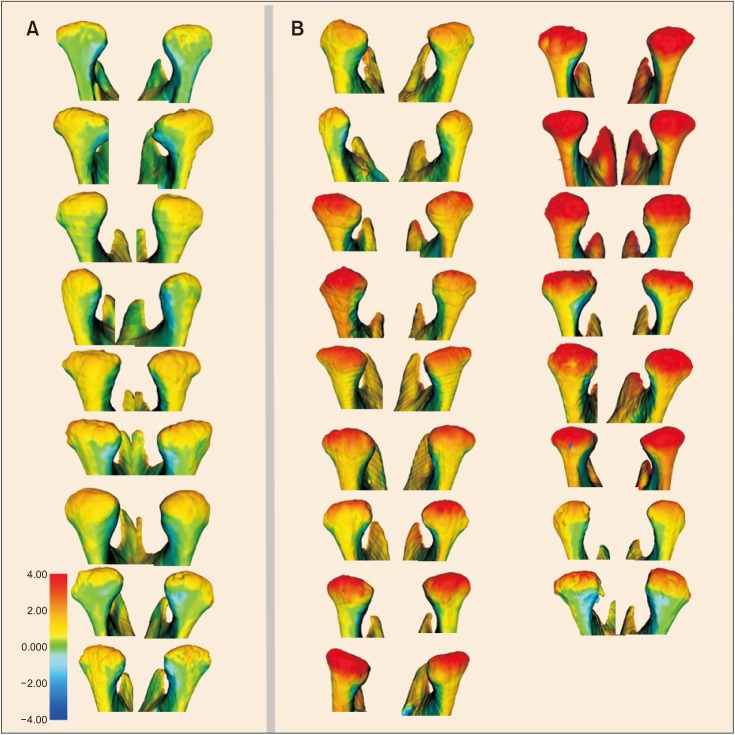
Figure 7 The posterior view of all condylar superimpositions in the contol (A) and experimental (B) groups (CG and EG, respectively). More obvious skeletal remodeling is observed in the posterior direction of the condyle in the EG than in the CG. All the skeletal superimpositions are visualized on a color-coded map and measured using the same scale, in which red color represents overgrowth and blue color represents resorption.
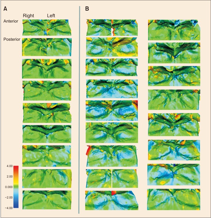
Figure 8 All superimpositions of the left and right glenoid fossa in the control (A) and experimental (B) groups (CG and EG, respecitvely). More obvious skeletal resorption is observed in the superior direction of the glenoid fossa in the EG than in the CG. All the skeletal superimpositions are visualized on a color-coded map and measured using the same scale, in which red color represents overgrowth and blue color represents resorption.
Reference
-
1. Baccetti T, Franchi L, Toth LR, McNamara JA Jr. Treatment timing for Twin-block therapy. Am J Orthod Dentofacial Orthop. 2000; 118:159–170. PMID: 10935956.
Article2. Jena AK, Duggal R, Parkash H. Skeletal and dentoalveolar effects of Twin-block and bionator appliances in the treatment of Class II malocclusion: a comparative study. Am J Orthod Dentofacial Orthop. 2006; 130:594–602. PMID: 17110256.
Article3. Lund DI, Sandler PJ. The effects of Twin Blocks: a prospective controlled study. Am J Orthod Dentofacial Orthop. 1998; 113:104–110. PMID: 9457025.
Article4. Morris DO, Illing HM, Lee RT. A prospective evaluation of Bass, Bionator and Twin Block appliances. Part II--The soft tissues. Eur J Orthod. 1998; 20:663–664. PMID: 9926634.5. Trenouth MJ. Cephalometric evaluation of the Twin-block appliance in the treatment of Class II Division 1 malocclusion with matched normative growth data. Am J Orthod Dentofacial Orthop. 2000; 117:54–59. PMID: 10629520.
Article6. Varlik SK, Gültan A, Tümer N. Comparison of the effects of Twin Block and activator treatment on the soft tissue profile. Eur J Orthod. 2008; 30:128–134. PMID: 18281262.
Article7. Bayram M, Kayipmaz S, Sezgin OS, Küçük M. Volumetric analysis of the mandibular condyle using cone beam computed tomography. Eur J Radiol. 2012; 81:1812–1816. PMID: 21680124.
Article8. Yildirim E, Karacay S, Erkan M. Condylar response to functional therapy with Twin-Block as shown by cone-beam computed tomography. Angle Orthod. 2014; 84:1018–1025. PMID: 24713070.
Article9. Elfeky HY, Fayed MS, Alhammadi MS, Soliman SAZ, El Boghdadi DM. Three-dimensional skeletal, dentoalveolar and temporomandibular joint changes produced by Twin Block functional appliance. J Orofac Orthop. 2018; 79:245–258. PMID: 29663034.
Article10. Liu B, Wang Y, Song F, Liu M, Duan Y, Zhou L. [Cone-beam CT evaluation of the changes in the temporomandibular joint of patients with class II division 1 subdivision malocclusion before and after twin-block treatment]. Hua Xi Kou Qiang Yi Xue Za Zhi. 2013; 31:610–614. Chinese. PMID: 24437298.11. Serbesis-Tsarudis C, Pancherz H. "Effective" TMJ and chin position changes in Class II treatment. Angle Orthod. 2008; 78:813–818. PMID: 18298212.
Article12. Koerich L, Weissheimer A, de Menezes LM, Lindauer SJ. Rapid 3D mandibular superimposition for growing patients. Angle Orthod. 2017; 87:473–479. PMID: 27767348.
Article13. Cevidanes LH, Styner MA, Proffit WR. Image analysis and superimposition of 3-dimensional cone-beam computed tomography models. Am J Orthod Dentofacial Orthop. 2006; 129:611–618. PMID: 16679201.
Article14. Cevidanes LH, Heymann G, Cornelis MA, DeClerck HJ, Tulloch JF. Superimposition of 3-dimensional cone-beam computed tomography models of growing patients. Am J Orthod Dentofacial Orthop. 2009; 136:94–99. PMID: 19577154.
Article15. Ruellas AC, Yatabe MS, Souki BQ, Benavides E, Nguyen T, Luiz RR, et al. 3D mandibular superimposition: comparison of regions of reference for voxelbased registration. PLoS One. 2016; 11:e0157625. PMID: 27336366.
Article16. Franchi L, Baccetti T, McNamara JA Jr. Mandibular growth as related to cervical vertebral maturation and body height. Am J Orthod Dentofacial Orthop. 2000; 118:335–340. PMID: 10982936.
Article17. Tecco S, Saccucci M, Nucera R, Polimeni A, Pagnoni M, Cordasco G, et al. Condylar volume and surface in Caucasian young adult subjects. BMC Med Imaging. 2010; 10:28. PMID: 21194477.
Article18. Kinzinger GS, Roth A, Gülden N, Bücker A, Diedrich PR. Effects of orthodontic treatment with fixed functional orthopaedic appliances on the condylefossa relationship in the temporomandibular joint: a magnetic resonance imaging study (Part I). Dentomaxillofac Radiol. 2006; 35:339–346. PMID: 16940482.
Article19. Ghafari J, Engel FE, Laster LL. Cephalometric superimposition on the cranial base: a review and a comparison of four methods. Am J Orthod Dentofacial Orthop. 1987; 91:403–413. PMID: 3472459.
Article20. Pancherz H. Treatment of class II malocclusions by jumping the bite with the Herbst appliance. A cephalometric investigation. Am J Orthod. 1979; 76:423–442. PMID: 291343.21. McNamara JA Jr, Hinton RJ, Hoffman DL. Histologic analysis of temporomandibular joint adaptation to protrusive function in young adult rhesus monkeys (Macaca mulatta). Am J Orthod. 1982; 82:288–298. PMID: 6961801.
Article22. Ruf S, Baltromejus S, Pancherz H. Effective condylar growth and chin position changes in activator treatment: a cephalometric roentgenographic study. Angle Orthod. 2001; 71:4–11. PMID: 11211297.23. Sharma AK, Sachdev V, Singla A, Kirtaniya BC. Skeletal and dentoalveolar changes concurrent to use of Twin Block appliance in class II division I cases with a deficient mandible: a cephalometric study. J Indian Soc Pedod Prev Dent. 2012; 30:218–226. PMID: 23263425.
Article24. Sun L, Zhao J, Wang H, Pan Y, Wang L, Zhang WB. Mechanical stress promotes matrix synthesis of mandibular condylar cartilage via the RKIP-ERK pathway. J Mol Histol. 2017; 48:437–446. PMID: 29119279.
Article25. Atresh A, Cevidanes LHS, Yatabe M, Muniz L, Nguyen T, Larson B, et al. Three-dimensional treatment outcomes in Class II patients with different vertical facial patterns treated with the Herbst appliance. Am J Orthod Dentofacial Orthop. 2018; 154:238–248.e1. PMID: 30075926.
Article26. Arici S, Akan H, Yakubov K, Arici N. Effects of fixed functional appliance treatment on the temporomandibular joint. Am J Orthod Dentofacial Orthop. 2008; 133:809–814. PMID: 18538243.
Article27. Mavreas D, Athanasiou AE. Tomographic assessment of alterations of the temporomandibular joint after orthognathic surgery. Eur J Orthod. 1992; 14:3–15. PMID: 1563472.
Article28. Wadhawan N, Kumar S, Kharbanda OP, Duggal R, Sharma R. Temporomandibular joint adaptations following two-phase therapy: an MRI study. Orthod Craniofac Res. 2008; 11:235–250. PMID: 18950321.
Article29. Sarnat BG. Some methods of assessing postnatal craniofaciodental growth: a retrospective of personal research. Cleft Palate Craniofac J. 1997; 34:159–172. PMID: 9138513.
Article30. Cevidanes LH, Bailey LJ, Tucker GR Jr, Styner MA, Mol A, Phillips CL, et al. Superimposition of 3D cone-beam CT models of orthognathic surgery patients. Dentomaxillofac Radiol. 2005; 34:369–375. PMID: 16227481.
Article31. Gazzani F, Ruellas ACO, Faltin K, Franchi L, Cozza P, Bigliazzi R, et al. 3D comparison of mandibular response to functional appliances: Balters Bionator versus Sander Bite Jumping. Biomed Res Int. 2018; 2018:2568235. PMID: 29854734.
Article32. Souki BQ, Vilefort PLC, Oliveira DD, Andrade I Jr, Ruellas AC, Yatabe MS, et al. Three-dimensional skeletal mandibular changes associated with Herbst appliance treatment. Orthod Craniofac Res. 2017; 20:111–118. PMID: 28414870.
Article33. Ivorra-Carbonell L, Montiel-Company JM, Almerich-Silla JM, Paredes-Gallardo V, Bellot-Arcís C. Impact of functional mandibular advancement appliances on the temporomandibular joint - a systematic review. Med Oral Patol Oral Cir Bucal. 2016; 21:e565–e572. PMID: 27475694.
Article34. Baccetti T, Franchi L, McNamara JA Jr, Tollaro I. Early dentofacial features of Class II malocclusion: a longitudinal study from the deciduous through the mixed dentition. Am J Orthod Dentofacial Orthop. 1997; 111:502–509. PMID: 9155809.
Article35. Burhan AS, Nawaya FR. Dentoskeletal effects of the Bite-Jumping Appliance and the Twin-Block Appliance in the treatment of skeletal Class II malocclusion: a randomized controlled trial. Eur J Orthod. 2015; 37:330–337. PMID: 25296729.
Article36. Proffit WR, Fields HW, Sarver DM. Contemporary orthodontics. 5th ed. India: Elsevier;2012.
- Full Text Links
-
- Actions
-
Cited
- CITED
-
- Close
- Share
-
- Similar articles
-
- Mandibular condyle position in cone beam computed tomography
- Three-dimensional imaging modalities in endodontics
- Three-dimensional cone-beam computed tomography based comparison of condylar position and morphology according to the vertical skeletal pattern
- Reliability of cone-beam computed tomography for temporomandibular joint analysis
- Osteoarthritic changes and condylar positioning of the temporomandibular joint in Korean children and adolescents
