Korean J Orthod.
2019 Mar;49(2):81-88. 10.4041/kjod.2019.49.2.81.
Reliability of cone-beam computed tomography for temporomandibular joint analysis
- Affiliations
-
- 1Department of Orthodontics, School of Dentistry, Hacettepe University, Ankara, Turkey. hande.gorucu@hotmail.com
- KMID: 2453971
- DOI: http://doi.org/10.4041/kjod.2019.49.2.81
Abstract
OBJECTIVE
The aim was to assess the intraobserver and interobserver reliabilities of temporomandibular joint linear measurements and condylar shape classifications performed with cone-beam computed tomography (CBCT).
METHODS
CBCT images of 30 patients were measured at two different time points by two orthodontists using the Dolphin 3D program (n = 60). Anterior, posterior, and superior joint space measurements and sagittal joint morphology classification in the sagittal view and medial and lateral joint space and mediolateral width measurements and coronal joint morphology classification in the coronal view were recorded. Intraclass-interclass correlation coefficients (ICC) and kappa statistics were used to assess intraobserver and interobserver reliability for the measurements and morphology classifications, respectively.
RESULTS
The ICC values were good for measurements of the posterior joint space by observer I and for measurements of the posterior, medial, and lateral joint spaces by observer II, while the other intraobserver measurements were excellent. Only the mediolateral width measurements showed excellent interobserver ICC values, while the other measurements showed good interobserver ICC values. Intraobserver agreement for the sagittal morphology classifications was moderate (κ = 0.479) and almost perfect (κ = 0.858) for observers I and II, respectively, while the corresponding agreement for the coronal morphology classifications was substantial for both observers. The interobserver agreement values for sagittal and coronal morphology classifications were slight (κ = 0.181) and fair (κ = 0.265), respectively.
CONCLUSIONS
Linear temporomandibular joint measurements were reproducible and reliable in both intraobserver and interobserver evaluations. However, interobserver agreement for assessments of condylar shape was low.
Keyword
MeSH Terms
Figure
-

Figure 1 Arrangement of the left condyle's long axis.
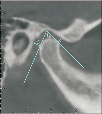
Figure 2 Measurement of the anterior (1), superior (2), and posterior (3) joint spaces.
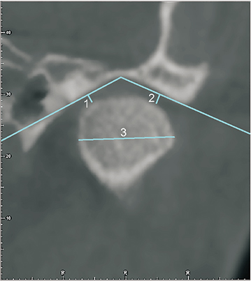
Figure 3 Measurement of the medial (1) and lateral (2) joint spaces and the mediolateral width (3).
Reference
-
1. Okeson JP. Orthodontic therapy and the patient with temporomandibular disorder. In : Graber LW, Vanarsdall RL, Vig KWL, Huang GJ, editors. Orthodontics: current principles and techniques. 6th ed. St. Louis, Missouri: Elsevier;2017. p. 353–366.2. Kaur A, Natt AS, Mehra SK, Maheshwari K, Singh G, Kaur A. Improved visualization and assessment of condylar position in the glenoid fossa for different occlusions: a CBCT study. J Contemp Dent Pract. 2016; 17:679–686.
Article3. Park IY, Kim JH, Park YH. Three-dimensional cone-beam computed tomography based comparison of condylar position and morphology according to the vertical skeletal pattern. Korean J Orthod. 2015; 45:66–73.
Article4. Al-Saleh MA, Alsufyani NA, Saltaji H, Jaremko JL, Major PW. MRI and CBCT image registration of temporomandibular joint: a systematic review. J Otolaryngol Head Neck Surg. 2016; 45:30.
Article5. Brooks SL, Brand JW, Gibbs SJ, Hollender L, Lurie AG, Omnell KA, et al. Imaging of the temporomandibular joint: a position paper of the American Academy of Oral and Maxillofacial Radiology. Oral Surg Oral Med Oral Pathol Oral Radiol Endod. 1997; 83:609–618.6. Barghan S, Tetradis S, Mallya S. Application of cone beam computed tomography for assessment of the temporomandibular joints. Aust Dent J. 2012; 57:Suppl 1. 109–118.
Article7. Adibi S, Zhang W, Servos T, O'Neill PN. Cone beam computed tomography in dentistry: what dental educators and learners should know. J Dent Educ. 2012; 76:1437–1442.
Article8. Kiljunen T, Kaasalainen T, Suomalainen A, Kortesniemi M. Dental cone beam CT: A review. Phys Med. 2015; 31:844–860.
Article9. Merigue LF, Conti AC, Oltramari-Navarro PV, Navarro Rde L, Almeida MR. Tomographic evaluation of the temporomandibular joint in malocclusion subjects: condylar morphology and position. Braz Oral Res. 2016; 30:S1806-83242016000100222. DOI: 10.1590/1807-3107BOR-2016.vol30.0017.
Article10. Coskuner HG, Ciger S. Three-dimensional assessment of the temporomandibular joint and mandibular dimensions after early correction of the maxillary arch form in patients with Class II division 1 or division 2 malocclusion. Korean J Orthod. 2015; 45:121–129.
Article11. Paknahad M, Shahidi S, Abbaszade H. Correlation between condylar position and different sagittal skeletal facial types. J Orofac Orthop. 2016; 77:350–356.
Article12. American Academy of Oral and Maxillofacial Radiology. Clinical recommendations regarding use of cone beam computed tomography in orthodontics. [corrected]. Position statement by the American Academy of Oral and Maxillofacial Radiology. Oral Surg Oral Med Oral Pathol Oral Radiol. 2013; 116:238–257.13. Dalili Z, Khaki N, Kia SJ, Salamat F. Assessing joint space and condylar position in the people with normal function of temporomandibular joint with cone-beam computed tomography. Dent Res J (Isfahan). 2012; 9:607–612.
Article14. Katsavrias EG. Morphology of the temporomandibular joint in subjects with Class II Division 2 malocclusions. Am J Orthod Dentofacial Orthop. 2006; 129:470–478.
Article15. Martins E, Silva JC, Pires CA, Ponces-Ramalhão MJ, Lopes JD. Coronal joint spaces of the temporomandibular joint: Systematic review and meta-analysis. J Clin Exp Dent. 2015; 7:e435–e440.
Article16. Leonardi R, Caltabiano M, Cavallini C, Sicurezza E, Barbato E, Spampinato C, et al. Condyle fossa relationship associated with functional posterior crossbite, before and after rapid maxillary expansion. Angle Orthod. 2012; 82:1040–1046.
Article17. Kinzinger G, Kober C, Diedrich P. Topography and morphology of the mandibular condyle during fixed functional orthopedic treatment --a magnetic resonance imaging study. J Orofac Orthop. 2007; 68:124–147.
Article18. Mattos CT, Cruz CV, da Matta TC, Pereira Lde A, Solon-de-Mello Pde A, Ruellas AC, et al. Reliability of upper airway linear, area, and volumetric measurements in cone-beam computed tomography. Am J Orthod Dentofacial Orthop. 2014; 145:188–197.
Article19. Landis JR, Koch GG. The measurement of observer agreement for categorical data. Biometrics. 1977; 33:159–174.
Article20. Paknahad M, Shahidi S, Iranpour S, Mirhadi S, Paknahad M. Cone-beam computed tomographic assessment of mandibular condylar position in patients with temporomandibular joint dysfunction and in healthy subjects. Int J Dent. 2015; 2015:301796.
Article21. Imanimoghaddam M, Madani AS, Mahdavi P, Bagherpour A, Darijani M, Ebrahimnejad H. Evaluation of condylar positions in patients with temporomandibular disorders: a cone-beam computed tomographic study. Imaging Sci Dent. 2016; 46:127–131.
Article22. Honey OB, Scarfe WC, Hilgers MJ, Klueber K, Silveira AM, Haskell BS, et al. Accuracy of cone-beam computed tomography imaging of the temporomandibular joint: comparisons with panoramic radiology and linear tomography. Am J Orthod Dentofacial Orthop. 2007; 132:429–438.
Article23. Ikeda R, Oberoi S, Wiley DF, Woodhouse C, Tallman M, Tun WW, et al. Novel 3-dimensional analysis to evaluate temporomandibular joint space and shape. Am J Orthod Dentofacial Orthop. 2016; 149:416–428.
Article24. Vitral RW, Telles Cde S, Fraga MR, de Oliveira RS, Tanaka OM. Computed tomography evaluation of temporomandibular joint alterations in patients with Class II division 1 subdivision malocclusions: condyle-fossa relationship. Am J Orthod Dentofacial Orthop. 2004; 126:48–52.
Article25. Rodrigues AF, Fraga MR, Vitral RW. Computed tomography evaluation of the temporomandibular joint in Class I malocclusion patients: condylar symmetry and condyle-fossa relationship. Am J Orthod Dentofacial Orthop. 2009; 136:192–198.
Article26. Rodrigues AF, Fraga MR, Vitral RW. Computed tomography evaluation of the temporomandibular joint in Class II Division 1 and Class III malocclusion patients: condylar symmetry and condylefossa relationship. Am J Orthod Dentofacial Orthop. 2009; 136:199–206.
Article27. Ikeda K, Kawamura A. Assessment of optimal condylar position with limited cone-beam computed tomography. Am J Orthod Dentofacial Orthop. 2009; 135:495–501.
Article28. Cholasueksa P, Warita H, Soma K. Alterations of the rat temporomandibular joint in functional posterior displacement of the mandible. Angle Orthod. 2004; 74:677–683.29. Isacsson G, Isberg A, Johansson AS, Larson O. Internal derangement of the temporomandibular joint: radiographic and histologic changes associated with severe pain. J Oral Maxillofac Surg. 1986; 44:771–778.
Article30. Pittayapat P, Bornstein MM, Imada TS, Coucke W, Lambrichts I, Jacobs R. Accuracy of linear measurements using three imaging modalities: two lateral cephalograms and one 3D model from CBCT data. Eur J Orthod. 2015; 37:202–208.
Article
- Full Text Links
-
- Actions
-
Cited
- CITED
-
- Close
- Share
-
- Similar articles
-
- Mandibular condyle position in cone beam computed tomography
- Osteoarthritic changes and condylar positioning of the temporomandibular joint in Korean children and adolescents
- Temporomandibular Joint Ankylosis Caused by Osteoarthritis: A Case Report Based on Cone Beam Computed Tomography Images
- Three-dimensional cone-beam computed tomography based comparison of condylar position and morphology according to the vertical skeletal pattern
- Comparison of bony changes between panoramic radiograph and cone beam computed tomographic images in patients with temporomandibular joint disorders
