Imaging Sci Dent.
2020 Mar;50(1):37-43. 10.5624/isd.2020.50.1.37.
Effect of slice inclination and object position within the field of view on the measurement accuracy of potential implant sites on cone-beam computed tomography
- Affiliations
-
- 1Dental Sciences Research Center, Department of Periodontics, School of Dentistry, Guilan University of Medical Sciences, Rasht, Iran.
- 2Dental Sciences Research Center, Department of Maxillofacial Radiology, School of Dentistry, Guilan University of Medical Sciences, Rasht, Iran. ngrkhosravi@yahoo.com
- 3Department of Maxillofacial Radiology, School of Dentistry, Guilan University of Medical Sciences, Rasht, Iran.
- KMID: 2471844
- DOI: http://doi.org/10.5624/isd.2020.50.1.37
Abstract
- PURPOSE
The purpose of this study was to evaluate the accuracy of linear measurements in the horizontal and vertical dimensions based on object position and slice inclination in cone-beam computed tomography (CBCT) images.
MATERIALS AND METHODS
Ten dry sheep hemi-mandibles, each with 4 sites (incisor, canine, premolar, and molar), were evaluated when either centrally or peripherally positioned within the field of view (FOV) with the image slices subjected to either oblique or orthogonal inclinations. Four types of images were created of each region: central/cross-sectional, central/coronal, peripheral/cross-sectional, and peripheral/coronal. The horizontal and vertical dimensions were measured for each region of each image type. Direct measurements of each region were obtained using a digital caliper in both horizontal and vertical dimensions. CBCT and direct measurements were compared using the Bland-Altman plot method. P values <0.05 were considered to indicate statistical significance.
RESULTS
The buccolingual dimension of the incisor and premolar areas and the height of the incisor, canine, and molar areas showed statistically significant differences on the peripheral/coronal images compared to the direct measurements (P<0.05). Molar area height in the central/coronal slices also differed significantly from the direct measurements (P<0.05). Cross-sectional images of either the central or peripheral position had no marked difference from the gold-standard values, indicating sufficient accuracy.
CONCLUSION
Peripheral object positioning within the FOV in combination with applying an orthogonal inclination to the slices resulted in significant inaccuracies in the horizontal and vertical measurements. The most undesirable effect was observed in the molar area and the vertical dimension.
MeSH Terms
Figure
-
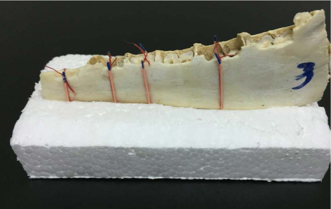
Fig. 1 A hemi-mandible fixed in the central slit of a plastic foam block. The 4 regions are defined by gutta-percha markers on the buccal and lingual sides.
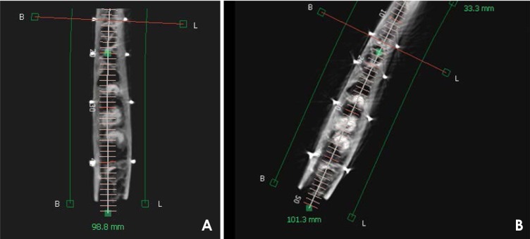
Fig. 2 Axial images of centrally (A) and peripherally positioned (B) exposures of a hemi-mandible.
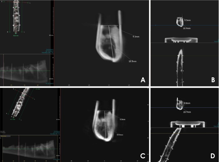
Fig. 3 Various image types of the same region with buccolingual and height measurements. A. Central/coronal. B. Central/cross-sectional. C. Peripheral/coronal. D. Peripheral/cross-sectional.
Reference
-
1. Lascala CA, Panella J, Marques MM. Analysis of the accuracy of linear measurements obtained by cone beam computed tomography (CBCT-NewTom). Dentomaxillofac Radiol. 2004; 33:291–294. PMID: 15585804.
Article2. Kosalagood P, Silkosessak OC, Pittayapat P, Pisarnturakit P, Pauwels R, Jacobs R. Linear measurement accuracy of eight cone beam computed tomography scanners. Clin Implant Dent Relat Res. 2015; 17:1217–1227. PMID: 24673845.
Article3. Hassan B, van der Stelt P, Sanderink G. Accuracy of three-dimensional measurements obtained from cone beam computed tomography surface-rendered images for cephalometric analysis: influence of patient scanning position. Eur J Orthod. 2009; 31:129–134. PMID: 19106265.
Article4. Timock AM, Cook V, McDonald T, Leo MC, Crowe J, Benninger BL, et al. Accuracy and reliability of buccal bone height and thickness measurements from cone-beam computed tomography imaging. Am J Orthod Dentofacial Orthop. 2011; 140:734–744. PMID: 22051495.
Article5. Mah J, Hatcher D. Three-dimensional craniofacial imaging. Am J Orthod Dentofacial Orthop. 2004; 126:308–309. PMID: 15356493.
Article6. Kobayashi K, Shimoda S, Nakagawa Y, Yamamoto A. Accuracy in measurement of distance using limited cone-beam computerized tomography. Int J Oral Maxillofac Implants. 2004; 19:228–231. PMID: 15101594.7. Frei C, Buser D, Dula K. Study on the necessity for cross-section imaging of the posterior mandible for treatment planning of standard cases in implant dentistry. Clin Oral Implants Res. 2004; 15:490–497. PMID: 15248885.
Article8. Ludlow JB, Laster WS, See M, Bailey LJ, Hershey HG. Accuracy of measurements of mandibular anatomy in cone beam computed tomography images. Oral Surg Oral Med Oral Pathol Oral Radiol Endod. 2007; 103:534–542. PMID: 17395068.
Article9. Sabban H, Mahdian M, Dhingra A, Lurie AG, Tadinada A. Evaluation of linear measurements of implant sites based on head orientation during acquisition: an ex vivo study using cone-beam computed tomography. Imaging Sci Dent. 2015; 45:73–80. PMID: 26125001.
Article10. Neves FS, Vasconcelos TV, Oenning AC, de Azevedo-Vaz SL, de Almeida SM, Freitas DQ. Oblique or orthoradial CBCT slices for preoperative implant planning: which one is more accurate? Braz J Oral Sci. 2014; 13:104–108.
Article11. Visconti MA, Verner FS, Assis NM, Devito KL. Influence of maxillomandibular positioning in cone beam computed tomography for implant planning. Int J Oral Maxillofac Surg. 2013; 42:880–886. PMID: 23566433.
Article12. Shokri A, Khajeh S. In vitro comparison of the effect of different slice thicknesses on the accuracy of linear measurements on cone beam computed tomography images in implant sites. J Craniofac Surg. 2015; 26:157–160. PMID: 25569395.
Article13. Nikbin A, Dalili Kajan Z, Taramsari M, Khosravifard N. Effect of object position in the field of view and application of a metal artifact reduction algorithm on the detection of vertical root fractures on cone-beam computed tomography scans: an in vitro study. Imaging Sci Dent. 2018; 48:245–254. PMID: 30607348.
Article14. Alkhader M, Hudieb M, Jarab F, Shaweesh A. The visibility of mandibular canal on orthoradial and oblique CBCT slices at molar implant sites. Biotechnol Biotechnol Equip. 2016; 30:770–776.
Article15. Kamburoğlu K, Murat S, Kılıç C, Yüksel S, Avsever H, Farman A, et al. Accuracy of CBCT images in the assessment of buccal marginal alveolar peri-implant defects: effect of field of view. Dentomaxillofac Radiol. 2014; 43:20130332. PMID: 24645965.
Article16. Dawood A, Brown J, Sauret-Jackson V, Purkayastha S. Optimization of cone beam CT exposure for pre-surgical evaluation of the implant site. Dentomaxillofac Radiol. 2012; 41:70–74. PMID: 22184628.
Article17. Moreira CR, Sales MA, Lopes PM, Cavalcanti MG. Assessment of linear and angular measurements on three-dimensional cone-beam computed tomographic images. Oral Surg Oral Med Oral Pathol Oral Radiol Endod. 2009; 108:430–436. PMID: 19386521.
Article18. Frongia G, Piancino MG, Bracco P. Cone-beam computed tomography: accuracy of three-dimensional cephalometry analysis and influence of patient scanning position. J Craniofac Surg. 2012; 23:1038–1043. PMID: 22777473.19. Rokn AR, Hashemi K, Akbari S, Kharazifard MJ, Barikani H, Panjnoosh M. Accuracy of linear measurements using cone beam computed tomography in comparison with clinical measurements. J Dent (Tehran). 2016; 13:333–339. PMID: 28127327.20. Moshfeghi M, Tavakoli MA, Hosseini ET, Hosseini AT, Hosseini IT. Analysis of linear measurement accuracy obtained by cone beam computed tomography (CBCT- NewTom VG). Dent Res J (Isfahan). 2012; 9(Suppl 1):S57–S62. PMID: 23814563.
- Full Text Links
-
- Actions
-
Cited
- CITED
-
- Close
- Share
-
- Similar articles
-
- Evaluation of linear measurements of implant sites based on head orientation during acquisition: An ex vivo study using cone-beam computed tomography
- Patient radiation dose and protection from cone-beam computed tomography
- Effect of object position in the field of view and application of a metal artifact reduction algorithm on the detection of vertical root fractures on cone-beam computed tomography scans: An in vitro study
- Effect of orthodontic treatment on temporomandibular joint’s articular eminence and condylar position
- Commentary on "Reliability of two different presurgical preparation methods for implant dentistry based on panoramic radiography and cone-beam computed tomography in cadavers"
