Korean J Radiol.
2020 Apr;21(4):413-421. 10.3348/kjr.2019.0703.
Development and Validation of a Simple Index Based on Non-Enhanced CT and Clinical Factors for Prediction of Non-Alcoholic Fatty Liver Disease
- Affiliations
-
- 1Department of Radiology and Research Institute of Radiology, Asan Medical Center, University of Ulsan College of Medicine, Seoul, Korea. seungsoolee@amc.seoul.kr
- 2Department of Clinical Epidemiology and Biostatistics, University of Ulsan College of Medicine, Seoul, Korea.
- 3Department of Diagnostic Pathology, Asan Medical Center, University of Ulsan College of Medicine, Seoul, Korea.
- KMID: 2471807
- DOI: http://doi.org/10.3348/kjr.2019.0703
Abstract
OBJECTIVE
A widely applicable, non-invasive screening method for non-alcoholic fatty liver disease (NAFLD) is needed. We aimed to develop and validate an index combining computed tomography (CT) and routine clinical data for screening for NAFLD in a large cohort of adults with pathologically proven NAFLD.
MATERIALS AND METHODS
This retrospective study included 2218 living liver donors who had undergone liver biopsy and CT within a span of 3 days. Donors were randomized 2:1 into development and test cohorts. CT(L-S) was measured by subtracting splenic attenuation from hepatic attenuation on non-enhanced CT. Multivariable logistic regression analysis of the development cohort was utilized to develop a clinical-CT index predicting pathologically proven NAFLD. The diagnostic performance was evaluated by analyzing the areas under the receiver operating characteristic curve (AUC). The cutoffs for the clinical-CT index were determined for 90% sensitivity and 90% specificity in the development cohort, and their diagnostic performance was evaluated in the test cohort.
RESULTS
The clinical-CT index included CT(L-S), body mass index, and aspartate transaminase and triglyceride concentrations. In the test cohort, the clinical-CT index (AUC, 0.81) outperformed CT(L-S) (0.74; p < 0.001) and clinical indices (0.73-0.75; p < 0.001) in diagnosing NAFLD. A cutoff of ≥ 46 had a sensitivity of 89% and a specificity of 41%, whereas a cutoff of ≥ 56.5 had a sensitivity of 57% and a specificity of 89%.
CONCLUSION
The clinical-CT index is more accurate than CT(L-S) and clinical indices alone for the diagnosis of NAFLD and may be clinically useful in screening for NAFLD.
MeSH Terms
Figure
-
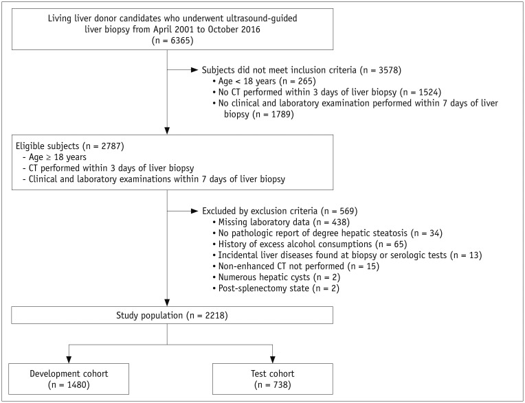
Fig. 1 Flow diagram for selection of study population.
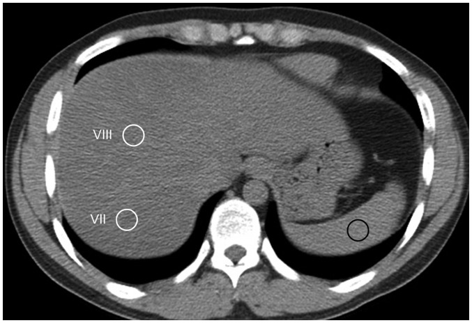
Fig. 2 Measurement of liver and spleen attenuation on non-enhanced axial CT images.Two 1.5-cm circular ROIs (white circles) were placed on hepatic segments VIII and VII, which were devoid of macroscopic vessels. 1.5-cm circular ROI was positioned on spleen (black circle). ROI = region of interest
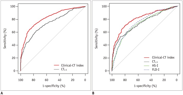
Fig. 3 Receiver operating characteristic curves of performance of clinical-CT index in diagnosing non-alcoholic fatty liver disease, compared with CTL-S in development cohort (A) and compared with CTL-S, HS-I, and FLD-I in test cohort (B).CTL-S = mean liver attenuation - mean spleen attenuation on non-enhanced CT, FLD-I = fatty liver disease index, HS-I = hepatic steatosis index
Reference
-
1. Wong VW, Wong GL, Yeung DK, Lau TK, Chan CK, Chim AM, et al. Incidence of non-alcoholic fatty liver disease in Hong Kong: a population study with paired proton-magnetic resonance spectroscopy. J Hepatol. 2015; 62:182–189. PMID: 25195550.
Article2. Younossi ZM, Blissett D, Blissett R, Henry L, Stepanova M, Younossi Y, et al. The economic and clinical burden of nonalcoholic fatty liver disease in the United States and Europe. Hepatology. 2016; 64:1577–1586. PMID: 27543837.
Article3. Adams LA, Sanderson S, Lindor KD, Angulo P. The histological course of nonalcoholic fatty liver disease: a longitudinal study of 103 patients with sequential liver biopsies. J Hepatol. 2005; 42:132–138. PMID: 15629518.
Article4. Teli MR, James OF, Burt AD, Bennett MK, Day CP. The natural history of nonalcoholic fatty liver: a follow-up study. Hepatology. 1995; 22:1714–1719. PMID: 7489979.
Article5. Gastaldelli A, Kozakova M, Højlund K, Flyvbjerg A, Favuzzi A, Mitrakou A, et al. Fatty liver is associated with insulin resistance, risk of coronary heart disease, and early atherosclerosis in a large European population. Hepatology. 2009; 49:1537–1544. PMID: 19291789.
Article6. Takyar V, Nath A, Beri A, Gharib AM, Rotman Y. How healthy are the “Healthy volunteers”? Penetrance of NAFLD in the biomedical research volunteer pool. Hepatology. 2017; 66:825–833. PMID: 28470683.
Article7. Lee SS, Park SH, Kim HJ, Kim SY, Kim MY, Kim DY, et al. Non-invasive assessment of hepatic steatosis: prospective comparison of the accuracy of imaging examinations. J Hepatol. 2010; 52:579–585. PMID: 20185194.
Article8. Lee SS, Park SH. Radiologic evaluation of nonalcoholic fatty liver disease. World J Gastroenterol. 2014; 20:7392–7402. PMID: 24966609.
Article9. Rogier J, Roullet S, Cornélis F, Biais M, Quinart A, Revel P, et al. Noninvasive assessment of macrovesicular liver steatosis in cadaveric donors based on computed tomography liver-to-spleen attenuation ratio. Liver Transpl. 2015; 21:690–695. PMID: 25761371.
Article10. Mellinger JL, Pencina KM, Massaro JM, Hoffmann U, Seshadri S, Fox CS, et al. Hepatic steatosis and cardiovascular disease outcomes: an analysis of the Framingham Heart Study. J Hepatol. 2015; 63:470–476. PMID: 25776891.
Article11. Pickhardt PJ, Hahn L, Muñoz del Rio A, Park SH, Reeder SB, Said A. Natural history of hepatic steatosis: observed outcomes for subsequent liver and cardiovascular complications. AJR Am J Roentgenol. 2014; 202:752–758. PMID: 24660702.
Article12. Bae JC, Lee WY, Yoon KH, Park JY, Son HS, Han KA, et al. Improvement of nonalcoholic fatty liver disease with carnitine-orotate complex in type 2 diabetes (CORONA): a randomized controlled trial. Diabetes Care. 2015; 38:1245–1252. PMID: 25877813.
Article13. Byun J, Lee SS, Sung YS, Shin Y, Yun J, Kim HS, et al. CT indices for the diagnosis of hepatic steatosis using non-enhanced CT images: development and validation of diagnostic cut-off values in a large cohort with pathological reference standard. Eur Radiol. 2019; 29:4427–4435. PMID: 30569183.
Article14. Fuyan S, Jing L, Wenjun C, Zhijun T, Weijing M, Suzhen W, et al. Fatty liver disease index: a simple screening tool to facilitate diagnosis of nonalcoholic fatty liver disease in the Chinese population. Dig Dis Sci. 2013; 58:3326–3334. PMID: 23900558.
Article15. Bedogni G, Bellentani S, Miglioli L, Masutti F, Passalacqua M, Castiglione A, et al. The fatty liver index: a simple and accurate predictor of hepatic steatosis in the general population. BMC Gastroenterol. 2006; 6:33. PMID: 17081293.
Article16. Lee JH, Kim D, Kim HJ, Lee CH, Yang JI, Kim W, et al. Hepatic steatosis index: a simple screening tool reflecting nonalcoholic fatty liver disease. Dig Liver Dis. 2010; 42:503–508. PMID: 19766548.
Article17. Choi YJ, Lee DH, Han KD, Yoon H, Shin CM, Park YS, et al. Is nonalcoholic fatty liver disease associated with the development of prostate cancer? A nationwide study with 10,516,985 Korean men. PLoS One. 2018; 13:e0201308. PMID: 30231041.
Article18. Neuschwander-Tetri BA, Caldwell SH. Nonalcoholic steatohepatitis: summary of an AASLD single topic conference. Hepatology. 2003; 37:1202–1219. PMID: 12717402.
Article19. Yip TC, Ma AJ, Wong VW, Tse YK, Chan HL, Yuen PC, et al. Laboratory parameter-based machine learning model for excluding non-alcoholic fatty liver disease (NAFLD) in the general population. Aliment Pharmacol Ther. 2017; 46:447–456. PMID: 28585725.
Article20. Kleiner DE, Brunt EM, Van Natta M, Behling C, Contos MJ, Cummings OW, et al. Design and validation of a histological scoring system for nonalcoholic fatty liver disease. Hepatology. 2005; 41:1313–1321. PMID: 15915461.
Article21. DeLong ER, DeLong DM, Clarke-Pearson DL. Comparing the areas under two or more correlated receiver operating characteristic curves: a nonparametric approach. Biometrics. 1988; 44:837–845. PMID: 3203132.
Article22. Moons KG, Altman DG, Reitsma JB, Ioannidis JP, Macaskill P, Steyerberg EW, et al. Transparent Reporting of a multivariable prediction model for Individual Prognosis or Diagnosis (TRIPOD): explanation and elaboration. Ann Intern Med. 2015; 162:W1–W73. PMID: 25560730.
Article23. Moons KG, Kengne AP, Woodward M, Royston P, Vergouwe Y, Altman DG, et al. Risk prediction models: I. Development, internal validation, and assessing the incremental value of a new (bio)marker. Heart. 2012; 98:683–690. PMID: 22397945.
Article24. Kotronen A, Peltonen M, Hakkarainen A, Sevastianova K, Bergholm R, Johansson LM, et al. Prediction of non-alcoholic fatty liver disease and liver fat using metabolic and genetic factors. Gastroenterology. 2009; 137:865–872. PMID: 19524579.
Article25. Zhang Z, Wang G, Kang K, Wu G, Wang P. Diagnostic accuracy and clinical utility of a new noninvasive index for hepatic steatosis in patients with hepatitis B virus infection. Sci Rep. 2016; 6:32875. PMID: 27597515.
Article26. Poynard T, Ratziu V, Naveau S, Thabut D, Charlotte F, Messous D, et al. The diagnostic value of biomarkers (SteatoTest) for the prediction of liver steatosis. Comp Hepatol. 2005; 4:10. PMID: 16375767.
Article27. Bedogni G, Kahn HS, Bellentani S, Tiribelli C. A simple index of lipid overaccumulation is a good marker of liver steatosis. BMC Gastroenterol. 2010; 10:98. PMID: 20738844.
Article28. Lin SC, Heba E, Wolfson T, Ang B, Gamst A, Han A, et al. Noninvasive diagnosis of nonalcoholic fatty liver disease and quantification of liver fat using a new quantitative ultrasound technique. Clin Gastroenterol Hepatol. 2015; 13:1337–1345.e6. PMID: 25478922.
- Full Text Links
-
- Actions
-
Cited
- CITED
-
- Close
- Share
-
- Similar articles
-
- Validation of Fatty Liver Index as a Marker for Non-Alcoholic Fatty Liver Disease
- Diagnosis of Non-Alcoholic Fatty Liver Disease Based on Clinical and Laboratory Data
- A Case of Hepatocellular Carcinoma in Non-alcoholic Fatty Liver Disease
- Should you advocate for hepatocellular carcinomasurveillance in patients with alcohol-related liverdisease or non-alcoholic fatty liver disease?
- Histologic Risk Factor for Mortality and Development of Severe Liver Disease in Biopsy-proven Non-alcoholic Fatty Liver Disease
