Ann Surg Treat Res.
2020 Mar;98(3):124-129. 10.4174/astr.2020.98.3.124.
A prospective study of discrepancy between clinical and pathological diagnosis of appendiceal mucinous neoplasm
- Affiliations
-
- 1Department of Surgery, Chonnam National University Hospital, Gwangju, Korea. drkwak@korea.ac.kr
- KMID: 2471177
- DOI: http://doi.org/10.4174/astr.2020.98.3.124
Abstract
- PURPOSE
Appendiceal tumoral lesions can occur as benign, malignant, or borderline disease. Determination of the extent of surgery through accurate diagnosis is important in these tumoral lesions. In this study, we assessed the accuracy of preoperative CT and identified the factors affecting diagnosis.
METHODS
Patients diagnosed or strongly suspected from July 2016 to June 2019 with appendiceal mucocele or mucinous neoplasm using abdominal CT were included in the study. All the patients underwent single-incision laparoscopic cecectomy with the margin of cecum secured at least 2 cm from the appendiceal base. To compare blood test results and CT findings, the patients were divided into a mucinous and a nonmucinous group according to pathology.
RESULTS
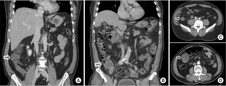
The total number of patients included in this study was 54 and biopsy confirmed appendiceal mucinous neoplasms in 39 of them. With CT, the accuracy of diagnosis was 89.7%. The mean age of the mucinous group was greater than that of the nonmucinous group (P = 0.035). CT showed that the maximum diameter of appendiceal tumor in the mucinous group was greater than that in the nonmucinous group (P < 0.001). Calcification was found only in the appendix of patients in the mucinous group (P = 0.012). Multivariate analysis revealed that lager tumor diameter was a factor of diagnosis for appendiceal mucinous neoplasm.
CONCLUSION
The accuracy of preoperative diagnosis of appendiceal mucinous neoplasms in this study was 89.7%. Blood test results did not provide differential diagnosis, and the larger the diameter of appendiceal tumor on CT, the more accurate the diagnosis.
Keyword
MeSH Terms
Figure
Reference
-
1. von Rokitansky C. A manual of pathological anatomy. Philadelphia (PA): Blanchard & Lea;1855.2. Carr NJ, Cecil TD, Mohamed F, Sobin LH, Sugarbaker PH, Gonzalez-Moreno S, et al. A consensus for classification and pathologic reporting of pseudomyxoma peritonei and associated appendiceal neoplasia: the results of the Peritoneal Surface Oncology Group International (PSOGI) Modified Delphi Process. Am J Surg Pathol. 2016; 40:14–26. PMID: 26492181.3. Leonards LM, Pahwa A, Patel MK, Petersen J, Nguyen MJ, Jude CM. Neoplasms of the appendix: pictorial review with clinical and pathologic correlation. Radiographics. 2017; 37:1059–1083. PMID: 28598731.
Article4. Hinson FL, Ambrose NS. Pseudomyxoma peritonei. Br J Surg. 1998; 85:1332–1339. PMID: 9782010.
Article5. Madwed D, Mindelzun R, Jeffrey RB Jr. Mucocele of the appendix: imaging findings. AJR Am J Roentgenol. 1992; 159:69–72. PMID: 1609724.
Article6. Isaacs KL, Warshauer DM. Mucocele of the appendix: computed tomographic, endoscopic, and pathologic correlation. Am J Gastroenterol. 1992; 87:787–789. PMID: 1590322.7. Won EJ, Ju JK, Cho YN, Jin HM, Park KJ, Kim TJ, et al. Clinical relevance of circulating mucosal-associated invariant T cell levels and their anti-cancer activity in patients with mucosal-associated cancer. Oncotarget. 2016; 7:76274–76290. PMID: 27517754.
Article8. Landen S, Bertrand C, Maddern GJ, Herman D, Pourbaix A, de Neve A, et al. Appendiceal mucoceles and pseudomyxoma peritonei. Surg Gynecol Obstet. 1992; 175:401–404. PMID: 1440166.9. Pickhardt PJ, Levy AD, Rohrmann CA Jr, Kende AI. Primary neoplasms of the appendix: radiologic spectrum of disease with pathologic correlation. Radiographics. 2003; 23:645–662. PMID: 12740466.
Article10. Caspi B, Cassif E, Auslender R, Herman A, Hagay Z, Appelman Z. The onion skin sign: a specific sonographic marker of appendiceal mucocele. J Ultrasound Med. 2004; 23:117–121. PMID: 14756359.11. Dachman AH, Lichtenstein JE, Friedman AC. Mucocele of the appendix and pseudomyxoma peritonei. AJR Am J Roentgenol. 1985; 144:923–929. PMID: 3885692.
Article12. Bennett GL, Tanpitukpongse TP, Macari M, Cho KC, Babb JS. CT diagnosis of mucocele of the appendix in patients with acute appendicitis. AJR Am J Roentgenol. 2009; 192:W103–W110. PMID: 19234237.
Article13. Van Hooser A, Williams TR, Myers DT. Mucinous appendiceal neoplasms: pathologic classification, clinical implications, imaging spectrum and mimics. Abdom Radiol (NY). 2018; 43:2913–2922. PMID: 29564494.
Article14. Carr NJ, McCarthy WF, Sobin LH. Epithelial noncarcinoid tumors and tumor-like lesions of the appendix. A clinicopathologic study of 184 patients with a multivariate analysis of prognostic factors. Cancer. 1995; 75:757–768. PMID: 7828125.
Article15. Marotta B, Chaudhry S, McNaught A, Quereshy F, Vajpeyi R, Chetty R, et al. Predicting underlying neoplasms in appendiceal mucoceles at CT: focal versus diffuse luminal dilatation. AJR Am J Roentgenol. 2019; 213:343–348. PMID: 30973782.
Article16. Randal Bollinger R, Barbas AS, Bush EL, Lin SS, Parker W. Biofilms in the large bowel suggest an apparent function of the human vermiform appendix. J Theor Biol. 2007; 249:826–831. PMID: 17936308.
Article17. Laurin M, Everett ML, Parker W. The cecal appendix: one more immune component with a function disturbed by post-industrial culture. Anat Rec (Hoboken). 2011; 294:567–579. PMID: 21370495.
Article18. Woodruff R. Benign and malignant cyst tumors of the appendix. Surg Gynecol Obstet. 1940; 71:750–755.19. Higa E, Rosai J, Pizzimbono CA, Wise L. Mucosal hyperplasia, mucinous cystadenoma, and mucinous cystadenocarcinoma of the appendix. A re-evaluation of appendiceal “mucocele”. Cancer. 1973; 32:1525–1541. PMID: 4757938.20. De Abreu Filho JG, De Lira EF. Mucocele of the appendix: appendectomy or colectomy? J Coloproctol (Rio J). 2011; 31:276–284.21. Miraliakbari R, Chapman WH 3rd. Laparoscopic treatment of an appendiceal mucocele. J Laparoendosc Adv Surg Tech A. 1999; 9:159–163. PMID: 10235354.
Article22. Gonzalez Moreno S, Shmookler BM, Sugarbaker PH. Appendiceal mucocele. Contraindication to laparoscopic appendectomy. Surg Endosc. 1998; 12:1177–1179. PMID: 9716778.23. Stocchi L, Wolff BG, Larson DR, Harrington JR. Surgical treatment of appendiceal mucocele. Arch Surg. 2003; 138:585–589. PMID: 12799327.
Article
- Full Text Links
- Actions
-
Cited
- CITED
-
- Close
- Share
- Similar articles
-
- Mucinous Tumors of the Appendix Associated with Mucinous Tumors of the Ovary and Pseudomyxoma Peritonei: A Clinicopathologic Analysis of 5 Cases Supporting an Appendiceal Origin
- A Case of Asymptomatic Appendiceal Intussusception by Mucinous Cystadenoma
- A rare presentation of low-grade appendiceal mucinous neoplasm within an amyand’s hernia: a case report
- Clinical Features and Prognostic Factors in Primary Adenocarcinoma of the Appendix
- Appendiceal Mucocele


