J Pathol Transl Med.
2025 Mar;59(2):139-146. 10.4132/jptm.2024.12.27.
Mucocele of the rectal stump: mucinous cystic neoplasm with low-grade dysplasia simulating low-grade appendiceal mucinous neoplasm
- Affiliations
-
- 1Department of Pathology and Laboratory Medicine, Albany Medical Center, Albany, NY, USA
- 2Department of Surgery, Albany Medical Center, Albany, NY, USA
- KMID: 2566164
- DOI: http://doi.org/10.4132/jptm.2024.12.27
Abstract
- Mucoceles, commonly observed in the appendix, are mucin-filled, dilated structures arising from a range of etiologies. Cases associated with dysplastic or neoplastic epithelium can rupture and disseminate within the abdominopelvic cavity. Similar lesions in other parts of the colon are exceedingly rare, with only 16 colonic mucoceles having been reported. The first case of a colonic mucinous neoplasm with dysplasia resembling a low-grade appendiceal mucinous neoplasm involving rectal stump was described in 2016. Here, we present the second such case arising in the rectal stump, identified in a 44-year-old male with extensive surgical history. Microscopic examination revealed low-grade dysplastic epithelium lining the cyst and mucin dissecting into the stroma, without evidence of rupture or extramural mucin. The patient was followed for 16 months without recurrence or peritoneal disease. The exact etiology and outcome of these rare lesions remain unknown, requiring close follow-up.
Keyword
Figure
-

Fig. 1. Abdominal computed tomography scan demonstrating a large, multiloculated cystic mass within the pelvis (coronal [A] and sagittal [B] planes respectively).

Fig. 2. Scanning view of the lumen lined by attenuated colorectal type mucosa (A), denuded area (B), colorectal mucosa (C), and anorectal transitional type mucosa (D).
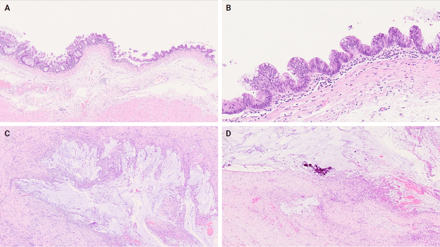
Fig. 3. (A) Scanning view of the lumen lined by attenuated colorectal type mucosa. (B) High-power view showing low-grade dysplasia. (C) Acellular mucin pools dissecting stroma. (D) Mucin pools with degenerative calcifications.
Reference
-
References
1. Nicosia JF, Abcarian H. Mucocele of the distal colonic segment, a late sequela of trauma to the perineum: report of a case. Dis Colon Rectum. 1974; 17:536–9. DOI: 10.1007/bf02587031. PMID: 4853279.2. Hitch DC, Patil UB, Panicek DM. Mucocele after endorectal pull-through for imperforate anus. J Pediatr Surg. 1987; 22:1023–4. DOI: 10.1016/s0022-3468(87)80506-7. PMID: 3430304.3. Creagh MF, Chan TY. Case report: rectal mucocele following Hartmann's procedure. Clin Radiol. 1991; 43:358–9. DOI: 10.1016/s0009-9260(05)80550-1. PMID: 2036766.4. Hsu KF, Hsieh CB, Yu JC, Chan DC, Wu CC, Jin JS, et al. Rare rectal mucocele mimic tumor following hemorrhoidectomy in an adult patient. Rev Esp Enferm Dig. 2011; 103:276–7. PMID: 21619395.5. Ali A, Krishnan A, Rehman S, Rao V, Pearson HJ. Giant colonic mucocele following palliative surgery for metastatic adenocarcinoma. J Surg Case Rep. 2011; 2011:1–4. DOI: 10.1093/jscr/2011.3.9.6. Ai XB, Feng JC, Gong FY, Wang A, Ren DH, Sui K. Endoscopic therapy of colonic liver flexure mucocele. Case Rep Gastroenterol. 2011; 5:433–7. DOI: 10.1159/000330484. PMID: 21960945.7. Teoh AY, Lee JF, Chong CC, Tang RS. Endoscopic ultrasonography-guided drainage of a rectal mucocele after total colectomy for Crohn's disease. Endoscopy. 2013; 45 Suppl 2 UCTN:E252–3. DOI: 10.1055/s-0033-1344415. PMID: 24008451.8. Appleton N, Day N, Walsh C. Rectal mucocoele following subtotal colectomy for colitis. Ann R Coll Surg Engl. 2014; 96:e13–4. DOI: 10.1308/003588414x13946184903009.9. Nakatani K, Tokuhara K, Sakaguchi T, Ryota H, Yoshioka K, Kon M. Low-grade mucinous neoplasia in a cecal diverticulum: A case report. Int J Surg Case Rep. 2015; 15:66–9. DOI: 10.1016/j.ijscr.2015.08.030. PMID: 26318130.10. Tanaka T, Kawai K, Abe H, Murono K, Otani K, Nishikawa T, et al. Giant mucocele of the colon at the distal stump due to low-grade mucinous neoplasia. Surg Case Rep. 2016; 2:117. DOI: 10.1186/s40792-016-0223-9. PMID: 27787812.11. Ishii D, Aoki T, Inaba S, Yabuki H. Rectal mucocele in the anterior wall of the rectum. BMJ Case Rep. 2018; 2018:bcr2018225097. DOI: 10.1136/bcr-2018-225097. PMID: 29776947.12. Schneider R, Kraljevic M, von Flue M, Fuglistaler I. Giant symptomatic rectal mucocele following subtotal colectomy. Case Rep Gastroenterol. 2018; 12:143–6. DOI: 10.1159/000488523. PMID: 29805357.13. Longchamp G, Colucci N, Ris F, Buchs NC. Rectal stump mucocele after a Hartmann's procedure causing mechanical ileus. BMJ Case Rep. 2021; 14:e237543. DOI: 10.1136/bcr-2020-237543. PMID: 33419748.14. Chen F, Harvey SE, Young ED, Liang TZ, Larman T, Voltaggio L. Extra-appendiceal mucinous neoplasms: a tumour with clinicopathologic similarities to low- and high-grade appendiceal counterpart. Hum Pathol. 2024; 148:23–31. DOI: 10.1016/j.humpath.2024.04.010. PMID: 38677555.15. Faure M, Salgado R, Op de Beeck B, Bellinck P, Termote JL, Parizel PM. Mucocele of the appendix: case report and review of literature. JBR-BTR. 2014; 97:217–21. DOI: 10.5334/jbr-btr.87. PMID: 25603629.16. Carr NJ, Cecil TD, Mohamed F, Sobin LH, Sugarbaker PH, Gonzalez-Moreno S, et al. A consensus for classification and pathologic reporting of pseudomyxoma peritonei and associated appendiceal neoplasia: the results of the Peritoneal Surface Oncology Group International (PSOGI) modified Delphi process. Am J Surg Pathol. 2016; 40:14–26. DOI: 10.1097/pas.0000000000000535. PMID: 26492181.17. Govaerts K, Lurvink RJ, De Hingh I, Van der Speeten K, Villeneuve L, Kusamura S, et al. Appendiceal tumours and pseudomyxoma peritonei: literature review with PSOGI/EURACAN clinical practice guidelines for diagnosis and treatment. Eur J Surg Oncol. 2021; 47:11–35. DOI: 10.1016/j.ejso.2020.02.012. PMID: 32199769.18. Guner M, Aydin C. Low-grade appendiceal mucinous neoplasm: what is the best treatment? Cureus. 2023; 15:e46591. DOI: 10.7759/cureus.46591. PMID: 37808597.19. Cristian DA, Grama FA, Becheanu G, Pop A, Popa I, Surlin V, et al. Low-grade appendiceal mucinous neoplasm mimicking an adnexal mass. Rom J Morphol Embryol. 2015; 56:837–42. PMID: 26429182.20. Pai RK, Longacre TA. Appendiceal mucinous tumors and pseudomyxoma peritonei: histologic features, diagnostic problems, and proposed classification. Adv Anat Pathol. 2005; 12:291–311. DOI: 10.1097/01.pap.0000194625.05137.51. PMID: 16330927.21. Misdraji J. Mucinous epithelial neoplasms of the appendix and pseudomyxoma peritonei. Mod Pathol. 2015; 28 Suppl 1:S67–79. DOI: 10.1038/modpathol.2014.129. PMID: 25560600.22. Gupta P, Mishra AK, Deo A, Yadav R, K CM, Bhattarai A. Complete small intestinal obstruction due to band formation in low-grade appendiceal mucinous neoplasm: a rare case report and literature review. Int J Surg Case Rep. 2023; 108:108422. DOI: 10.1016/j.ijscr.2023.108422. PMID: 37348199.
- Full Text Links
-
- Actions
-
Cited
- CITED
-
- Close
- Share
-
- Similar articles
-
- A rare presentation of low-grade appendiceal mucinous neoplasm within an amyand’s hernia: a case report
- Asymptomatic Appendiceal Mucocele with Typical Onion Skin Sign on Ultrasound
- Appendiceal mucocele masquerading as an epithelial borderline ovarian tumor: a case report and literature review
- Appendiceal Mucinous Neoplasm Detected due to the Protrusion of Mucin, in the Absence of Appendiceal Distension: A Case Report
- A prospective study of discrepancy between clinical and pathological diagnosis of appendiceal mucinous neoplasm
