Orthodontic protraction of the third molars to the posterior teeth missing area
- Affiliations
-
- 1Department of Orthodontics, School of Dentistry, Institute of Oral Biosciences, Jeonbuk National University, Jeonju, Republic of Korea. young@jbnu.ac.kr
- 2Biomedical Research Institute of Jeonbuk National University Hospital, Jeonju, Republic of Korea.
- KMID: 2470632
- DOI: http://doi.org/10.14368/jdras.2019.35.4.260
Abstract
- The prolonged neglect of the posterior teeth missing area may cause mesial drift, extrusion, unexpected movement of the adjacent teeth and alveolar bone loss with occlusion collapse. Therefore it is recommended to treat that area by the prosthesis as soon as possible after tooth missing. However, if orthodontic treatment is applied to move the remained teeth, it can create improved biomechanical dentoalveolar environment. The use of the third molars in teeth missing area provides advantages as optimizing of prosthesis size. However, crown shape, location, soundness of the third molar and possible of eruption failure should be considered. In this case report, two patients closed a second teeth missing site and reduced the size of the first and second teeth missing area for an implant by protraction of impacted third molars. This case reports the considerations for closing or reducing the posterior teeth space with protracting the third molars by comparing two patients.
Figure
-

Fig. 1 Pre-treatment clinical photographs of Case I. She has hopeless posterior teeth with deep caries and root rest but well aligned anterior teeth.
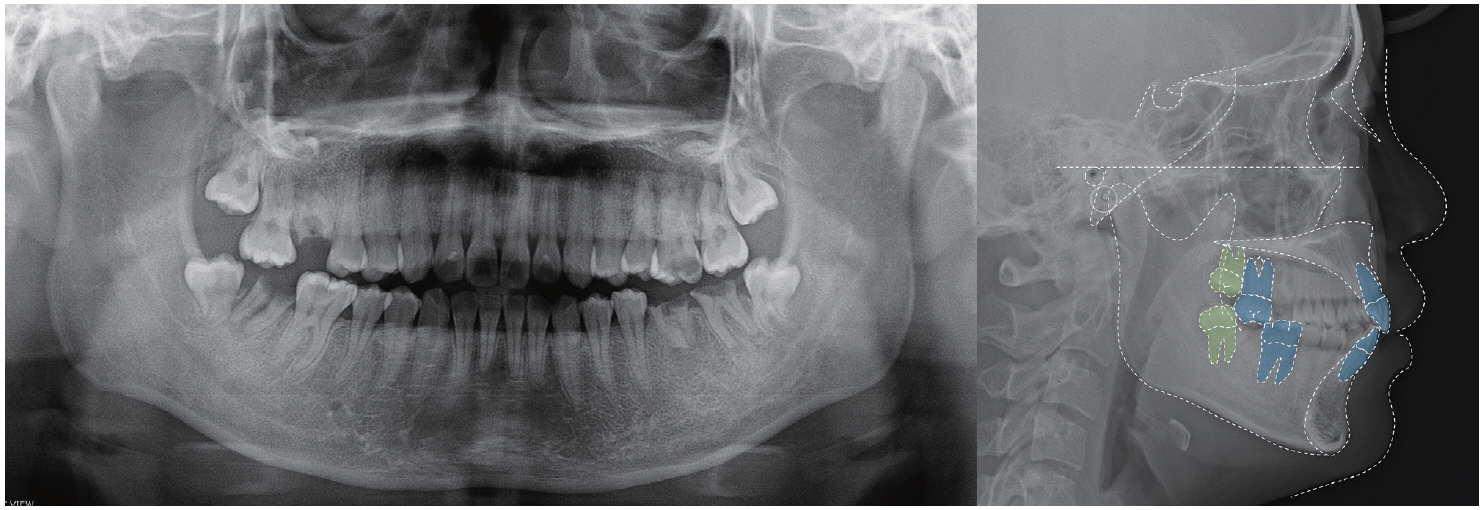
Fig. 2 Pre-treatment panoramic and lateral cephalometric radiographs of Case I. Maxillary second molar and mandibular first molar was traced because treatment plan included maxillary second molar protraction.
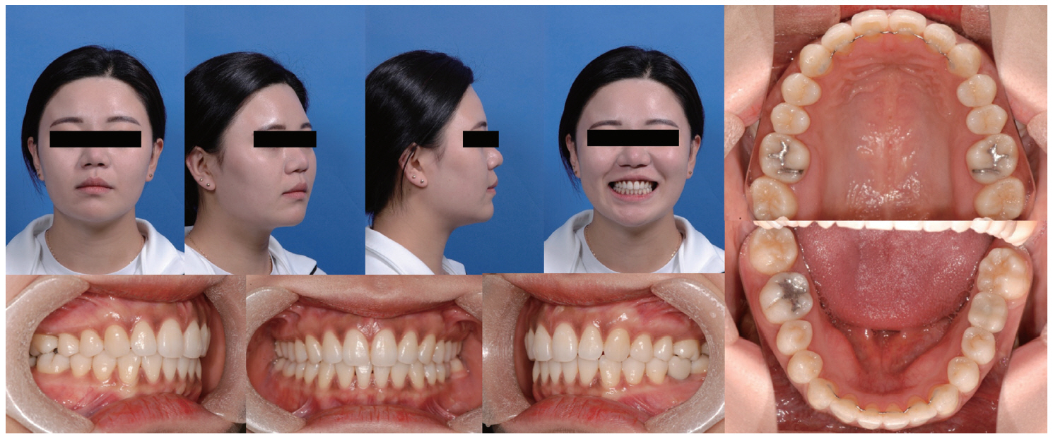
Fig. 3 Post-treatment clinical photographs of Case I. The maxillary first molars were replaced by second molars. The mandibular second molars were replaced by third molars and mandibular left first molar was replaced by a prosthetic implant.
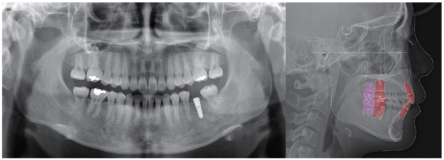
Fig. 4 Post-treatment panoramic and lateral cephalometric radiographs of Case I.
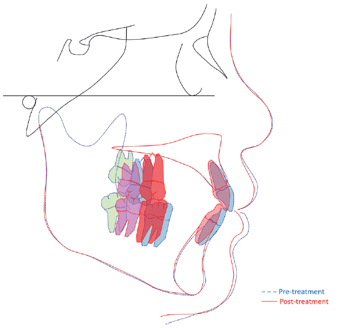
Fig. 5 Superimposition of pre- and post-treatment lateral cephalograms of Case I. Blue line means pretreatment tracing and red line means post-treatment tracing. Blue and red teeth indicate maxillary second molars and mandibular first molar’s position before and after treatment. Green and pink teeth means maxillary and mandibular third molar’s position before and after orthodontic treatment.
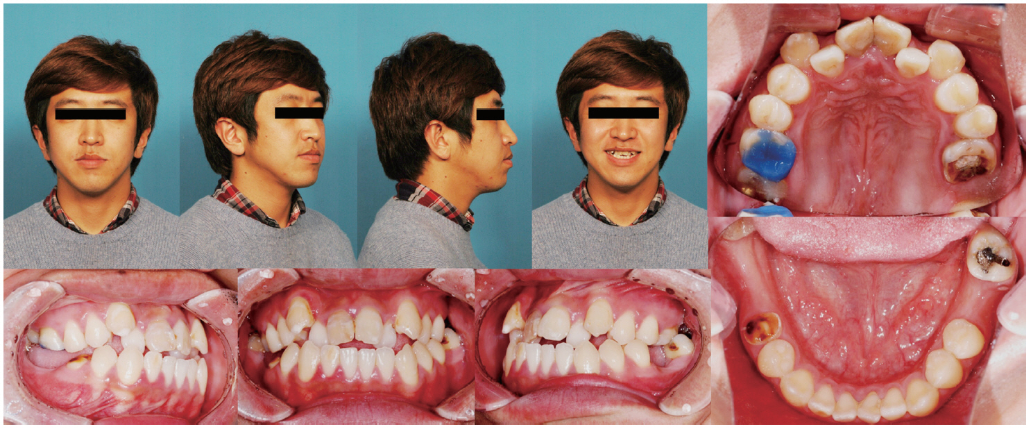
Fig. 6 Pre-treatment clinical photographs of Case II. He has teeth missing and hopeless teeth with deep caries and root rest on posterior area but also anterior crowding.
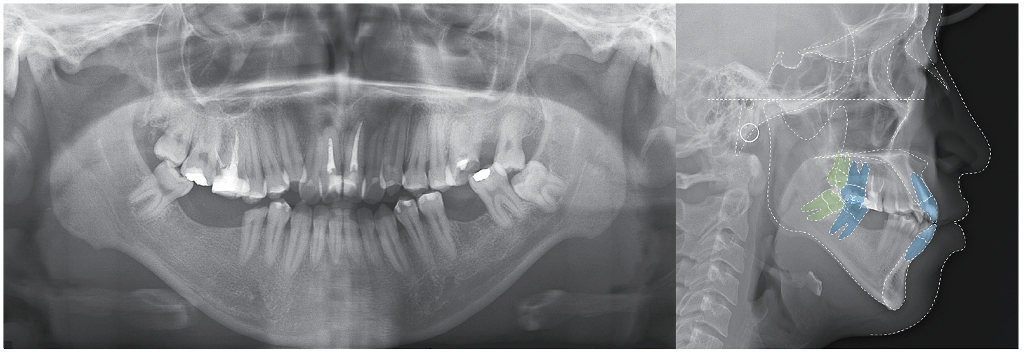
Fig. 7 Pre-treatment panoramic and lateral cephalometric radiographs of Case II. Maxillary and mandibular second molar was traced because the upper and lower first molars were defective or missing and treatment plan included second molar protraction.
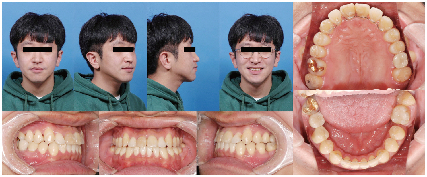
Fig. 8 Post-treatment clinical photographs of Case II. The right maxillary and left mandibular first molars were replaced by second molars and left maxillary and right mandibular first molars were replaced by prosthetic implants.

Fig. 9 Post-treatment panoramic and lateral cephalometric radiographs of Case II.
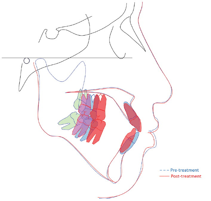
Fig. 10 Superimposition of pre- and post-treatment lateral cephalograms of Case II. Blue line means pretreatment tracing and red line means post-treatment tracing. Blue and red teeth indicate maxillary and mandibular second molar's position before and after treatment. Green and pink teeth means maxillary and mandibular third molar's position before and after orthodontic treatment.

Fig. 11 Comparison of (A) case I and (B) case II patient’s pre-treatment alveolar bone level. (A) Redline indicates alveolar bone level of patient I. (B) Red color area indicates absorbed alveolar bone level.
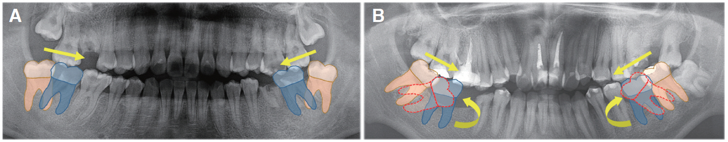
Fig. 12 Protraction force (direction as yellow straight arrow) make distal tipped third molars mesial tipping. (A) Mesial traction force makes leveling easier in patient with distal tipped third molars, (B) but it needs additional time for tipping control as like round yellow arrow in patient with mesial tipped third molars because mesial traction force makes molar more mesial tipping as dotted red teeth.
Reference
-
References
1. Graber LW, Vanarsdall RL, Vig KW, Huang GJ. 2017. Orthodontics current principles and technique. 6th ed. Elsevier;Seoul: p. 644–97.2. Kokich VG, Spear FM. 1997; Guidelines for managing the orthodontic-restorative patient. Semin Orthod. 3:3–20. DOI: 10.1016/S1073-8746(97)80036-9. PMID: 9206469.3. Nanda R. 2015. Esthetics and biomechanics in orthodontics. 2nd ed. Elsevier;Seoul: p. 567–87.4. Tomonari H, Yagi T, Kuninori T, Ikemori T, Miyawaki S. 2015; Replacement of a first molar and 3 second molars by the mesial inclination of 4 impacted third molars in an adult with a Class II division 1 malocclusion. Am J Orthod Dentofacial Orthop. 147:755–65. DOI: 10.1016/j.ajodo.2014.05.030. PMID: 26038080.5. Gooris CG, Artun J, Joondeph DR. 1990; Eruption of mandibular third molars after second-molar extraction: a radiographic study. Am J Orthod Dentofacial Orthop. 98:161–7. DOI: 10.1016/0889-5406(90)70010-A. PMID: 2378320.6. Orton-Gibbs S, Crow V, Orton HS. 2001; Eruption of third permanent molars after the extraction of second permanent molars. Par1: Assessment of third molar position and size. 119:226–38. DOI: 10.1067/mod.2001.111556. PMID: 11244416.7. Orton-Gibbs S, Orton S, Orton H. 2001; Eruption of third permanent molars after the extraction of second permanent molars. Part 2: Functional occlusion and periodontal status. 119:239–44. DOI: 10.1067/mod.2001.111555. PMID: 11244417.8. De-la-Rosa-Gay C, Valmaseda-Castellon E, Gay-Escoda C. 2006; Spontaneous third-molar eruption after second-molar extraction in orthodontic patients. Am J Orthod Dentofacial Orthop. 129:337–44. DOI: 10.1016/j.ajodo.2005.11.002. PMID: 16527628.9. De-la-Rosa-Gay C, Valmaseda-Castellon E, Gay-Escoda C. 2010; Predictive model of third molar eruption after second molar extraction. Am J Orthod Dentofacial Orthop. 137:346–53. DOI: 10.1016/j.ajodo.2008.02.026. PMID: 20197171.10. Richardson ME, Richardson A. 1993; Lower third molar development subsequent to second molar extraction. Am J Orthod Dentofacial Orthop. 104:566–74. DOI: 10.1016/S0889-5406(05)80440-8. PMID: 8249932.
- Full Text Links
-
- Actions
-
Cited
- CITED
-
- Close
- Share
-
- Similar articles
-
- Protraction of mandibular molars through a severely atrophic edentulous space in a case of juvenile periodontitis
- The second molars in orthodontics
- Analysis of the Characteristics of First Permanent Molars with Delayed Eruption
- Comprehensive Orthodontic Treatment in a Middle-Aged Patient with Missing Maxillary Left First Premolar: A Case Report
- Non-Surgical Correction of Collapsed Posterior Occlusion in a Unilateral Cleft Lip and Palate Patient with Multiple Missing Teeth: A Case Report
