Korean J Orthod.
2020 Mar;50(2):145-154. 10.4041/kjod.2020.50.2.145.
Protraction of mandibular molars through a severely atrophic edentulous space in a case of juvenile periodontitis
- Affiliations
-
- 1Department of Stomatology, The Second Affiliated Hospital, School of Medicine, Zhejiang University, Hangzhou, China. paulwu010203@zju.edu.cn
- 2Department of Orthodontics, Affiliated Hospital of Stomatology, School of Medicine, Zhejiang University, Hangzhou, China.
- KMID: 2471875
- DOI: http://doi.org/10.4041/kjod.2020.50.2.145
Abstract
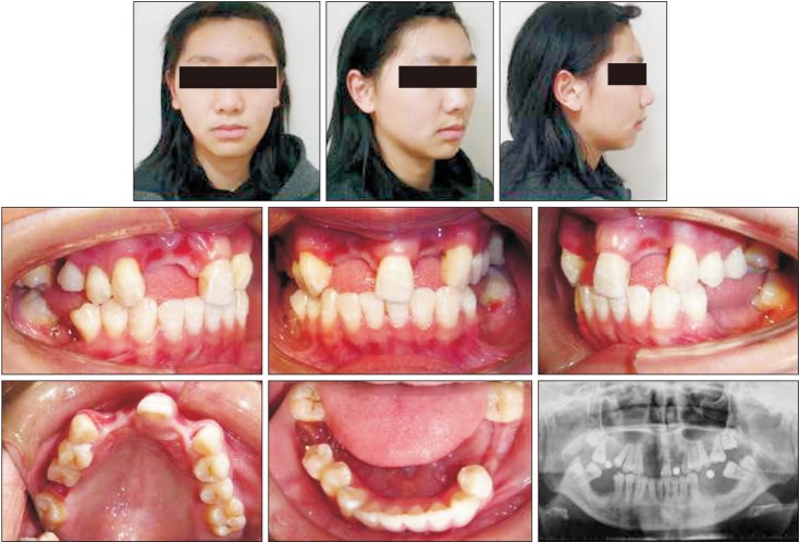
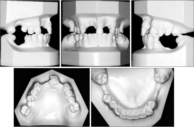
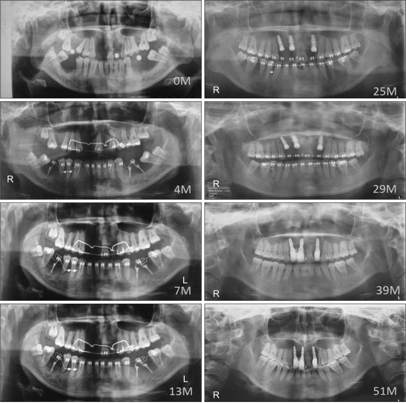
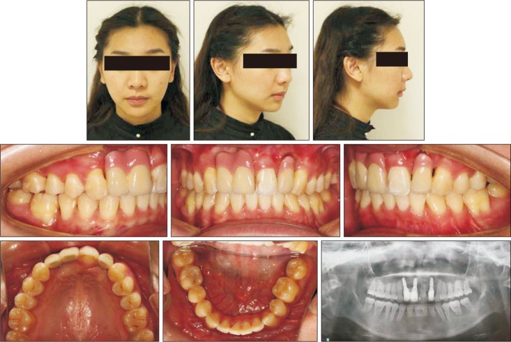
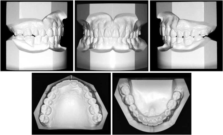
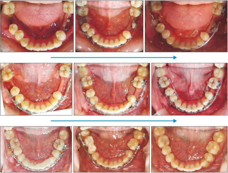
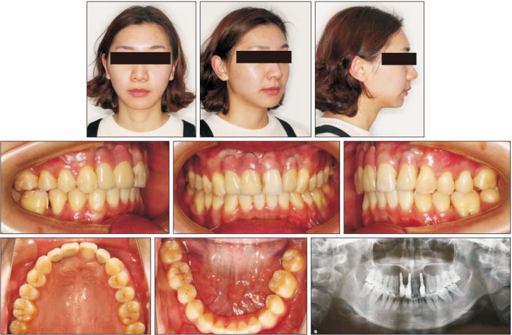
- Moving the mandibular posterior teeth into a severely atrophic edentulous space is a challenge. A carefully designed force-and-moment system that results in bodily protraction of the posterior teeth with balanced bone resorption and apposition is needed in such cases. This report describes the treatment of a 19-year-old woman with missing mandibular first molars due to juvenile periodontitis. Miniscrews were used as absolute anchorage during protraction of the mandibular second and third molars. Bodily mesial movement of the mandibular second and third molars was achieved over a distance of 11 to 17 mm after 39 months of orthodontic treatment.
Keyword
Figure
Reference
-
1. Baik UB, Chun YS, Jung MH, Sugawara J. Protraction of mandibular second and third molars into missing first molar spaces for a patient with an anterior open bite and anterior spacing. Am J Orthod Dentofacial Orthop. 2012; 141:783–795. PMID: 22640680.
Article2. Roberts WE. Bone physiology, metabolism and biomechanics in orthodontic practice. In : Graber TM, Vanarsdall RL, editors. Orthodontics: current principles and techniques. St Louis: Mosby;1994.3. Baik UB, Kim MR, Yoon KH, Kook YA, Park JH. Orthodontic uprighting of a horizontally impacted third molar and protraction of mandibular second and third molars into the missing first molar space for a patient with posterior crossbites. Am J Orthod Dentofacial Orthop. 2017; 151:572–582. PMID: 28257742.
Article4. Cacciafesta V, Melsen B. Mesial bodily movement of maxillary and mandibular molars with segmented mechanics. Clin Orthod Res. 2001; 4:182–188. PMID: 11553103.
Article5. Kim SJ, Sung EH, Kim JW, Baik HS, Lee KJ. Mandibular molar protraction as an alternative treatment for edentulous spaces: focus on changes in root length and alveolar bone height. J Am Dent Assoc. 2015; 146:820–829. PMID: 26514887.6. Saga AY, Maruo IT, Maruo H, Guariza Filho O, Camargo ES, Tanaka OM. Treatment of an adult with several missing teeth and atrophic old mandibular first molar extraction sites. Am J Orthod Dentofacial Orthop. 2011; 140:869–878. PMID: 22133953.
Article7. Hom BM, Turley PK. The effects of space closure of the mandibular first molar area in adults. Am J Orthod. 1984; 85:457–469. PMID: 6587780.
Article8. Nagaraj K, Upadhyay M, Yadav S. Titanium screw anchorage for protraction of mandibular second molars into first molar extraction sites. Am J Orthod Dentofacial Orthop. 2008; 134:583–591. PMID: 18929277.
Article9. Kim HJ, Choi YS, Jeong MJ, Kim BO, Lim SH, Kim DK, et al. Expression of UNCL during development of periodontal tissue and response of periodontal ligament fibroblasts to mechanical stress in vivo and in vitro. Cell Tissue Res. 2007; 327:25–31. PMID: 17004066.
Article10. Bolcato-Bellemin AL, Elkaim R, Abehsera A, Fausser JL, Haikel Y, Tenenbaum H. Expression of mRNAs encoding for alpha and beta integrin subunits, MMPs, and TIMPs in stretched human periodontal ligament and gingival fibroblasts. J Dent Res. 2000; 79:1712–1716. PMID: 11023268.11. Basdra EK, Papavassiliou AG, Huber LA. Rab and rho GTPases are involved in specific response of periodontal ligament fibroblasts to mechanical stretching. Biochim Biophys Acta. 1995; 1268:209–213. PMID: 7662710.
Article12. Oh H, Herchold K, Hannon S, Heetland K, Ashraf G, Nguyen V, et al. Orthodontic tooth movement through the maxillary sinus in an adult with multiple missing teeth. Am J Orthod Dentofacial Orthop. 2014; 146:493–505. PMID: 25263152.
Article13. Kessler M. Interrelationships between orthodontics and periodontics. Am J Orthod. 1976; 70:154–172. PMID: 1066052.
Article14. Melsen B. Biological reaction of alveolar bone to orthodontic tooth movement. Angle Orthod. 1999; 69:151–158. PMID: 10227556.15. Giancotti A, Greco M, Mampieri G, Arcuri C. The use of titanium miniscrews for molar protraction in extraction treatment. Prog Orthod. 2004; 5:236–247. PMID: 15546014.
- Full Text Links
- Actions
-
Cited
- CITED
-
- Close
- Share
- Similar articles
-
- Surgical Management of Edentulous Atrophic Mandible Fractures in the Elderly
- A study of distribution, prevalence and relationship of the localized periodontitis of first and second molar root fusion
- The remodeling of the posterior edentulous mandible as illustrated by computed tomography
- Functional impression technique using temporary denture for rehabilitation of severely atrophic maxillary and mandibular ridges
- Orthodontic Traction of the Impacted Mandibular Third Molars to Replace Severely Resorbed Mandibular Second Molars

