J Dent Rehabil Appl Sci.
2019 Dec;35(4):214-219. 10.14368/jdras.2019.35.4.214.
Relationship between inter-condylar width and inter-maxillary first molar width
- Affiliations
-
- 1Department of Prosthodontics, College of Dentistry, Wonkwang University, Iksan, Republic of Korea. scoh@wku.ac.kr
- KMID: 2470626
- DOI: http://doi.org/10.14368/jdras.2019.35.4.214
Abstract
- PURPOSE
The aim of this study was to evaluate the correlation between inter-condylar width and inter-maxillary first molar width to present the criteria for prosthetic reconstruction of dental arch width in maxillary and mandibular fully edentulous patients.
MATERIALS AND METHODS
120 Koreans (60 males and 60 females) who underwent the cone beam computerized tomography (Cone-beam CT) were selected. The Cone-beam CT images were analysed using Invivo 5.1. After reorientation of axis, inter-maxillary first molar width was measured by clicking both mesio-buccal cusp tip of maxillary first molar. And inter-condylar width was measured by clicking both middle points of condyles. The collected data were analysed with SPSS Version 20.0 and statistical significance of the correlation between inter-condylar width and inter-maxillary first molar width was verified by Pearson's correlation analysis.
RESULTS
The mean inter-condylar width of Korean was 105.9 mm, and that of male (108.3 mm) was statistically significantly wider than the female (103.4 mm). The inter-maxillary first molar width of Korean was 57.1 mm, and that of male (57.9 mm) was statistically significantly wider than the female (56.2 mm). Pearson's correlation analysis between inter-condylar width and inter-maxillary first molar width showed a Pearson correlation coefficient of 0.614 and statistically significantly positive correlation.
CONCLUSION
Inter-condylar width and inter-maxillary first molar width showed positive correlation and the average ratio of inter-condylar with and inter-maxillary first molar width was 1:0.54. Based on the results of this limited study, inter-condylar width can be used as a guide for setting up dental arch width in fully edentulous patient.
Figure
-
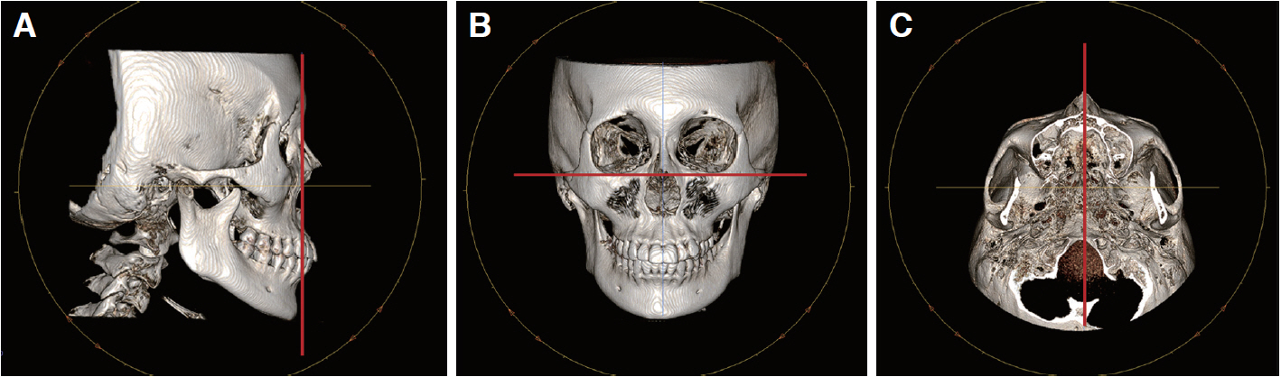
Fig. 1 Reorientation of axis. (A) Sagittal view, (B) Coronal view, (C) Axial view.
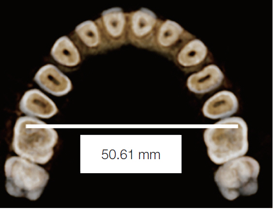
Fig. 2 Inter-maxillary first molar width.
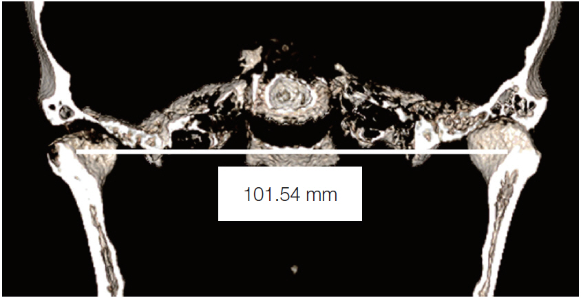
Fig. 3 Inter-condylar width.
Reference
-
References
1. Watt DM, McGregor AR. 1986. Designing complete denture. 2nd ed. WB Saunders Co.;Philadelphia: p. 22–8.2. Pound E. 1957; Recapturing esthetic tooth position in the edentulous patient. J Am Dent Assoc. 55:181–91. DOI: 10.14219/jada.archive.1957.0171. PMID: 13448859.3. Lang BR. 2004; Complete denture occlusion. Dent Clin North Am. 48:641–65. DOI: 10.1016/j.cden.2004.03.006. PMID: 15261798.4. Debnath N, Gupta R, Meenakshi A, Kumar S, Hota S, Rawat P. 2014; Relationship of inter-condylar distance with inter-dental distance of maxillary arch and occlusal vertical dimension: a clinical anthropometric study. J Clin Diagn Res. 8:ZC39–43. DOI: 10.7860/JCDR/2014/10194.5289. PMID: 25654029. PMCID: PMC4316335.5. Lee KY, Lee DJ. 1993; A comparative study of teeth and dental arch of Korean and Caucasian. Oral Biol Res. 71:1–15. DOI: 10.1043/0003-3219(2001)071<0195:ACSOCA>2.0.CO;2. PMID: 11407772.6. Song WC, Yun KH, Koh KS. 2003; Facial flatness of Korean: using facial depth. Korean J Anat. 36:499–506.7. Kook YA, Nojima K, Moon HB, McLaughlin RP, Sinclair PM. 2004; Comparison of arch forms between Korean and north American white populations. Am J Orthod Dentofacial Orthop. 126:680–6. DOI: 10.1016/j.ajodo.2003.10.038. PMID: 15592215.8. Hwang HS, Kim WS, McNamara JA Jr. 2002; Ethnic differences in the soft tissue profile of Korean and European-American adults with normal occlusions and well-balanced faces. Angle Orthod. 72:72–80. DOI: 10.1043/0003-3219(2002)072<0072:EDITST>2.0.CO;2. PMID: 11843277.9. Ferrario VF, Sforza C, Miani A Jr, Colombo A, Tartaglia G. 1992; Mathematical definition of the curve of Spee in permanent healthy dentitions in man. Arch Oral Biol. 37:691–4. DOI: 10.1016/0003-9969(92)90073-H. PMID: 1417519.10. Ferrario VF, Sforza C, Miani A Jr, Tartaglia G. 1994; Mathematical definition of the shape of dental arches in human permanent healthy dentitions. Eur J Orthod. 16:287–94. DOI: 10.1093/ejo/16.4.287. PMID: 7957653.11. Bayome M, Park JH, Kook YA. 2013; New three-dimensional cephalometric analyses among adults with a skeletal Class I pattern and normal occlusion. Korean J Orthod. 43:62–73. DOI: 10.4041/kjod.2013.43.2.62. PMID: 23671831. PMCID: PMC3650215.12. Alam MK, Shahid F, Purmal K, Ahmad B, Khamis MF. 2014; Tooth size and dental arch dimension measurement through cone beam computed tomography: Effect of age and gender. Res J Recent Sci. 3:85–94.13. Naji P, Alsufyani NA, Lagravere MO. 2014; Reliability of anatomic structures as landmarks in three-dimensional cephalometric analysis using CBCT. Angle Orthod. 84:762–72. DOI: 10.2319/090413-652.1. PMID: 24364751.14. Boucher CO. 1964. Swenson's complete dentures. 5th ed. Mosby Company;St. Louis: p. 246–51.15. Lunquist DO, Luther WW. 1970; Occlusal plane determination. J Prosthet Dent. 23:489–98. DOI: 10.1016/0022-3913(70)90198-8. PMID: 4908801.16. Boucher CO. 1974. Current clinical dental terminology. 2nd ed. Mosby Company;St. Louis: p. 229.17. Lee SH. 2007. Fixed prosthodontics. 2nd ed. Jeeseung Publisuing co.;Seoul: p. 102.18. Keshvad A, Winstanley RB, Hooshmand T. 2000; Intercondylar width as a guide to setting up complete denture teeth. J Oral Rehabil. 27:217–26. DOI: 10.1046/j.1365-2842.2000.00509.x. PMID: 10784334.19. Taylor R. 1990; Interpretation of the correlation coefficient: A basic review. JDMS. 6:35–9. DOI: 10.1177/875647939000600106.
- Full Text Links
-
- Actions
-
Cited
- CITED
-
- Close
- Share
-
- Similar articles
-
- An evaluation of the adequacy of PONT's index
- A study on maxillary basal bone morphology in skeletal Class III malocclusion requiring orthognathic surgery
- Model analysis in orthodontic treatment changes of the maxillary dental arch
- Short-term treatment effects produced by rapid maxillary expansion evaluated with computed tomography: A systematic review with meta-analysis
- A study of golden proportion application in Korean normal dentition
