Korean J Orthod.
2020 Sep;50(5):314-323. 10.4041/kjod.2020.50.5.314.
Short-term treatment effects produced by rapid maxillary expansion evaluated with computed tomography: A systematic review with meta-analysis
- Affiliations
-
- 1Department of Medical-Surgical Specialties, Section of Orthodontics, School of Dentistry, University of Catania, Policlinico Universitario V. Emanuele, Catania, Italy
- 2Department of Biomedical and Dental Sciences and Morphofunctional Imaging, Section of Orthodontics, School of Dentistry, University of Messina, Policlinico Universitario G. Martino, Messina, Italy
- KMID: 2506466
- DOI: http://doi.org/10.4041/kjod.2020.50.5.314
Abstract
Objective
To identify the available evidence on the effects of rapid maxillary expansion (RME) with three-dimensional imaging and provide meta-analytic data from studies assessing the outcomes using computed tomography.
Methods
Eleven electronic databases were searched, and prospective case series were selected. Two authors screened all titles and abstracts and assessed full texts of the remaining articles. Seventeen case series were included in the quantitative synthesis. Seven outcomes were investigated: nasal cavity width, maxillary basal bone width, alveolar buccal crest width, alveolar palatal crest width, inter-molar crown width, inter-molar root apex width, and buccopalatal molar inclination. The outcomes were investigated at two-time points: postexpansion (2–6 weeks) and post-retention (4–8 months). Mean differences and 95% confidence intervals were used to summarize and combine the data.
Results
All the investigated outcomes showed significant differences postexpansion (maxillary basal bone width, +2.46 mm; nasal cavity width, +1.95 mm; alveolar buccal crest width, +3.90 mm; alveolar palatal crest width, +3.09 mm; intermolar crown width, +5.69 mm; inter-molar root apex width, +2.85 mm; and dental tipping, +3.75°) and post-retention (maxillary basal bone width, +2.21 mm; nasal cavity width, +1.55 mm; alveolar buccal crest width, +3.57 mm; alveolar palatal crest width, +3.32 mm; inter-molar crown width, +5.43 mm; inter-molar root apex width, +4.75 mm; and dental tipping, 2.22°) compared to pre-expansion.
Conclusions
After RME, skeletal expansion of the nasomaxillary complex was greater in most caudal structures. Maxillary basal bone showed 10% post-retention relapse. During retention period, uprighting of maxillary molars occurred.
Figure
-
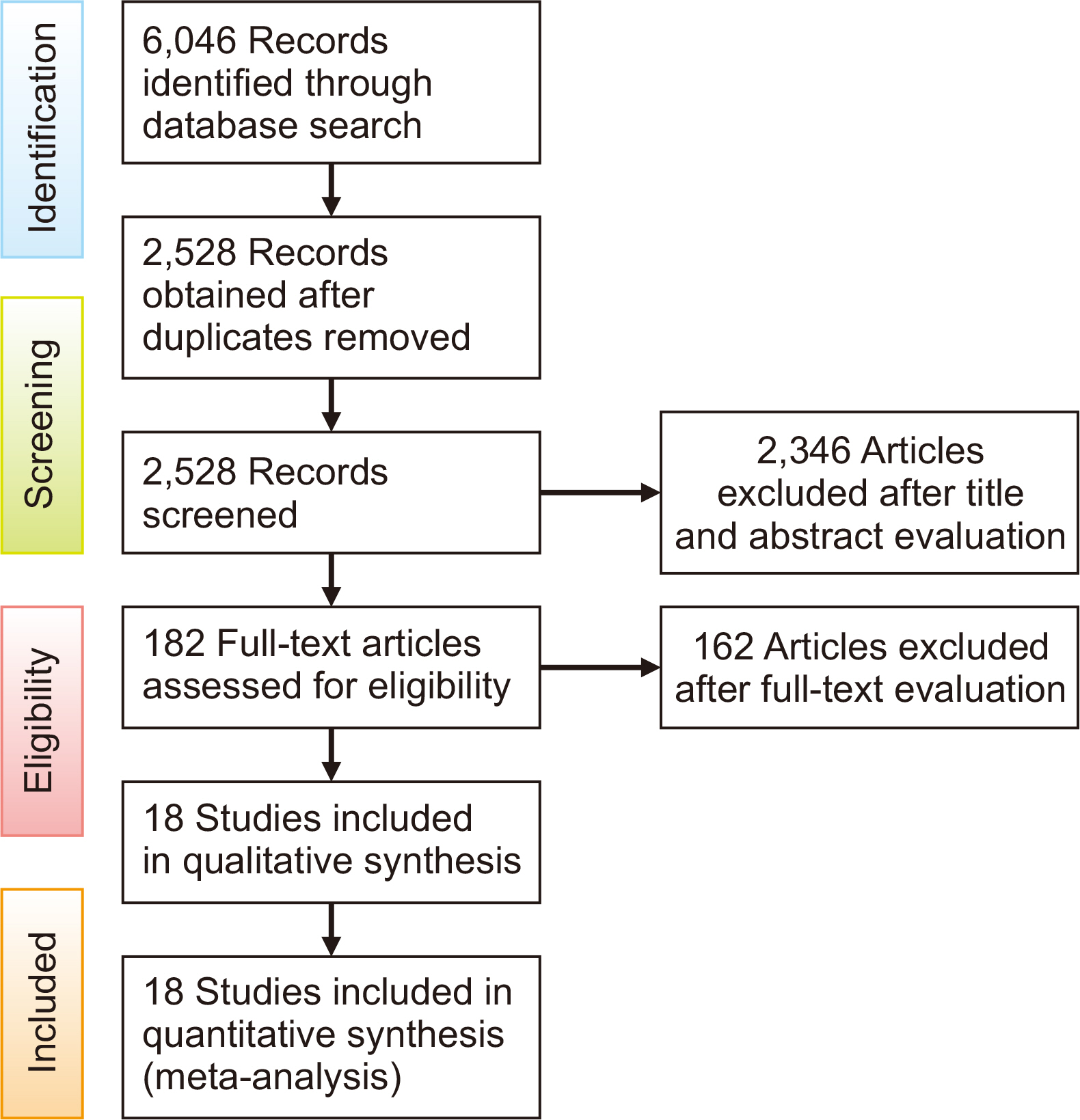
Figure 1 Flow diagram of included studies according to the PRISMA guidelines.
Reference
-
1. Angell DH. 1860; Treatment of irregularity of the permanent or adult teeth. Dent Cosmos. 1:540–4.2. Haas AJ. 1970; Palatal expansion: just the beginning of dentofacial orthopedics. Am J Orthod. 57:219–55. DOI: 10.1016/0002-9416(70)90241-1. PMID: 5263785.
Article3. Lo Giudice A, Fastuca R, Portelli M, Militi A, Bellocchio M, Spinuzza P, et al. 2017; Effects of rapid vs slow maxillary expansion on nasal cavity dimensions in growing subjects: a methodological and reproducibility study. Eur J Paediatr Dent. 18:299–304. DOI: 10.23804/ejpd.2017.18.04.07. PMID: 29380616.4. Lo Giudice A, Barbato E, Cosentino L, Ferraro CM, Leonardi R. 2018; Alveolar bone changes after rapid maxillary expansion with tooth-born appliances: a systematic review. Eur J Orthod. 40:296–303. DOI: 10.1093/ejo/cjx057. PMID: 29016774.
Article5. Wertz RA. 1970; Skeletal and dental changes accompanying rapid midpalatal suture opening. Am J Orthod. 58:41–66. DOI: 10.1016/0002-9416(70)90127-2. PMID: 5269181.
Article6. Leonardi R, Annunziata A, Caltabiano M. 2008; Landmark identification error in posteroanterior cephalometric radiography. A systematic review. Angle Orthod. 78:761–5. DOI: 10.2319/0003-3219(2008)078[0761:LIEIPC]2.0.CO;2. PMID: 18302479.7. Timms DJ, Preston CB, Daly PF. 1982; A computed tomographic assessment of maxillary movement induced by rapid expansion - a pilot study. Eur J Orthod. 4:123–7. DOI: 10.1093/ejo/4.2.123. PMID: 7049711.
Article8. Podesser B, Williams S, Bantleon HP, Imhof H. 2004; Quantitation of transverse maxillary dimensions using computed tomography: a methodological and reproducibility study. Eur J Orthod. 26:209–15. DOI: 10.1093/ejo/26.2.209. PMID: 15130045.
Article9. Ballanti F, Lione R, Fanucci E, Franchi L, Baccetti T, Cozza P. 2009; Immediate and post-retention effects of rapid maxillary expansion investigated by computed tomography in growing patients. Angle Orthod. 79:24–9. DOI: 10.2319/012008-35.1. PMID: 19123695.
Article10. Baratieri C, Nojima LI, Gonçalves Nojima M. Gomes de Souza MM. 2010; Transverse effects of rapid maxillary expansion in Class II malocclusion patients: a Cone-Beam Computed Tomography study. Dental Press J Orthod. 15:89–97. DOI: 10.1590/S2176-94512010000500011.11. Baysal A, Veli I, Ucar FI, Eruz M, Ozer T, Uysal T. 2011; Changes in mandibular transversal arch dimensions after rapid maxillary expansion procedure assessed through Cone-Beam Computed Tomography. Korean J Orthod. 41:200–10. DOI: 10.4041/kjod.2011.41.3.200.
Article12. Christie KF, Boucher N, Chung CH. 2010; Effects of bonded rapid palatal expansion on the transverse dimensions of the maxilla: a Cone-Beam Computed Tomography study. Am J Orthod Dentofacial Orthop. 137(4 Suppl):S79–85. DOI: 10.1016/j.ajodo.2008.11.024. PMID: 20381765.
Article13. Cordasco G, Nucera R, Fastuca R, Matarese G, Lindauer SJ, Leone P, et al. 2012; Effects of orthopedic maxillary expansion on nasal cavity size in growing subjects: a low dose computer tomography clinical trial. Int J Pediatr Otorhinolaryngol. 76:1547–51. DOI: 10.1016/j.ijporl.2012.07.008. PMID: 22840779.
Article14. Dias LS. 2010. Avaliação pós-expansão rápida da maxila com aparelhos do tipo Haas e Hyrax por meio de tomografia computadorizada cone beam [MD thesis]. Pontifícia Universidade Católica do Rio Grande do Sul;Porto Alegre:
Article15. Dogra N, Sidhu MS, Dabas A, Grover S, Gupta M. 2016; Cone-Beam Computed Tomography evaluation of dental, skeletal, and alveolar bone changes associated with bonded rapid maxillary expansion. J Indian Orthod Soc. 50:19–25. DOI: 10.4103/0301-5742.175709.
Article16. Görgülü S, Gokce SM, Olmez H, Sagdic D, Ors F. 2011; Nasal cavity volume changes after rapid maxillary expansion in adolescents evaluated with 3-dimensional simulation and modeling programs. Am J Orthod Dentofacial Orthop. 140:633–40. DOI: 10.1016/j.ajodo.2010.12.020. PMID: 22051483.
Article17. Helmkamp ME. 2016. Three-dimensional evaluation of implant-supported rapid maxillary expansion vs. traditional tooth-borne rapid maxillary expansion using Cone-Beam Computed Tomography [MD thesis]. St. Louis University;St. Louis:18. Kanomi R, Deguchi T, Kakuno E, Takano-Yamamoto T, Roberts WE. 2013; CBCT of skeletal changes following rapid maxillary expansion to increase arch-length with a development-dependent bonded or banded appliance. Angle Orthod. 83:851–7. DOI: 10.2319/082012-669.1. PMID: 23488528.
Article19. Li L, Qi S, Wang H, Ren S, Ban J. 2015; Cone-Beam Computed Tomography analysis of effects of rapid maxillary expansion on cranio maxillo-facial bones and upper airway. Honghua Xi Kou Qiang Yi Xue Za Zhi. 50:403–7.20. Luebbert J, Ghoneima A, Lagravère MO. 2016; Skeletal and dental effects of rapid maxillary expansion assessed through three-dimensional imaging: A multicenter study. Int Orthod. 14:15–31. DOI: 10.1016/j.ortho.2015.12.013. PMID: 26850998.
Article21. Martins LP. 2011. Análise dos resultados do tratamento da mordida cruzada posterior funcional com o expansor fixo hyrax [MD thesis]. Universidade Estadual Paulista;San Pablo:22. Mosleh MI, Kaddah MA, Abd ElSayed FA, ElSayed HS. 2015; Comparison of transverse changes during maxillary expansion with 4-point bone-borne and tooth-borne maxillary expanders. Am J Orthod Dentofacial Orthop. 148:599–607. DOI: 10.1016/j.ajodo.2015.04.040. PMID: 26432315.
Article23. Pangrazio-Kulbersh V, Wine P, Haughey M, Pajtas B, Kaczynski R. 2012; Cone Beam Computed Tomography evaluation of changes in the naso-maxillary complex associated with two types of maxillary expanders. Angle Orthod. 82:448–57. DOI: 10.2319/072211-464.1. PMID: 22032536.
Article24. Podesser B, Williams S, Crismani AG, Bantleon HP. 2007; Evaluation of the effects of rapid maxillary expansion in growing children using computer tomography scanning: a pilot study. Eur J Orthod. 29:37–44. DOI: 10.1093/ejo/cjl068. PMID: 17290015.
Article25. Rocco MA. 2012. Avaliação dos efeitos da expansão rápida da maxila no volume aéreo nasal, por meio da tomografia computadorizada de feixe cônico [PhD dissertation]. Universidade Estadual Paulista;San Pablo:
Article26. Weissheimer A, de Menezes LM, Mezomo M, Dias DM, de Lima EM, Rizzatto SM. 2011; Immediate effects of rapid maxillary expansion with c-type and hyrax-type expanders: a randomized clinical trial. Am J Orthod Dentofacial Orthop. 140:366–76. DOI: 10.1016/j.ajodo.2010.07.025. PMID: 21889081.27. Schiffman PH, Tuncay OC. 2001; Maxillary expansion: a meta analysis. Clin Orthod Res. 4:86–96. DOI: 10.1034/j.1600-0544.2001.040205.x. PMID: 11553090.
Article28. Lagravère MO, Heo G, Major PW, Flores-Mir C. 2006; Meta-analysis of immediate changes with rapid maxillary expansion treatment. J Am Dent Assoc. 137:44–53. DOI: 10.14219/jada.archive.2006.0020. PMID: 16456998.
Article29. Zhou Y, Long H, Ye N, Xue J, Yang X, Liao L, et al. 2014; The effectiveness of non-surgical maxillary expansion: a meta-analysis. Eur J Orthod. 36:233–42. DOI: 10.1093/ejo/cjt044. PMID: 23828862.
Article30. Liberati A, Altman DG, Tetzlaff J, Mulrow C, Gøtzsche PC, Ioannidis JP, et al. 2009; The PRISMA statement for reporting systematic reviews and meta-analyses of studies that evaluate health care interventions: explanation and elaboration. J Clin Epidemiol. 62:e1–34. DOI: 10.1016/j.jclinepi.2009.06.006. PMID: 19631507.
Article31. Korn EL, Baumrind S. 1990; Transverse development of the human jaws between the ages of 8.5 and 15.5 years, studied longitudinally with use of implants. J Dent Res. 69:1298–306. DOI: 10.1177/00220345900690061501. PMID: 2355125.
Article32. Chambers D, Rodgers M, Woolacott N. 2009; Not only randomized controlled trials, but also case series should be considered in systematic reviews of rapidly developing technologies. J Clin Epidemiol. 62:1253–60.e4. DOI: 10.1016/j.jclinepi.2008.12.010. PMID: 19349144.
Article33. Dalziel K, Round A, Stein K, Garside R, Castelnuovo E, Payne L. 2005; Do the findings of case series studies vary significantly according to methodological characteristics? Health Technol Assess. 9:iii–iv. 1–146. DOI: 10.3310/hta9020. PMID: 15588556.
Article34. Fitzpatrick-Lewis D, Ciliska D, Thomas H. 2009. The methods for the synthesis of studies without control groups. National Collaborating Centre for Methods and Tools;Hamilton:35. Guo B, Moga C, Harstall C, Schopflocher D. 2016; A principal component analysis is conducted for a case series quality appraisal checklist. J Clin Epidemiol. 69:199–207.e2. DOI: 10.1016/j.jclinepi.2015.07.010. PMID: 26307459.
Article36. Guyatt GH, Oxman AD, Vist GE, Kunz R, Falck-Ytter Y, Alonso-Coello P, et al. 2008; GRADE: an emerging consensus on rating quality of evidence and strength of recommendations. BMJ. 336:924–6. DOI: 10.1136/bmj.39489.470347.AD. PMID: 18436948. PMCID: PMC2335261.
Article37. Newman MG, Weyant R, Hujoel P. 2007; JEBDP improves grading system and adopts strength of recommendation taxonomy grading (SORT) for guidelines and systematic reviews. J Evid Based Dent Pract. 7:147–50. DOI: 10.1016/j.jebdp.2007.09.014. PMID: 18155075.38. Baratieri C, Bolognese AM, Nojima MC, Nojima LI. 2014; Changes in skeletal and dental relationship in Class II Division I malocclusion after rapid maxillary expansion: a prospective study. Dental Press J Orthod. 19:75–81. DOI: 10.1590/2176-9451.19.3.075-081.oar. PMID: 25162569.
Article39. Brandt S. 1973; JCO interviews Dr. Andrew J. Haas. J Clin Orthod. 7:227–34.
- Full Text Links
-
- Actions
-
Cited
- CITED
-
- Close
- Share
-
- Similar articles
-
- A meta analysis of maxillary expansion: comparisons of intercanine/intermolar expansion and rapid/slow expansion
- Effectiveness of miniscrew assisted rapid palatal expansion using cone beam computed tomography: A systematic review and meta-analysis
- A posteroanterior cephalometric study on the change of maxilla by rapid palatal expansion
- A study on the effect of rapid maxillary expansion and its relapse
- Does mini-implant-supported rapid maxillary expansion cause less root resorption than traditional approaches? A micro-computed tomography study
