Implant-assisted removable partial denture using freely removable abutment in a fully edentulous patient: A case report
- Affiliations
-
- 1Department of Prosthodontics, School of Dentistry, Pusan National University, Yangsan, Republic of Korea. neoplasia96@hanmail.net
- KMID: 2469922
- DOI: http://doi.org/10.4047/jkap.2020.58.1.58
Abstract
- Implant-Assisted Removable Partial Dentures (IARPDs) treatment is being performed in a fully edentulous patient using implant surveyed prosthesis as an abutment. Implant-supported prosthesis as an abutment of IARPDs is classified into screw-retained and cement-retained type according to the retention type, and each has advantages and disadvantages. The EZ crown system (Samwon DMP, Yangsan, Korea) has a cylinder combined with abutment, and the nickel-titanium spring in this cylinder provides a constant force on the zirconia ball to obtain retention in EZ crown system. In this patient, the natural abutment teeth of the mandibular overdenture was hopeless. We planned implant assisted removable partial denture using anterior implant surveyed prosthesis considering functional and esthetical rehabilitation, cost and patient's needs. When fabricating IARPDs using implant as abutment, we could compensate for the shortcomings of existing implant-supported prosthesis retention type and made the design of removable partial denture easy due to using EZ crown system.
Keyword
MeSH Terms
Figure
-
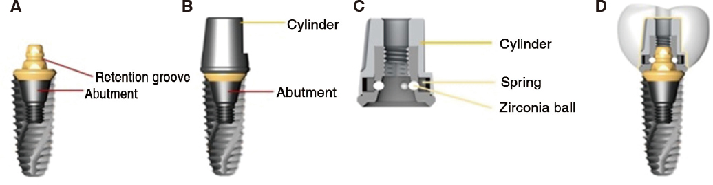
Fig. 1 Schematic Design of EZ crown system (Samwon DMP, Yangsan, Korea). (A) Abutment connected to implant fixture, (B) Cylinder connected to abutment, (C) Cylinder inner structure, (D) All components of EZ crown system connected.
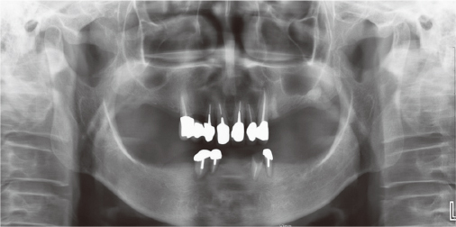
Fig. 2 Panoramic radiograph before treatment.
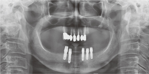
Fig. 3 Panoramic radiograph after implantation.
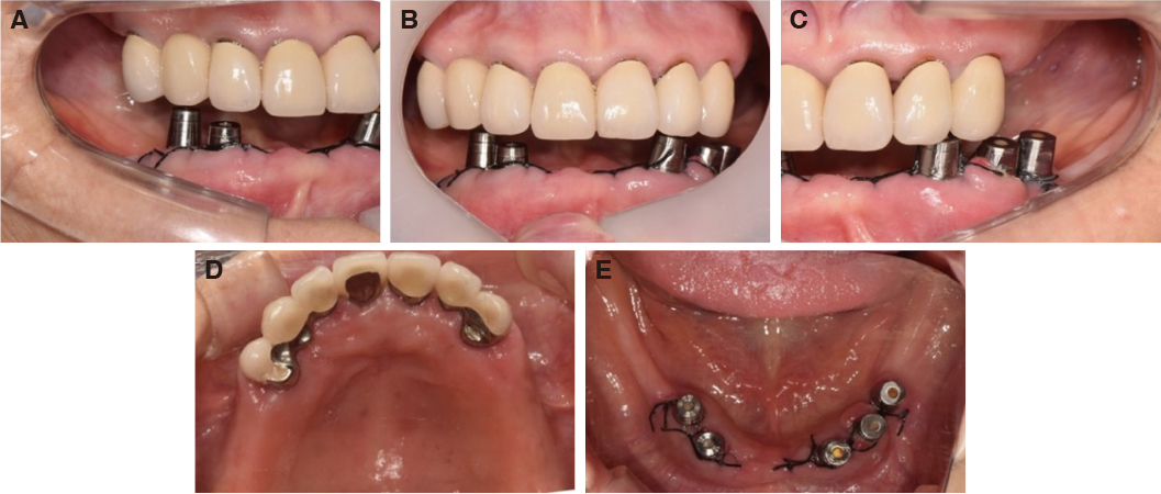
Fig. 4 Intraoral photographs after implantation. (A) Right view, (B) Frontal view, (C) Left view, (D) Occlusal view (Maxilla), (E) Occlusal view (Mandible).
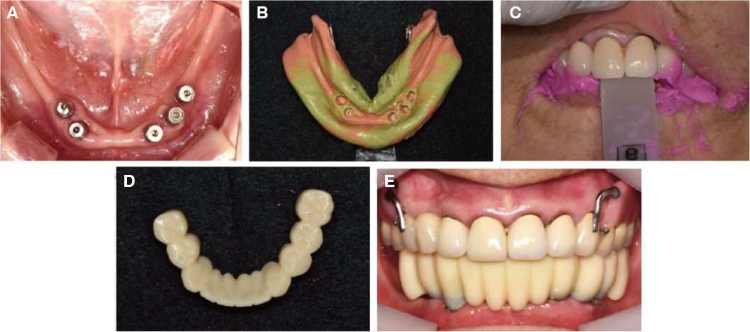
Fig. 5 Fabrication of provisional restoration. (A) Intraoral photographs after cap delivered (Occlusal view, Mandible), (B) Impression taking of mandible, (C) Registration of inter-occlusal relationship with leaf gauge, (D) Occlusal view of provisional restoration, (E) Frontal view of provisional restoration.
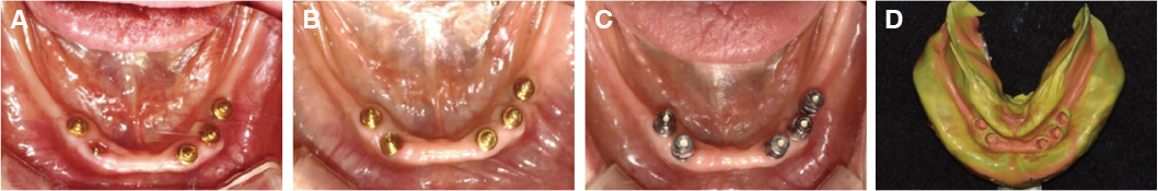
Fig. 6 Fabrication of implant surveyed prosthesis. (A) Occlusal view of abutment before gingivectomy, (B) Occlusal view of abutment after gingivectomy, (C) Occlusal view of cap delivered, (D) Final impression taking for implant surveyed prosthesis.
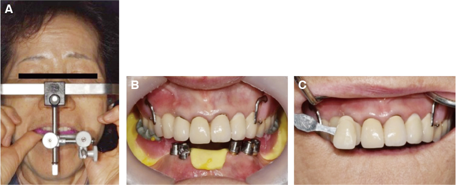
Fig. 7 Fabrication of implant surveyed prosthesis. (A) Facebow transfer, (B) Registration for inter-occlusal relationship with resin base and wax rim, (C) Shade taking.
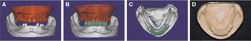
Fig. 8 Computer-aided design for zirconia implant surveyed prosthesis. (A) Frontal view, (B) Frontal view of implant surveyed prosthesis design, (C) Occlusal view of implant surveyed prosthesis design, (D) Occlusal view of implant surveyed prosthesis on working model.
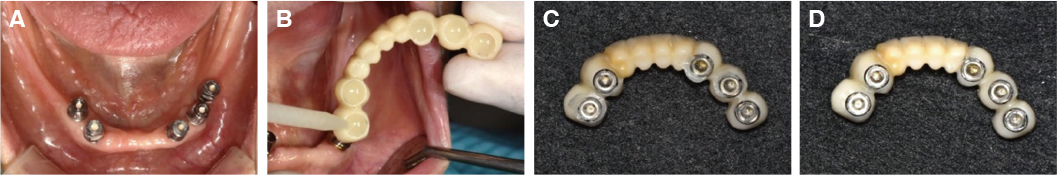
Fig. 9 Cementation process of implant surveyed prosthesis. (A) Occlusal view of cap attached, (B) Cementation (G-cem linkace), (C) Excess cement around cap, (D) Bottom surface of implant surveyed prosthesis after removing excess cement.
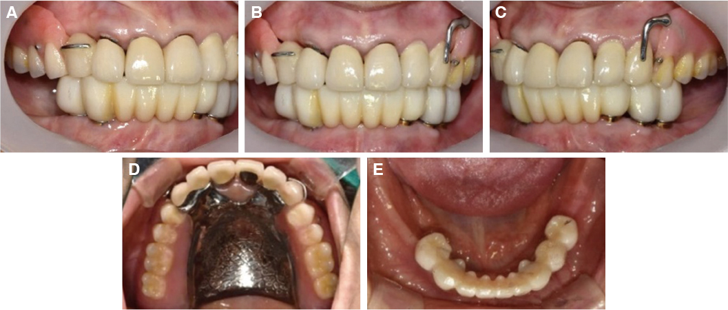
Fig. 10 Intraoral photographs after implant surveyed prosthesis placement. (A) Right view, (B) Frontal view, (C) Left view, (D) Occlusal view (Maxilla), (E) Occlusal view (Mandible).
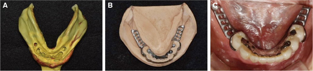
Fig. 11 Fabrication of metal framework. (A) Final impression for removable partial denture, (B) Metal framework on working model, (C) Intraoral try-in of metal framework.
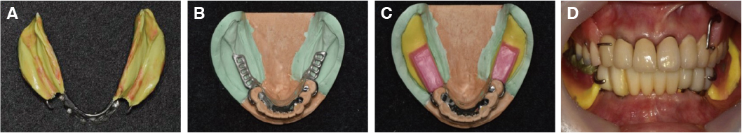
Fig. 12 (A) Functional impression for removable partial denture, (B) Altered cast fabrication, (C) Metal framework and occlusal rim, (D) Bite registration.
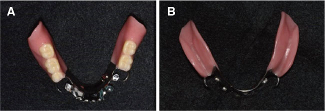
Fig. 13 Definitive prosthesis.
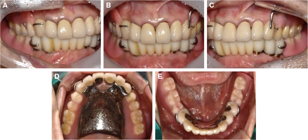
Fig. 14 Intraoral photographs after definitive prosthesis delivery. (A) Right view, (B) Frontal view, (C) Left view, (D) Occlusal view (Maxilla), (E) Occlusal view (Mandible).
Reference
-
1. Jorge JH, Quishida CC, Vergani CE, Machado AL, Pavarina AC, Giampaolo ET. Clinical evaluation of failures in removable partial dentures. J Oral Sci. 2012; 54:337–342.
Article2. Cho YJ, Suh BH. The use of an implant-supported fixed partial denture as abutment teeth for removable partial denture in edentulous patient: A cases report. Korean Acad Oral Maxillofac Implantol. 2017; 21:96–108.
Article3. Lee YJ, Bae EB, Jeong CM, Lee JJ, Jim JY, Huh JB. Removable partial denture using anterior implant-supported fixed prostheses for edentulous patients: A case report. J Korean Dent Sci. 2017; 10:87–95.4. Na HJ, Kang DW, Son MK. Distal-extension removable partial denture with anterior implant prostheses: Case report. J Dent Rehabil Appl Sci. 2011; 27:437–447.5. Pellecchia M, Pellecchia R, Emtiaz S. Distal extension mandibular removable partial denture connected to an anterior fixed implant-supported prosthesis: A clinical report. J Prosthet Dent. 2000; 83:607–612.
Article6. Kourtis SG, Sotiriadou S, Voliotis S, Challas A. Private practice results of dental implants. Part I: survival and evaluation of risk factors-Part II: surgical and prosthetic complications. Implant Dent. 2004; 13:373–385.
Article7. Phillips K, Wong KM. Space requirements for implant-retained bar-and-clip overdentures. Compend Contin Educ Dent. 2001; 22:516–518. 5205228. Wittneben JG, Joda T, Weber HP, Brägger U. Screw retained vs. cement retained implant-supported fixed dental prosthesis. Periodontol 2000. 2017; 73:141–151.
Article9. Kang SH, Kim SK, Heo SJ, Koak JY, Lee JH, Park JM. Clinical evaluation of the removable partial dentures with implant fixed prostheses. J Korean Acad Prosthodont. 2016; 54:239–245.
Article10. Choi JW, Choi KH, Chae HJ, Chae SK, Bae EB, Lee JJ, Lee SH, Jeong CM, Huh JB. Load-bearing capacity and retention of newly developed micro-locking implant prosthetic system: An in vitro pilot study. Materials (Basel). 2018; 11:564.
Article11. Choi JW, Lee JJ, Bae EB, Huh JB. Implant-supported fixed dental prosthesis with a microlocking implant prosthetic system: A clinical report. J Prosthet Dent. 2019; 05. S0022-3913(19)30014-9.
Article12. Choi JW, Song CH, Huh JB. Implant-supported fixed dental prostheses with new retention type using zirconia ball and nickel-titanium spring. Implantology. 2019; 23:16–24.
Article13. Wang TM, Leu LJ, Wang J, Lin LD. Effects of prosthesis materials and prosthesis splinting on peri-implant bone stress around implants in poor-quality bone: a numeric analysis. Int J Oral Maxillofac Implants. 2002; 17:231–237.14. Park JM, Koak JY, Kim SK, Alexander J, Heon SJ. Consideration for the combination treatment of removable partial denture and implant. Implantology. 2015; 19:104–111.15. Shahmiri R, Das R, Aarts JM, Bennani V. Finite element analysis of an implant-assisted removable partial denture during bilateral loading: occlusal rests position. J Prosthet Dent. 2014; 112:1126–1133.
Article16. Shahmiri R, Das R. Finite element analysis of implant-assisted removable partial dentures: Framework design considerations. J Prosthet Dent. 2017; 118:177–186.
Article17. Ryu BG, Choi WS, Cho HJ, Kim KK. The use of an implant fixed partial denture as abutment teeth for removable partial denture in unilateral edentulous patient: A cases report. Implantology. 2016; 20:24–34.18. Cochran DL. The evidence for immediate loading of implants. J Evid Based Dent Pract. 2006; 6:155–163.
Article19. Gallucci GO, Benic GI, Eckert SE, Papaspyridakos P, Schimmel M, Schrott A, Weber HP. Consensus statements and clinical recommendations for implant loading protocols. Int J Oral Maxillofac Implants. 2013; 08. 15.
Article
- Full Text Links
-
- Actions
-
Cited
- CITED
-
- Close
- Share
-
- Similar articles
-
- Implant-assisted removable partial denture restoration in a partially edentulous patient with a single remaining tooth: a case report
- Rehabilitation of maxillary partial edentulous patients using implant assisted removable partial denture
- Restoration of bilateral distal extension removable partial denture using a fixed implant prosthesis in unilateral partial edentulous patient: A case report
- The use of implant-assisted removable partial denture in the partially edentulous maxilla with a few unilateral remaining teeth and implant overdenture in the mandible: A case report
- Implant assisted removable partial denture with implant surveyed crown: A 20-month follow-up case report
