Comprehensive orthodontic treatment of a young girl with an odontogenic keratocyst and impacted teeth in the mandible
- Affiliations
-
- 1Department of Orthodontics, Yonsei University College of Dentistry, Seoul, Korea. selfexam@yuhs.ac
- 2Institute of Craniofacial Deformity, Yonsei University College of Dentistry, Seoul, Korea.
- KMID: 2468642
- DOI: http://doi.org/10.4041/kjod.2020.50.1.63
Abstract
- Odontogenic keratocysts (OKCs) are one of the most aggressive cysts in the oral and maxillofacial area because of their high recurrence rate and infiltrative behavior. In growing patients with OKCs, a radical treatment approach might cause numerous complications, including the disturbance of jaw growth and loss of the involved tooth. This case report describes successful comprehensive orthodontic treatment combined with marsupialization of the cyst in a young girl who exhibited an OKC with impacted teeth. The 10-year-old girl presented with an OKC extending from the mandibular symphysis through the left mandibular body, with ectopic impaction of the mandibular left canine and first premolar, as well as congenitally missing bilateral mandibular second premolars. Interestingly, spontaneous improvement of the positions of the ectopic impacted teeth, along with a reduction in the size of the cyst, occurred during marsupialization. The sequential use of removable and fixed appliances enabled orthodontic traction of the impacted teeth. The treatment outcome was stable at 2.5 years after the end of the treatment. We speculate that comprehensive orthodontic treatment combined with marsupialization can be an effective treatment strategy for patients with OKCs, especially when they are encountered in young, growing patients with impacted teeth.
MeSH Terms
Figure
-
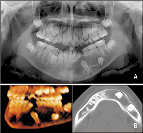
Figure 1 A, Panoramic radiograph acquired before marsupialization of the cyst. B, Pretreatment computed tomographic images acquired before marsupialization of the cyst.
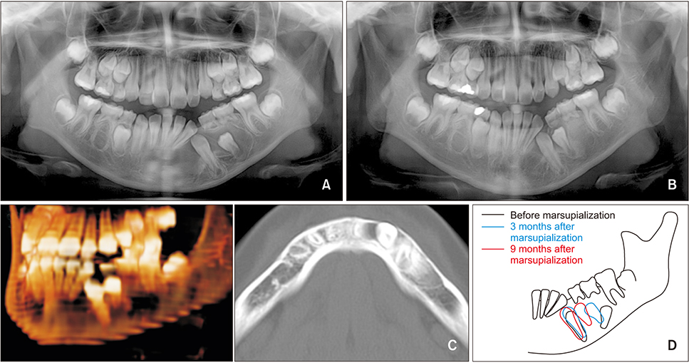
Figure 2 A, Panoramic radiographs acquired 3 months after the initiation of marsupialization of the cyst. B, Panoramic radiographs acquired 9 months after the initiation of marsupialization of the cyst. C, Computed tomographic images acquired 9 months after the initiation of marsupialization showing a reduction in the size of the cyst. D, Schematic illustration of the impacted teeth during marsupialization of the cyst.
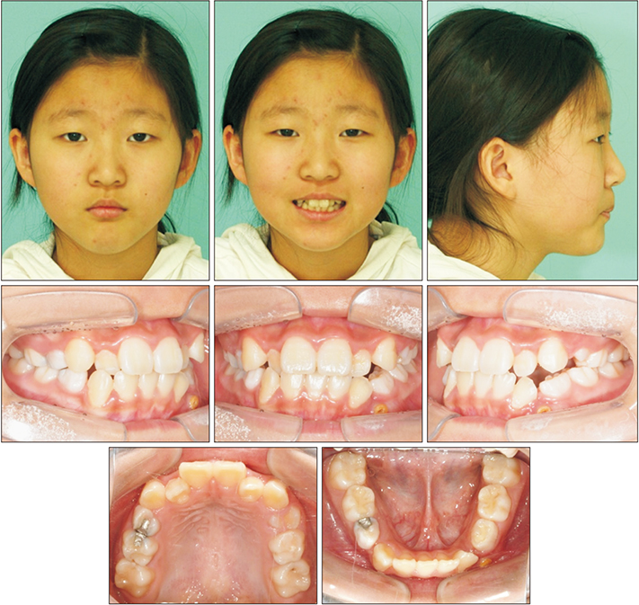
Figure 3 Pre-orthodontic treatment facial and intraoral photographs.
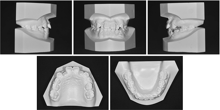
Figure 4 Pre-orthodontic treatment cast models.
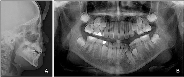
Figure 5 Pre-orthodontic treatment radiographs. A, Lateral cephalogram; B, panoramic radiograph.
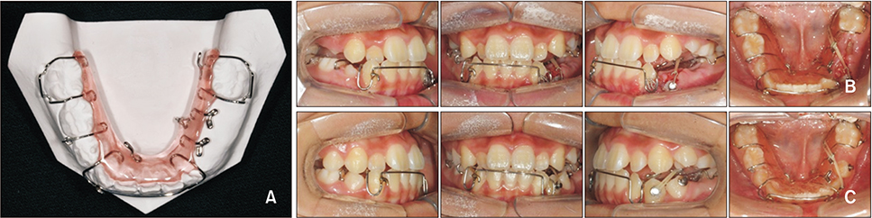
Figure 6 A, A removable appliance design. Six hooks were added to the appliance to control the direction of the force. B, Intraoral photographs showing treatment progress with a removable appliance. C, After 6 months of wearing the removable appliance.
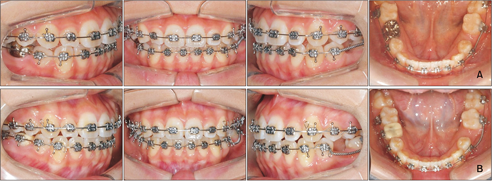
Figure 7 A, Intraoral photographs showing treatment progress with fixed orthodontic appliances. B, After correcting the axis of the distally tipped mandibular incisors and obtaining space for the restoration of the mandibular left second premolar.
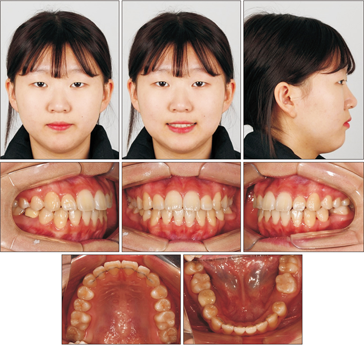
Figure 8 Posttreatment facial and intraoral photographs.
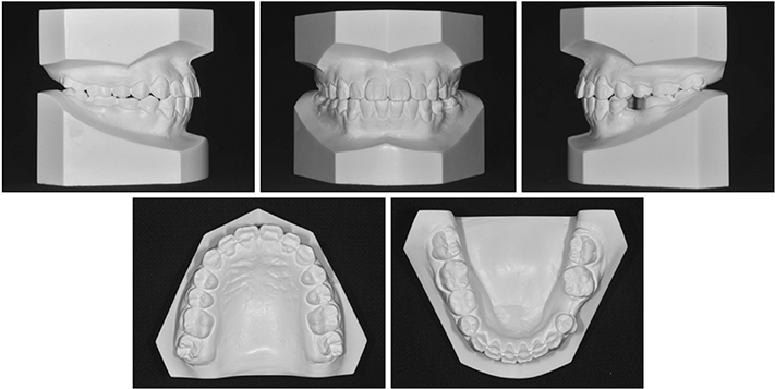
Figure 9 Posttreatment cast models.
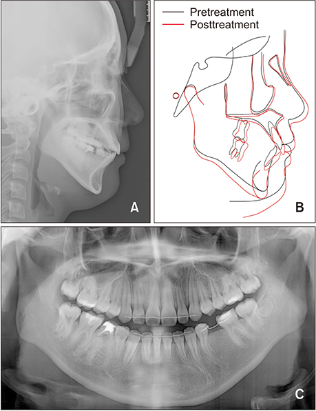
Figure 10 A, Posttreatment lateral cephalogram. B, Superimpositions before and after orthodontic treatment. C, Posttreatment panoramic radiograph.
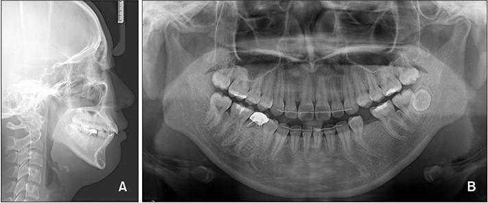
Figure 11 Post-retention radiographs acquired 2.5 years after debonding. A, Lateral cephalogram; B, panoramic radiograph.
Reference
-
1. Cha YH, Cho ES, Kang HE, Ko J, Nam W, Kim HJ, et al. Frequent oncogenic BRAF V600E mutation in odontogenic keratocyst. Oral Oncol. 2017; 74:62–67.
Article2. Chirapathomsakul D, Sastravaha P, Jansisyanont P. A review of odontogenic keratocysts and the behavior of recurrences. Oral Surg Oral Med Oral Pathol Oral Radiol Endod. 2006; 101:5–9. discussion 10.
Article3. Johnson NR, Gannon OM, Savage NW, Batstone MD. Frequency of odontogenic cysts and tumors: a systematic review. J Investig Clin Dent. 2014; 5:9–14.
Article4. Neville BW, Damm DD, Allen CM. Odontogenic cysts and tumors. In : Neville BW, Damm DD, Allen CM, Bouquot JE, editors. Oral and maxillofacial pathology. 2nd ed. Philadelphia, PA: WB Saunders Co.;2002. p. 590–610.5. Madras J, Lapointe H. Keratocystic odontogenic tumour: reclassification of the odontogenic keratocyst from cyst to tumour. J Can Dent Assoc. 2008; 74:165–165h.6. Maurette PE, Jorge J, de Moraes M. Conservative treatment protocol of odontogenic keratocyst: a preliminary study. J Oral Maxillofac Surg. 2006; 64:379–383.
Article7. Blanas N, Freund B, Schwartz M, Furst IM. Systematic review of the treatment and prognosis of the odontogenic keratocyst. Oral Surg Oral Med Oral Pathol Oral Radiol Endod. 2000; 90:553–558.
Article8. Boffano P, Ruga E, Gallesio C. Keratocystic odontogenic tumor (odontogenic keratocyst): preliminary retrospective review of epidemiologic, clinical, and radiologic features of 261 lesions from University of Turin. J Oral Maxillofac Surg. 2010; 68:2994–2999.
Article9. Hyun HK, Hong SD, Kim JW. Recurrent keratocystic odontogenic tumor in the mandible: a case report and literature review. Oral Surg Oral Med Oral Pathol Oral Radiol Endod. 2009; 108:e7–e10.
Article10. Berti Sde A, Pompermayer AB, Couto Souza PH, Tanaka OM, Westphalen VP, Westphalen FH. Spontaneous eruption of a canine after marsupialization of an infected dentigerous cyst. Am J Orthod Dentofacial Orthop. 2010; 137:690–693.
Article11. Nakamura N, Mitsuyasu T, Mitsuyasu Y, Taketomi T, Higuchi Y, Ohishi M. Marsupialization for odontogenic keratocysts: long-term follow-up analysis of the effects and changes in growth characteristics. Oral Surg Oral Med Oral Pathol Oral Radiol Endod. 2002; 94:543–553.
Article12. Deboni MC, Brozoski MA, Traina AA, Acay RR, Naclério-Homem Mda G. Surgical management of dentigerous cyst and keratocystic odontogenic tumor in children: a conservative approach and 7-year follow-up. J Appl Oral Sci. 2012; 20:282–285.
Article13. Morankar R, Bhatia SK, Goyal A, Gulia P. Conservative management of keratocystic odontogenic tumour in a young child with decompression and an intraoral appliance: 5-year follow-up. BMJ Case Rep. 2018; 2018:bcr-2017-221563.
Article14. Barnes L, Eveson JW, Reichart P, Sidransky D. WHO classification of tumours; pathology and genetics of head and neck tumours. Lyon: IARC Press;2005. p. 306–307.15. Wright JM, Vered M. Update from the 4th edition of the World Health Organization classification of head and neck tumours: odontogenic and maxillofacial bone tumors. Head Neck Pathol. 2017; 11:68–77.
Article16. Shear M. The aggressive nature of the odontogenic keratocyst: is it a benign cystic neoplasm? Part 1. Clinical and early experimental evidence of aggressive behaviour. Oral Oncol. 2002; 38:219–226.
Article17. Bataineh AB, al Qudah M. Treatment of mandibular odontogenic keratocysts. Oral Surg Oral Med Oral Pathol Oral Radiol Endod. 1998; 86:42–47.
Article18. Wilson C, Murphy M. Conservative management of multiple keratocystic odontogenic tumours in a child with Gorlin-Goltz syndrome: a case report. Eur J Paediatr Dent. 2008; 9:195–198.19. Miyawaki S, Hyomoto M, Tsubouchi J, Kirita T, Sugimura M. Eruption speed and rate of angulation change of a cyst-associated mandibular second premolar after marsupialization of a dentigerous cyst. Am J Orthod Dentofacial Orthop. 1999; 116:578–584.
Article20. Hu X, Zhao Y, Man QW, Li RF, Liu B, Zhao YF. The effects of marsupialization on bone regeneration adjacent to keratocystic odontogenic tumors, and the mechanisms involved. J Oral Sci. 2017; 59:475–481.
Article21. Wushou A, Zhao YJ, Shao ZM. Marsupialization is the optimal treatment approach for keratocystic odontogenic tumour. J Craniomaxillofac Surg. 2014; 42:1540–1544.
Article
- Full Text Links
-
- Actions
-
Cited
- CITED
-
- Close
- Share
-
- Similar articles
-
- A Case of Squamous Cell Carcinoma arising from an Odontogenic Keratocyst
- Orthodontic Traction of the Permanent Molar Using Skeletal Anchorage: A Case Report
- Orthodontic treatment of molar teeth impacted by local factors
- A Large Dentigerous Cyst Found in the Mandible
- Clinicoradiologic Differential Diagnosis of Odontogenic Keratocyst and Ameloblastoma
