Susceptibility-Weighted MR Imaging for the Detection of Isolated Cortical Vein Thrombosis in a Patient with Spontaneous Intracranial Hypotension
- Affiliations
-
- 1Department of Radiology, Gyeongsang National University Hospital and Gyeongsang National University School of Medicine, Jinju, Korea. choids@gnu.ac.kr
- KMID: 2468056
- DOI: http://doi.org/10.13104/imri.2019.23.4.381
Abstract
- Spontaneous intracranial hypotension (SIH) can be a rare risk factor of cerebral venous thrombosis. We describe a case of isolated cortical vein thrombosis (CVT) secondary to SIH and discuss the value of susceptibility-weighted imaging for the detection of isolated CVT.
Keyword
MeSH Terms
Figure
-
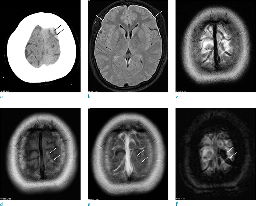
Fig. 1 An age 26 female with a headache. (a) The non-contrast CT scan shows a hyperdense cortical vein at the left cerebral vertex (arrows). (b) The axial fluid-attenuated inversion recovery image reveals small subdural fluid collection at both anterior cerebral convexities (arrows). (c) The T2-weighted image of the cerebral vertex shows no definite abnormality. (d) The T1-weighted image reveals a hyperintense lesion in the cortical vein of the left cerebral vertex (arrows). (e) On the contrast-enhanced T1-weighted image, the thrombosed cortical vein is not enhanced (arrows). (f) The susceptibility-weighted minimum intensity projection (minIP) image shows a blooming artifact due to thrombus in and beyond the cortical vein (arrows).
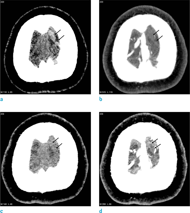
Fig. 2 An age 26 female with a headache. (a, b) The initial CT venography. (c, d) The follow-up CT venography, 10 days later. (a) The noncontrast CT scan shows a hyperdense cortical vein at the left cerebral vertex (arrows). (b) On the source image of the CT venography, the cortical vein is not opacified (arrows). (c) The follow-up non-contrast CT scan reveals the thrombosed cortical vein as iso-density to the cerebral cortex (arrows). (d) On the follow-up CT venography, there is a filling defect in the thrombosed cortical vein (arrows).
Reference
-
1. Coutinho JM, Gerritsma JJ, Zuurbier SM, Stam J. Isolated cortical vein thrombosis: systematic review of case reports and case series. Stroke. 2014; 45:1836–1838.2. Alvis-Miranda HR, Milena Castellar-Leones S, Alcala-Cerra G, Rafael Moscote-Salazar L. Cerebral sinus venous thrombosis. J Neurosci Rural Pract. 2013; 4:427–438.3. Jang J, Choi DS, Shin HS, et al. Clinical and radiological manifestations of cerebral venous thrombosis: are there any differences according to presence or absence of cortical vein involvement? Iran J Radiol. 2018; 15:e68250.4. Lee S, Cho SB, Choi DS, et al. Susceptibility vessel sign for the detection of hyperacute MCA occlusion: evaluation with susceptibility-weighted MR imaging. Investig Magn Reson Imaging. 2016; 20:105–113.5. Sinnaeve L, Vanopdenbosch L, Paemeleire K. Association of cerebral venous thrombosis and intracranial hypotension: review of 3 cases. J Stroke Cerebrovasc Dis. 2017; 26:e165–e169.6. Schievink WI, Maya MM. Cerebral venous thrombosis in spontaneous intracranial hypotension. Headache. 2008; 48:1511–1519.7. Seo H, Choi DS, Shin HS, Cho JM, Koh EH, Son S. Bone subtraction 3D CT venography for the evaluation of cerebral veins and venous sinuses: imaging techniques, normal variations, and pathologic findings. AJR Am J Roentgenol. 2014; 202:W169–W175.8. Costa P, Del Zotto E, Giossi A, et al. Headache due to spontaneous intracranial hypotension and subsequent cerebral vein thrombosis. Headache. 2012; 52:1592–1596.9. Yoon KW, Cho MK, Kim YJ, Lee SK. Sinus thrombosis in a patient with intracranial hypotension: a suggested hypothesis of venous stasis. a case report. Interv Neuroradiol. 2011; 17:248–225.10. Singh R, Cope WP, Zhou Z, De Witt ME, Boockvar JA, Tsiouris AJ. Isolated cortical vein thrombosis: case series. J Neurosurg. 2015; 123:427–433.11. Zhang D, Wang J, Zhang Q, He F, Hu X. Cerebral venous thrombosis in spontaneous intracranial hypotension: a report on 4 cases and a review of the literature. Headache. 2018; 58:1244–1255.12. Haacke EM, Mittal S, Wu Z, Neelavalli J, Cheng YC. Susceptibility-weighted imaging: technical aspects and clinical applications, part 1. AJNR Am J Neuroradiol. 2009; 30:19–30.
- Full Text Links
-
- Actions
-
Cited
- CITED
-
- Close
- Share
-
- Similar articles
-
- Spontaneous Intracranial Hypotension Complicated by Cortical Vein Thrombosis
- A Case of Spontaneous Intracranial Hypotension: Detection of Cerebrospinal Fluid Leakage by Early Dynamic Radionuclide Cisternography
- A Case of Cortical Vein Thrombosis in Wegener's Granulomatosis
- Spontaneous Intracranial Hypotension Secondary to Lumbar Disc Herniation
- Spontaneous Intracranial Hypotension Complicated by Cerebral Venous Thrombosis Relieved by Epidural Blood Patch
