Collison Tumor of Adenocarcinoma and Diffuse Large B-Cell Lymphoma in the Rectum: a Case Report and Literature Review
- Affiliations
-
- 1Departmet of Radiology, School of Medicine, Kyungpook National University, Kyungpook National University Hospital, Daegu, Korea. kimseehyung72@outlook.kr
- KMID: 2468055
- DOI: http://doi.org/10.13104/imri.2019.23.4.374
Abstract
- Collision tumor is a synchronous neoplasm wherein two histologically distinct tumors co-exist within the same anastomosis site. Collision tumor can occur in any organ, but the incidence is markedly rare. Additionally, preoperative diagnosis can be challenging to the radiologist. Herein, we report an age 60 male with collision tumor of rectal adenocarcinoma and diffuse large B-cell lymphoma, presented as a semi-annular wall thickening and bulky exophytic mass on MR imaging.
Keyword
MeSH Terms
Figure
-
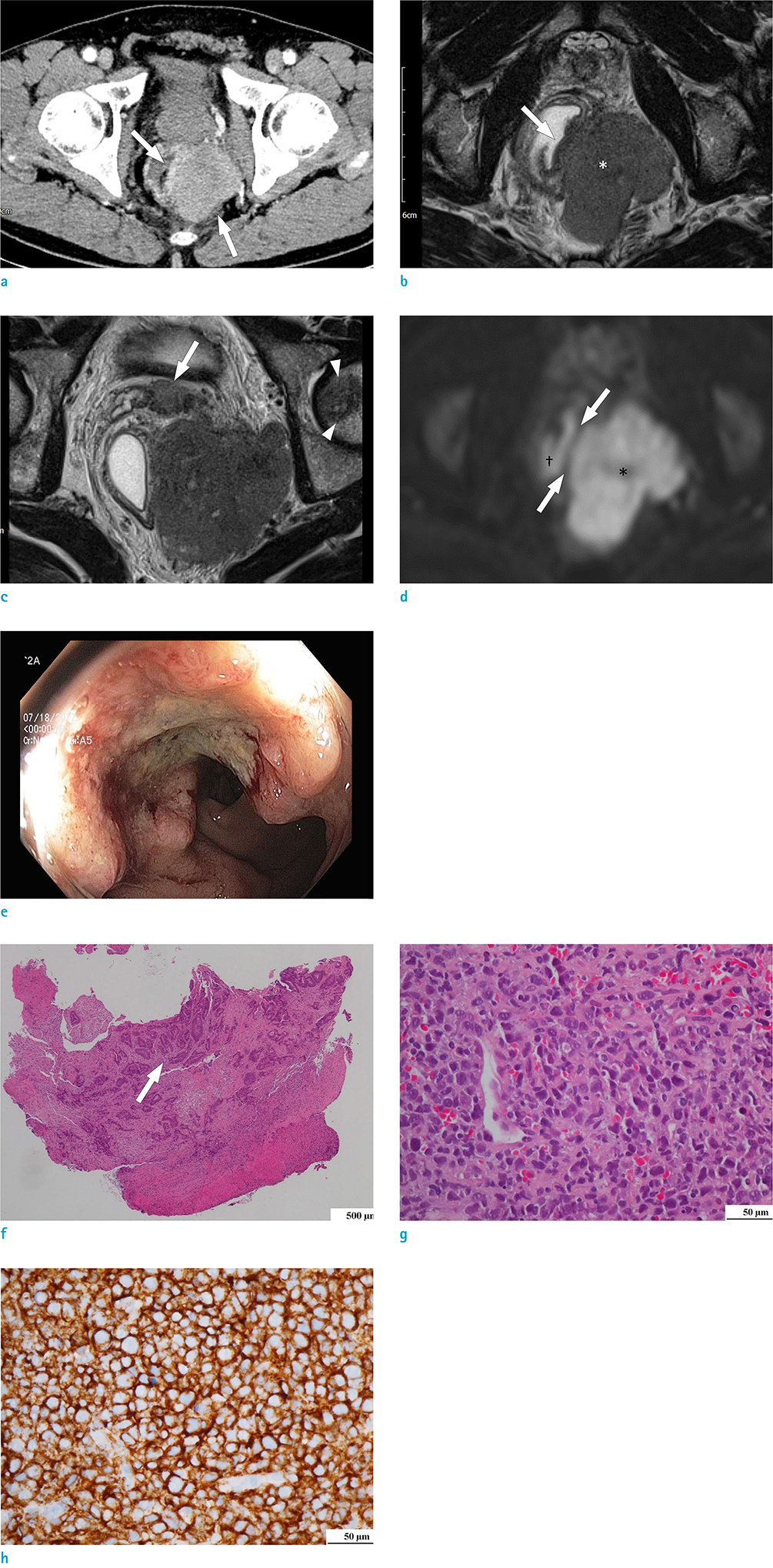
Fig. 1 An age 62 male with collision tumor of rectal adenocarcinoma and DLBCL. (a) Axial CT image obtained from portal venous phase shows a lobulated, relatively homogeneous enhancing mass (arrows) in the left mesorectum. The mass is compressing the distal rectum. (b) On a T2-weighted MR image, the mesorectal mass (asterisk) shows homogeneous, intermediate signal intensity. Additionally, there is a semi-annular wall thickening (arrow) in the left quadrant of the compressed distal rectum, which shows lower signal intensity than that of the mesorectal mass. (c) On a T2-weighted MR image, two nodular lesions, similar to the signal intensity of the mesorectal mass, are noted in the seminal vesicle (arrow) and the left femoral head (arrowheads). (d) On diffusion-weighted image, there is a linear area of non-restricted diffusion (arrows) between the semi-annular wall thickening (obelisk) and the mesorectal mass (asterisk). (e) On colonoscopy, the semi-annular wall thickening on MR image appears as an ulceroinfiltrative lesion with easy contact bleeding. (f) A photograph of colonoscopic biopsy specimen (Hematoxylin and Eosin [H&E] stanning) shows moderately differentiated adenocarcinoma with glandular formation (arrow). (g) H&E stanning from percutaneous needle biopsy specimen demonstrates an abnormal diffuse infiltrate of small to intermediate-sized lymphoid cells. (h) CD20 B-cell marker are positive.
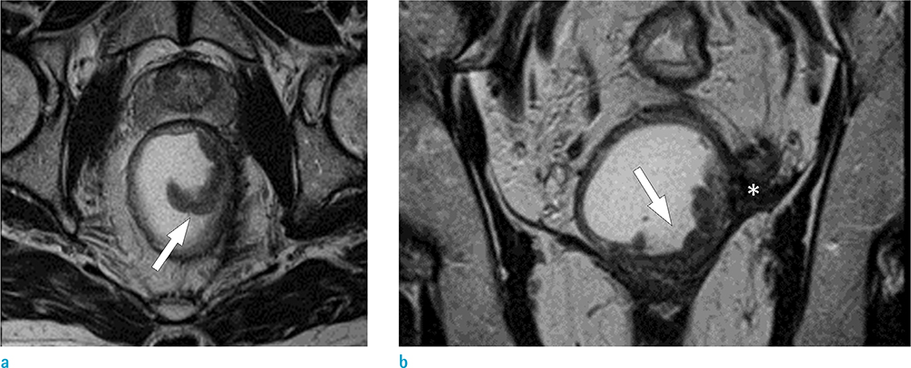
Fig. 2 MR images after six-cycle CHOP chemotherapy. (a, b) On a T2-weighted image, a semi-annular wall thickening (arrows) in the left quadrant of the distal rectum is noted. Previously seen mesorectal mass disappeared and remained as a thin low signal intensity lesion (asterisk), indicating a complete response.
Reference
-
1. Korea Central Cancer Registry. National Cancer Center Web site. Accessed November 6, 2019. http://ncc.re.kr.2. Kus T, Aktas G, Kalender ME, Sari I, Ulker E, Camci C. Collision tumor consisting of primary follicular lymphoma and adenocarcinoma in the cecum: a case report and literature review. Oncol Lett. 2016; 11:2801–2805.3. Sasaki S, Hatanaka K, Sahara N, et al. Collision tumor of primary malignant lymphoma and adenocarcinoma in the colon: report of a case. Surg Today. 2010; 40:975–981.4. Lin HH, Jiang JK, Lin JK. Collision tumor of low-grade B-cell lymphoma and adenocarcinoma with tuberculosis in the colon: a case report and literature review. World J Surg Oncol. 2014; 12:147.5. Lee DY, Hong SW, Chang YG, Lee WY, Lee B, Kang YK. Synchronous T-cell lymphoma in patient with colon cancer: a case report. J Korean Surg Soc. 2012; 83:60–64.6. Soto AR, Vazquez EG, Grigg-Gutierrez NM, Magno-Pagatzaurtundua P, Caceres W, Toro DH. Conundrum of a large bowel neoplasm: Collision tumor. ACG Case Rep J. 2018; 5:e13.7. Shigeno T, Fujimori K, Tsuruta F, Nozawa Y, Nagaya T, Maejima T. Ileocecal collision tumor composed of adenocarcinoma and primary malignant lymphoma. Clin J Gastroenterol. 2011; 4:79–84.8. Mannweiler S, Dinges HP, Beham-Schmid C, Hauser H, Starlinger M, Regauer S. Colliding / concomitant tumors of the intestine: report of 3 cases. Pathol Oncol Res. 2003; 9:188–192.9. Chang H, Chuang WY, Shih LY, Tang TC. Collision in the colon: concurrent adenocarcinoma and diffuse large B-cell lymphoma in the same tumour. Acta Clin Belg. 2011; 66:302–304.10. Miyamoto R, Kikuchi K, Uchida A, et al. Collision tumor consisting of a colorectal adenocarcinoma and dissemination of a gastric adenocarcinoma. SAGE Open Med Case Rep. 2018; 6:2050313X17751839.
- Full Text Links
-
- Actions
-
Cited
- CITED
-
- Close
- Share
-
- Similar articles
-
- A Case of Primary Rectal Diffuse Large B Cell LymphomaPresented as Multiple Polypoid Lesions
- Primary Diffuse Large B Cell Lymphoma Developing at the Ileocolonic Anastomosis Site after Right Hemicolectomy for Adenocarcinoma: A Case Report
- A Case of Advanced Gastric Adenocarcinoma with Synchronous Gastric Diffuse Large B Cell Lymphoma
- Diffuse Large B-Cell Lymphoma Transformed from a Rectal Mucosa-Associated Lymphoid Tissue Lymphoma
- Relapse of Ocular Lymphoma following Primary Testicular Diffuse Large B-cell Lymphoma
